. 2017 Sep 11;18(1):392.
doi: 10.1186/s12891-017-1752-5.

Plasminogen activator inhibitor-1 deficiency enhances subchondral osteopenia after induction of osteoarthritis in mice

Affiliations

Plasminogen activator inhibitor-1 deficiency enhances subchondral osteopenia after induction of osteoarthritis in mice

Akihiro Moritake et al. BMC Musculoskelet Disord. .

Abstract

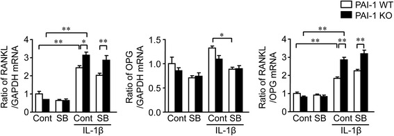
Background: Subchondral osteopenia is important for the pathophysiology of osteoarthritis (OA). Although previous studies suggest that plasminogen activator inhibitor-1 (PAI-1), an inhibitor of fibrinolysis, is related to bone metabolism, its role in OA remains unknown. We therefore investigated the roles of PAI-1 in the subchondral bone in OA model mice.

Methods: Wild type (WT) and PAI-1-deficient (KO) mice were ovariectomized (OVX), and then destabilization of the medial meniscus (DMM) surgery was performed.

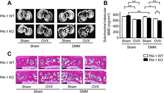
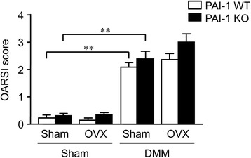
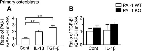
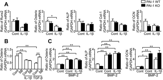
Results: DMM and OVX significantly decreased the trabecular bone mineral density of the subchondral bone evaluated by quantitative computed tomography in PAI-1 KO mice. The effects of OVX and/or PAI-1 deficiency on the OARSI score for the evaluation of the progression of knee degeneration were not significant. PAI-1 deficiency significantly augmented receptor activator nuclear factor κB ligand mRNA levels enhanced by IL-1β in mouse primary osteoblasts, although it did not affect osteoblast differentiation. Moreover, PAI-1 deficiency significantly increased osteoclast formation from mouse bone marrow cells.

Conclusion: We showed that PAI-1 deficiency accelerates the subchondral osteopenia after induction of OA in mice. PAI-1 might suppress an enhancement of bone resorption and subsequent subchondral osteopenia after induction of OA in mice.

Keywords: Osteoarthritis; Osteoclast; Ovariectomy; Plasminogen activator inhibitor-1; Subchondral bone.


Conflict of interest statement

Ethics approval

All animal experiments were approved by the Committee for the Care and Use of Laboratory Animals at Kindai University (KAME-28-19). All procedures for animal experiments were performed according to the guidelines of the National Institutes of Health and the institutional rules for the use and care of laboratory animals at Kindai University.

Consent for publication

Not applicable

Competing interests

The authors declare there are no competing interests.

Publisher’s Note

Springer Nature remains neutral with regard to jurisdictional claims in published maps and institutional affiliations.

Figures

Fig. 1
Fig. 1
Effects of DMM, OVX and PAI-1 deficiency on the BMD of the tibial subchondral bone in mice. a qCT images of the subchondral bone in WT and PAI-1 KO mice 8 weeks after DMM or sham surgery with or without OVX. b The trabecular BMD of the subchondral bone was assessed by qCT in WT and PAI-1 KO mice 8 weeks after DMM or sham surgery with or without OVX. Regions of interest were defined as 240-μm (10-slice) segments from 72 μm proximal to the end of the proximal growth plate towards the joint. Data represent the mean ± SEM of 10–11 mice. **p < 0.01 and *p < 0.05. c Hematoxylin and Eosin-stained sections from the subchondral bone in WT and PAI-1 KO mice 8 weeks after DMM or sham surgery with or without OVX. Scale bars = 200 μm
Fig. 2
Fig. 2
Effects of DMM, OVX and PAI-1 deficiency on articular cartilage degeneration in mice. Cartilage degeneration was assessed using the OARSI histological scoring system in WT and PAI-1 KO mice 8 weeks after DMM or sham surgery with or without OVX. Each knee was evaluated using 6 grades for the progression of arthropathy. Data represent the mean ± SEM of 10–11 mice. **p < 0.01
Fig. 3
Fig. 3
Effects of IL-1β on the expressions of PAI-1 and TGF-β1 in primary mouse osteoblasts. a Primary osteoblasts were obtained from 3-day-old female WT mice. Total RNA was extracted from primary osteoblasts cultured with or without 10 ng/mL IL-1β or 1 ng/ml TGF-β1 for 24 h. The levels of PAI-1 or GAPDH mRNA were assessed by real-time PCR. Data represent the mean ± SEM. n = 6 for each group. **p < 0.01. b Primary osteoblasts were obtained from 3-day-old female WT and PAI-1 KO mice. Total RNA was extracted from primary osteoblasts cultured with or without 10 ng/mL IL-1β for 24 h. Then, the levels of TGF-β1 or GAPDH mRNA were assessed by real-time PCR. Data represent the mean ± SEM. n = 6 for each group
Fig. 4
Fig. 4
Effects of PAI-1 deficiency on osteoblast differentiation in mouse osteoblasts and bone marrow cells. a Primary osteoblasts were obtained from 3-day-old female WT and PAI-1 KO mice. Total RNA was extracted from primary osteoblasts cultured with or without 10 ng/mL IL-1β or 1 ng/ml TGF-β1 for 24 h. The levels of Runx2, Osterix, ALP, Col-1, OCN or GAPDH mRNA were assessed by real-time PCR. Data represent the mean ± SEM. n = 6 for each group. *p < 0.05. b Primary osteoblasts were obtained from 3-day-old female WT mice. Total RNA was extracted from primary osteoblasts cultured with or without 10 ng/mL IL-1β or 1 ng/ml TGF-β1 in the presence or absence of 1 μM SB421542 for 24 h. The levels of Osterix or GAPDH mRNA were assessed by real-time PCR. Data represent the mean ± SEM. n = 6 for each group. **p < 0.01. c Bone marrow cells were obtained from 8-week-old female WT and PAI-1 KO mice. Total RNA was extracted from the bone marrow cells cultured with or without 200 ng/ml BMP-2 or 10 ng/mL IL-1β for 48 h. The levels of Osterix, ALP, OCN or GAPDH mRNA were assessed by real-time PCR. Data represent the mean ± SEM. n = 5 for each group. **p < 0.01 and *p < 0.05
Fig. 5
Fig. 5
Effects of PAI-1 deficiency on the expression of RANKL and OPG in mouse osteoblasts. Primary osteoblasts were obtained from 3-day-old female WT and PAI-1 KO mice. Total RNA was extracted from primary osteoblasts cultured with or without 10 ng/mL IL-1β or 1 ng/ml TGF-β1 in the presence or absence of 1 μM SB421542 for 24 h. The levels of RANKL, OPG or GAPDH mRNA were assessed by real-time PCR. Data represent the mean ± SEM. n = 6 for each group. **p < 0.01 and *p < 0.05
Fig. 6
Fig. 6
Effects of PAI-1 deficiency on osteoclast formation from mouse bone marrow cells. After bone marrow cells obtained from WT and PAI-1 KO mice cultured with M-CSF (50 ng/ml) for 3 days, cells were treated with or without IL-1β in the presence of M-CSF (50 ng/ml) and RANKL (50 ng/ml) for 3 days. Then, TRAP-positive MNCs were counted. Data represent the mean ± SEM. n = 6 for each group. **p < 0.01

Similar articles

Cited by

References

    1. Rangger C, Kathrein A, Klestil T, Glötzer W. Partial meniscectomy and osteoarthritis. Implications for treatment of athletes. Sports Med. 1997;23:61–68. doi: 10.2165/00007256-199723010-00006. - DOI - PubMed
    1. Srikanth VK, Fryer JL, Zhai G, Winzenberg TM, Hosmer D, Jones G. A meta-analysis of sex differences prevalence, incidence and severity of osteoarthritis. Osteoarthritis Cartilge. 2005;13:769–781. doi: 10.1016/j.joca.2005.04.014. - DOI - PubMed
    1. Helmick CG, Felson DT, Lawrence RC, Gabriel S, Hirsch R, Kwoh CK, et al. Estimates of the prevalence of arthritis and other rheumatic conditions in the United States. Part I. Arthritis Rheum. 2008;58:15–25. doi: 10.1002/art.23177. - DOI - PubMed
    1. Roman-Blas JA, Castañeda S, Largo R, Herrero-Beaumont G. Osteoarthritis associated with estrogen deficiency. Arthritis Res Ther. 2009;11:241. doi: 10.1186/ar2791. - DOI - PMC - PubMed
    1. Wang CJ, Huang CY, Hsu SL, Chen JH, Cheng JH. Extracorporeal shockwave therapy in osteoporotic osteoarthritis of the knee in rats; an experiment in animals. Arthritis Res Ther. 2014;16:R139. doi: 10.1186/ar4601. - DOI - PMC - PubMed