. 2017 Sep 11;16(1):151.
doi: 10.1186/s12943-017-0719-3.

Circular RNA_LARP4 inhibits cell proliferation and invasion of gastric cancer by sponging miR-424-5p and regulating LATS1 expression

Affiliations

Circular RNA_LARP4 inhibits cell proliferation and invasion of gastric cancer by sponging miR-424-5p and regulating LATS1 expression

Jing Zhang et al. Mol Cancer. .

Abstract

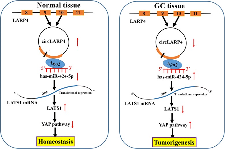
Background: Non-coding RNAs (ncRNAs) have been shown to regulate gene expression involved in tumor progression of multiple malignancies. Our previous studies indicated that large tumor suppressor kinase 1 (LATS1), a core part of Hippo signaling pathway, functions as a tumor suppressor in gastric cancer (GC). But, the underlying molecular mechanisms by which ncRNAs modulate LATS1 expression in GC remain undetermined.

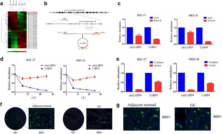
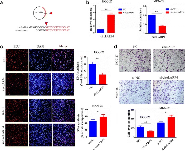
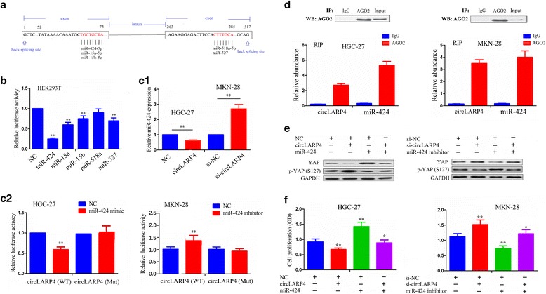
Methods: The correlation of LATS1 and has-miR-424-5p (miR-424) expression with clinicopathological characteristics and prognosis of GC patients was analyzed by TCGA RNA-sequencing data. A novel circular RNA_LARP4 (circLARP4) was identified to sponge miR-424 by circRNA expression profile and bioinformatic analysis. The binding site between miR-424 and LATS1 or circLARP4 was verified using dual luciferase assay and RNA immunoprecipitation (RIP) assay. The expression and localization of circLARP4 in GC tissues were investigated by fluorescence in situ hybridization (FISH). MTT, colony formation, Transwell and EdU assays were performed to assess the effects of miR-424 or circLARP4 on cell proliferation and invasion.

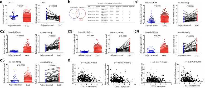
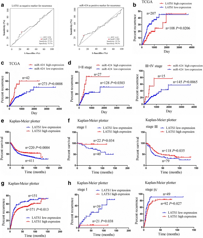
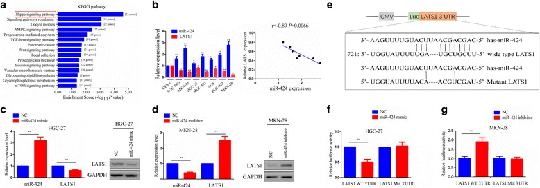
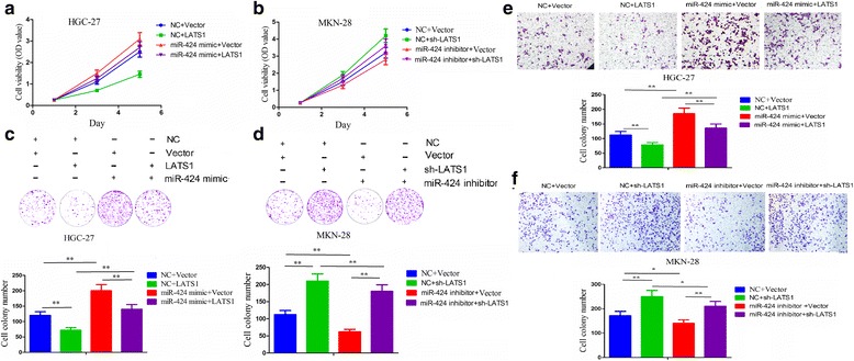
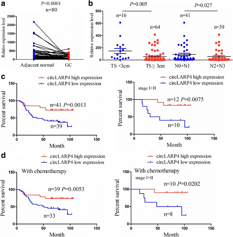
Results: Increased miR-424 expression or decreased LATS1 expression was associated with pathological stage and unfavorable prognosis of GC patients. Ectopic expression of miR-424 promoted proliferation and invasion of GC cells by targeting LATS1 gene. Furthermore, circLARP4 was mainly localized in the cytoplasm and inhibited biological behaviors of GC cells by sponging miR-424. The expression of circLARP4 was downregulated in GC tissues and represented an independent prognostic factor for overall survival of GC patients.

Conclusion: circLARP4 may act as a novel tumor suppressive factor and a potential biomarker in GC.

Keywords: Circular RNA_LARP4; Gastric cancer; Invasion; LATS1; miR-424-5p.


Conflict of interest statement

Ethics approval and consent to participate

The present study was approved by the Hospital’s Protection of Human Subjects Committee.

Competing interests

The authors declare that they have no competing interests.

Publisher’s Note

Springer Nature remains neutral with regard to jurisdictional claims in published maps and institutional affiliations.

Figures

Fig. 1
Fig. 1
MiR-424-5p was negatively correlated with LATS1 expression in GC. a TCGA RNA sequencing database analysis of the relatively differential expression level of LATS1 in human GC and pair-matched normal tissues. b Bioinformatic software identification of the miRNAs which target LATS1 gene in human cancer tissues. c1–5 TCGA analysis of the relatively differential expression levels of miR-16-5p, miR-15a-5p, miR-15b-5p, miR-590-3p and miR-424-5p between human GC and pair-matched normal tissues. d Pearson correlation analysis of the correlation of miR-16-5p, miR-15a-5p, miR-590-3p or miR-424-5p with LATS1 expression in human GC tissues
Fig. 2
Fig. 2
Correlation of LATS1 and miR-424 expression with clinicopathological characteristics and prognosis of GC patients. a Receiver operating characteristic (ROC) curve analysis of the cutoff value, sensitivity, specificity and AUC of LATS1 and miR-424 in GC patients. b and c Kaplan Meier analysis of the correlation of LATS1 and miR-424 expression levels with the recurrence of GC patients. d Kaplan Meier analysis of the correlation of miR-424 expression level with the recurrence of GC patients with early stage (stage I + II) and late stage (stage III + IV). Online bioinformatics tool Kaplan-Meier plotter analysis of the correlation of LATS1 expression level with e overall survival (OS) and g recurrence of GC patients. f Kaplan-Meier plotter analysis of the correlation of LATS1 expression level with OS of GC patients with stage I and stage III. h Kaplan-Meier plotter analysis of the correlation of LATS1 expression level with recurrence of GC patients with stage I and stage IV
Fig. 3
Fig. 3
Validation of LATS1 as a target gene of miR-424 in GC cells. a KEGG enrichment analysis of the association of miR-424 expression with enriched signaling pathways by using mirPath v.3 software. b Relative expression levels of miR-424 and LATS1 in different GC cell lines and GES-1 cells and spearman correlation analysis of their correlation in GC cells. c and d qRT-PCR and western blot analysis of the expression levels of miR-424 and LATS1 after transfection with miR-424 mimic or inhibitor in HGC-27 or MKN28 cell lines. e The binding sites of wild type or mutant LATS1 3’UTR with miR-424. f and g The luciferase activity of wild type LATS1 3’UTR or mutant LATS1 3’UTR after transfection with miR-424 mimic or inhibitor in HGC-27 or MKN28 cell lines. Data are the means ± SEM of three experiments. **P < 0.01
Fig. 4
Fig. 4
Ectopic expression of miR-424 promotes GC cell growth and invasion by targeting LATS1 gene. a, b Cell proliferation activity, c, d colony formation capacity and e, f invasive potential were respectively measured by MTT, cell colony formation and Transwell assays after transfection with miR-424 mimic+LATS1 overexpression vector or miR-424 inhibitor+sh-LATS1 vector in HGC-27 or MKN-28 cell lines. Data are the means ± SEM of three experiments. *P < 0.05; **P < 0.01
Fig. 5
Fig. 5
Identification and characteristics of circLARP4 in GC cells. a CircRNA microarray chip was used to identify the differentially expressed circRNAs between GC and adjacent normal tissues. b The genomic loci of the LATP4 gene and circLARP4. Arrows represent divergent primers that bind to the genomic region of circLARP4. c qRT-PCR analysis of circLARP4 and LARP4 RNA after treatment with RNase R in HGC-27 and MKN-28 cells. d qRT-PCR analysis of circLARP4 and LARP4 RNA after treatment with Actinomycin D at the indicated time points in HGC-27 and MKN-28 cells. e qRT-PCR analysis of circLARP4 and LARP4 RNA in the cytoplasm or the nucleus in HGC-27 and MKN-28 cells. e The expression level of circLARP4 was decreased in GC tissues compared the normal tissues. f The cellular localization of circLARP4 in GC tissue cells and adjacent normal tissue cells. The nuclei were stained with DAPI for blue color, and the cytoplasmic circLARP4 was stained for green color
Fig. 6
Fig. 6
The effects of circLARP4 on GC cell proliferation and invasion. a Schematic representation of target sequences of the siRNAs specific to the back-splicing junction of circLARP4. b qRT-PCR analysis of the transfection efficiency of circLARP4 overexpression or si-circLARP4 vectors after transfection for 48 h in HGC-27 or MKN-28 cells. c Observation of DNA synthesis of HGC-27 or MKN-28 cells transfected with circLARP4 overexpression or si-circLARP4 vectors by EdU assay. d Determination of cell invasive potential of HGC-27 or MKN-28 cells transfected with circLARP4 overexpression or si-circLARP4 vectors by Transwell assay. Data are the means ± SEM of three experiments. *P < 0.05; **P < 0.01
Fig. 7
Fig. 7
circLARP4 acts as a miRNA sponge for miR-424 in GC cells. a Schematic representation of potential binding sites of miRNAs with circLARP4. b The luciferase activity of circLARP4 after transfection with pMIR-Luc-circLARP4 combined with each miRNA mimic in HEK-293 T cells. c1 The effects of circLARP4 overexpression of knockdown on the expression level of miR-424 in HCG-27 or MKN-28 cell line indicated by qRT-PCR. c2 The luciferase activity of wild type circLARP4 3’UTR or mutant circLARP4 3’UTR after transfection with miR-424 mimic or inhibitor in HGC-27 or MKN-28 cell lines. d AGO2 RNA immunoprecipitation (RIP) assay for the amount of circLARP4 and miR-424 in HGC-27 or MKN-28 cells. Ago2 was detected using IP-western blot (up panel), and circLARP4 and miR-424 expression levels were detected using qRT-PCR (down panel). e Western blot analysis of the expression levels of YAP and p-YAP (S127) after transfection with circLARP4 + miR-424 in HGC-27 cells or si-circLARP4 + miR-424 inhibitor in MKN-28 cells. f MTT analysis of cell proliferation of HGC-27 or MKN-28 cells after transfection with circLARP4 + miR-424 or si-circLARP4 + miR-424 inhibitor. Data are the means ± SEM of three experiments. *P < 0.05; **P < 0.01
Fig. 8
Fig. 8
circLARP4 acts as an independent prognostic factor for OS of GC patients. a Schematic representation of the low expression level of circLARP4 in GC tissues compared with adjacent normal tissues by FISH analysis. b The expression levels of circLARP4 in GC patients with TS >3 cm or ≤3 cm and those with stage N2 + N3 or stage N0 + N1. c Kaplan-Meier analysis of the correlation of circLARP4 expression level with OS of GC patients or early stage ones (stageI + II). d Kaplan-Meier analysis of the correlation of circLARP4 expression level with therapeutic outcomes of GC patients or early stage ones (stageI + II) treated with adjuvant chemotherapy of oxaliplatin and 5-Fu
Fig. 9
Fig. 9
Schematic representation of the proposed mechanism of circLARP4 in GC cells. circLARP4 acted as a miR-424 sponge to regulate the miR-424/LATS1/YAP pathway in GC cells. Decreased circLARP4 expression in GC increased the activity of miR-424, which downregulated the expression of LATS1 and upregulated LATS1 downstream effector, thereby promoting gastric tumorigenesis and progression

Similar articles

Cited by

References

    1. Torre LA, Bray F, Siegel RL, Ferlay J, Lortet-Tieulent J, Jemal A. Global cancer statistics, 2012. CA Cancer J Clin. 2015;65(2):87–108. doi: 10.3322/caac.21262. - DOI - PubMed
    1. Allemani C, Weir HK, Carreira H, Harewood R, Spika D, Wang XS, Bannon F, Ahn JV, Johnson CJ, Bonaventure A, et al. Global surveillance of cancer survival 1995e2009: analysis of individual data for 25,676,887 patients from 279 population-based registries in 67 countries (CONCORD-2) Lancet. 2015;385(9972):977–101. doi: 10.1016/S0140-6736(14)62038-9. - DOI - PMC - PubMed
    1. Ehmer U, Sage J. Control of proliferation and cancer growth by the hippo signaling pathway. Mol Cancer Res. 2016;14(2):127–140. doi: 10.1158/1541-7786.MCR-15-0305. - DOI - PMC - PubMed
    1. Yimlamai D, Fowl BH, Camargo FD. Emerging evidence on the role of the hippo/YAP pathway in liver physiology and cancer. J Hepatol. 2015;63(6):1491–1501. doi: 10.1016/j.jhep.2015.07.008. - DOI - PMC - PubMed
    1. Janse van Rensburg HJ, Yang X. The roles of the hippo pathway in cancer metastasis. Cell Signal. 2016;28(11):1761–1772. doi: 10.1016/j.cellsig.2016.08.004. - DOI - PubMed

MeSH terms