

Myocardial performance index in female athletes
- PMID: 28893266
- PMCID: PMC5594499
- DOI: 10.1186/s12947-017-0112-9
Myocardial performance index in female athletes
Abstract
Background: Long-term intensive training leads to morphological and mechanical changes in the heart generally known as "athlete's heart". Previous studies have suggested that the diastolic and systolic function of the ventricles is unaltered in athletes compared to sedentary. The purpose of this study was to investigate myocardial performance index (MPI) by pulsed wave Doppler (PWD) and by tissue Doppler imaging (TDI) in female elite athletes compared to sedentary controls.
Methods: The study consisted of 32 athletes (mean age 20 ± 2 years) and 34 sedentary controls (mean age 23 ± 2 years). MPI by PWD and TDI were measured in the left (LV) and right ventricle (RV) in both groups. Moreover, comparisons of MPI by the two methods and between the LV and RV within the two groups were made.
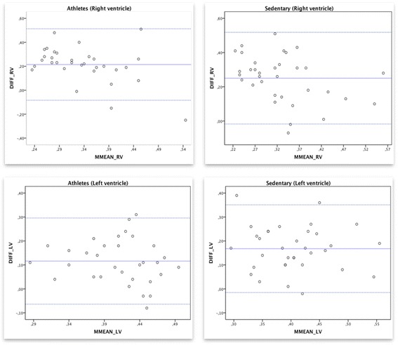
Results: There were no significant differences in MPI between athletes and controls (p > 0.05), whereas the LV had significantly higher MPI compared to RV (p < 0.001, in athletes and controls). The agreement and the correlation between the two methods measuring MPI showed low agreement and no correlation (athletes RV r = -0.027, LV r = 0.12; controls RV r = 0.20, LV r = 0.30).
Conclusion: The global function of the LV and RV measured by MPI with PWD and TDI is similar in female athletes compared to sedentary controls. Conversely, both MPI by PWD and by TDI shows a significant difference between the LV and RV. However, the agreement and correlation between conventional methods of measuring MPI by PWD compared to MPI by TDI is very poor in both these populations.
Keywords: Athlete’s heart; Diastolic function; Echocardiography; Left ventricle; Myocardial performance index; Right ventricle; Systolic function.
Conflict of interest statement
Ethics approval and consent to participate
This study was approved by the Regional Ethical Review Board, Lund, Sweden. All participants gave their written informed agreement to take part in the study. Reference number 2012/77.
Consent for publication
Not applicable.
Competing interests
The authors declare that they have no competing interests.
Publisher’s Note
Springer Nature remains neutral with regard to jurisdictional claims in published maps and institutional affiliations.
Figures
References
-
- Wernstedt P, Sjostedt C, Ekman I, Du H, Thuomas KA, Areskog NH, et al. Adaptation of cardiac morphology and function to endurance and strength training. A comparative study using MR imaging and echocardiography in males and females. Scand J Med Sci Sports. 2002;12(1):17–25. doi: 10.1034/j.1600-0838.2002.120104.x. - DOI - PubMed
Publication types
MeSH terms
LinkOut - more resources
Full Text Sources
Other Literature Sources
