

Diagnostic performance of image navigated coronary CMR angiography in patients with coronary artery disease
- PMID: 28893296
- PMCID: PMC5594598
- DOI: 10.1186/s12968-017-0381-3
Diagnostic performance of image navigated coronary CMR angiography in patients with coronary artery disease
Abstract
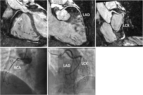
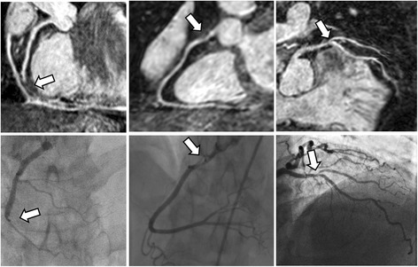
Background: The use of coronary MR angiography (CMRA) in patients with coronary artery disease (CAD) remains limited due to the long scan times, unpredictable and often non-diagnostic image quality secondary to respiratory motion artifacts. The purpose of this study was to evaluate CMRA with image-based respiratory navigation (iNAV CMRA) and compare it to gold standard invasive x-ray coronary angiography in patients with CAD.
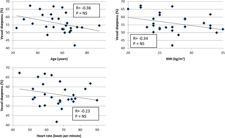
Methods: Consecutive patients referred for CMR assessment were included to undergo iNAV CMRA on a 1.5 T scanner. Coronary vessel sharpness and a visual score were assigned to the coronary arteries. A diagnostic reading was performed on the iNAV CMRA data, where a lumen narrowing >50% was considered diseased. This was compared to invasive x-ray findings.
Results: Image-navigated CMRA was performed in 31 patients (77% male, 56 ± 14 years). The iNAV CMRA scan time was 7 min:21 s ± 0 min:28 s. Out of a possible 279 coronary segments, 26 segments were excluded from analysis due to stents or diameter less than 1.5 mm, resulting in a total of 253 coronary segments. Diagnostic image quality was obtained for 98% of proximal coronary segments, 94% of middle segments, and 91% of distal coronary segments. The sensitivity and specificity was 86% and 83% per patient, 80% and 92% per vessel and 73% and 95% per segment.
Conclusion: In this study, iNAV CMRA offered a very good diagnostic performance when compared against invasive x-ray angiography. Due to the short and predictable scan time it can add clinical value as a part of a comprehensive CAD assessment protocol.
Keywords: Coronary MR angiography; Coronary artery disease; Image navigators; Respiratory motion correction.
Conflict of interest statement
Ethical approval and consent to participate
This study was approved by the Institutional Review Board at St Thomas’ Hospital (15/NS/0030). All patients provided written informed consent for participation in the study.
Consent for publication
Written informed consent was obtained from patients for publication of their individual details and accompanying images in this manuscript. The consent form is held in the patients’ clinical notes and is available for review by the Editor-in-Chief.
Competing interests
The authors declare that they have no competing interests.
Publisher’s Note
Springer Nature remains neutral with regard to jurisdictional claims in published maps and institutional affiliations.
Figures


Similar articles
-
Clinical comparison of sub-mm high-resolution non-contrast coronary CMR angiography against coronary CT angiography in patients with low-intermediate risk of coronary artery disease: a single center trial.J Cardiovasc Magn Reson. 2021 May 17;23(1):57. doi: 10.1186/s12968-021-00758-9. J Cardiovasc Magn Reson. 2021. PMID: 33993890 Free PMC article.
-
Feasibility of contrast-enhanced coronary artery magnetic resonance angiography using compressed sensing.J Cardiovasc Magn Reson. 2020 Feb 13;22(1):15. doi: 10.1186/s12968-020-0601-0. J Cardiovasc Magn Reson. 2020. PMID: 32050982 Free PMC article.
-
Visualization of coronary arteries in paediatric patients using whole-heart coronary magnetic resonance angiography: comparison of image-navigation and the standard approach for respiratory motion compensation.J Cardiovasc Magn Reson. 2019 Feb 25;21(1):13. doi: 10.1186/s12968-019-0525-8. J Cardiovasc Magn Reson. 2019. PMID: 30798789 Free PMC article.
-
Diagnostic Performance of Hybrid Cardiac Imaging Methods for Assessment of Obstructive Coronary Artery Disease Compared With Stand-Alone Coronary Computed Tomography Angiography: A Meta-Analysis.JACC Cardiovasc Imaging. 2018 Apr;11(4):589-599. doi: 10.1016/j.jcmg.2017.05.020. Epub 2017 Aug 16. JACC Cardiovasc Imaging. 2018. PMID: 28823745 Free PMC article.
-
Diagnostic performance of coronary magnetic resonance angiography as compared against conventional X-ray angiography: a meta-analysis.J Am Coll Cardiol. 2004 Nov 2;44(9):1867-76. doi: 10.1016/j.jacc.2004.07.051. J Am Coll Cardiol. 2004. PMID: 15519021 Review.
Cited by
-
Coronary Magnetic Resonance Angiography in Chronic Coronary Syndromes.Front Cardiovasc Med. 2021 Aug 17;8:682924. doi: 10.3389/fcvm.2021.682924. eCollection 2021. Front Cardiovasc Med. 2021. PMID: 34485397 Free PMC article. Review.
-
Automated detection of cardiac rest period for trigger delay calculation for image-based navigator coronary magnetic resonance angiography.J Cardiovasc Magn Reson. 2023 Oct 2;25(1):52. doi: 10.1186/s12968-023-00962-9. J Cardiovasc Magn Reson. 2023. PMID: 37779192 Free PMC article.
-
Motion compensated whole-heart coronary cardiovascular magnetic resonance angiography using focused navigation (fNAV).J Cardiovasc Magn Reson. 2021 Mar 29;23(1):33. doi: 10.1186/s12968-021-00717-4. J Cardiovasc Magn Reson. 2021. PMID: 33775246 Free PMC article.
-
Journal of Cardiovascular Magnetic Resonance: 2017/2018 in review.J Cardiovasc Magn Reson. 2019 Dec 30;21(1):79. doi: 10.1186/s12968-019-0594-8. J Cardiovasc Magn Reson. 2019. PMID: 31884956 Free PMC article. Review.
-
Optimization of the acceleration of compressed sensing in whole-heart contrast-free coronary magnetic resonance angiography.J Cardiovasc Magn Reson. 2025 Summer;27(1):101845. doi: 10.1016/j.jocmr.2025.101845. Epub 2025 Jan 27. J Cardiovasc Magn Reson. 2025. PMID: 39864742 Free PMC article.
References
Publication types
MeSH terms
Grants and funding
LinkOut - more resources
Full Text Sources
Other Literature Sources
Medical
Miscellaneous
