. 2017 Sep 12:6:e27814.
doi: 10.7554/eLife.27814.

Localization of spontaneous bursting neuronal activity in the preterm human brain with simultaneous EEG-fMRI

Affiliations

Localization of spontaneous bursting neuronal activity in the preterm human brain with simultaneous EEG-fMRI

Tomoki Arichi et al. Elife. .

Abstract

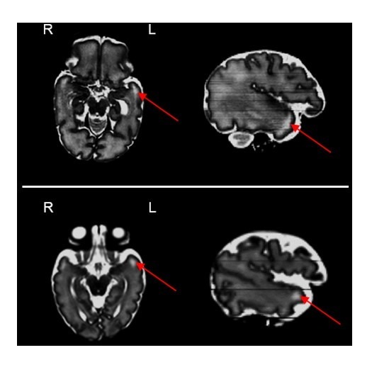
Electroencephalographic recordings from the developing human brain are characterized by spontaneous neuronal bursts, the most common of which is the delta brush. Although similar events in animal models are known to occur in areas of immature cortex and drive their development, their origin in humans has not yet been identified. Here, we use simultaneous EEG-fMRI to localise the source of delta brush events in 10 preterm infants aged 32-36 postmenstrual weeks. The most frequent patterns were left and right posterior-temporal delta brushes which were associated in the left hemisphere with ipsilateral BOLD activation in the insula only; and in the right hemisphere in both the insular and temporal cortices. This direct measure of neural and hemodynamic activity shows that the insula, one of the most densely connected hubs in the developing cortex, is a major source of the transient bursting events that are critical for brain maturation.

Keywords: EEG; brain development; fMRI; human; human biology; medicine; neonate; neuroscience; prematurity; spontaneous neuronal activity.


Conflict of interest statement

No competing interests declared.

Figures

Figure 1.
Figure 1.. Delta brushes occur with distinct topographies.
Segments of EEG recordings showing individual examples of delta brushes with the most common topographical distributions (occurred at least three times in a given subject). These rarely involved frontal and midline electrodes which are therefore omitted for illustration clarity. Right and left posterior-temporal delta brushes occurred in 10/10 and 9/10 subjects respectively, while other delta brushes were recorded in no more than two subjects. EEG traces and recording electrodes where delta brush activity was maximal are marked in red. Shaded areas represent the time of occurrence of each event.
Figure 2.
Figure 2.. Localization of posterior-temporal delta brushes.
In a group of 10 preterm infants (35 + 0 weeks PMA, range 32 + 2 to 36 + 2 weeks), right posterior-temporal delta brush activity (blue) was significantly associated with BOLD clusters in the right temporal pole (z50, z55), right superior temporal lobe (x28), and the right insular cortex (z60, z62, z64, z66). Left posterior-temporal delta brush activity (red-yellow) was significantly associated with BOLD clusters in the left posterior insula (z64, z66) and left parietal operculum (z66, z68). Images show the results of a one-sample t-test (p<0.05) performed using permutation testing and corrected for family-wise error overlaid on an age-specific T2-weighted brain atlas (Serag et al., 2012).
Figure 2—figure supplement 1.
Figure 2—figure supplement 1.. Example of subject level analysis.
Hemodynamic activity correlated to right posterior-temporal delta brushes in a single infant of 35 + 1 weeks PMA. (a) Right posterior-temporal delta brush occurrence as identified by EEG (dashed vertical lines) was convolved with an age-appropriate hemodynamic response function to generate a general linear model regressor (blue) to fit to the acquired BOLD time series (red) for fMRI analysis. (b) Right posterior-temporal delta brush occurrence was significantly associated with well localized clusters of BOLD activity (red-yellow) in the right temporal pole and superior temporal lobe (z49); the left parietal operculum (z55); the anterior cingulate and right fronto-parietal lobes (z65 and z69). Images show the thresholded z-statistical map with a corrected cluster significance of p<0.05 overlaid on the subject’s T2-weighted image.
Figure 2—figure supplement 2.
Figure 2—figure supplement 2.. T2-weighted MR images from two infants in our study sample imaged at 32 + 2 weeks PMA (top row) and 35 + 2 weeks PMA (bottom row).
As described in the literature, the subplate can be clearly seen as an area of high signal intensity (long T2) on the temporal poles bilaterally (red arrows).
Figure 3.
Figure 3.. Localization of delta brush events in a single preterm infant.
Example of the significant hemodynamic activity correlated to less frequent delta brushes in a single preterm subject at 35 + 6 weeks PMA. (a) The occurrence of bilateral posterior-temporal delta brushes was significantly associated with well localized clusters of BOLD activity (red-yellow) in the bilateral superior temporal lobe and insulae (z55); while (b) bilateral occipital delta brushes were associated with a cluster in the medial occipital region (z33). Images show the thresholded z-statistical map with a corrected cluster significance of p<0.05 overlaid on the subject’s T2-weighted image.

Comment in

References

    1. Allen PJ, Polizzi G, Krakow K, Fish DR, Lemieux L. Identification of EEG events in the MR scanner: the problem of pulse artifact and a method for its subtraction. NeuroImage. 1998;8:229–239. doi: 10.1006/nimg.1998.0361. - DOI - PubMed
    1. Allievi AG, Arichi T, Tusor N, Kimpton J, Arulkumaran S, Counsell SJ, Edwards AD, Burdet E. Maturation of Sensori-Motor Functional Responses in the Preterm Brain. Cerebral Cortex. 2016;26:402–413. doi: 10.1093/cercor/bhv203. - DOI - PMC - PubMed
    1. American Clinical Neurophysiology Society Critical Care Monitoring Committee. Tsuchida TN, Wusthoff CJ, Shellhaas RA, Abend NS, Hahn CD, Sullivan JE, Nguyen S, Weinstein S, Scher MS, Riviello JJ, Clancy RR. American clinical neurophysiology society standardized EEG terminology and categorization for the description of continuous EEG monitoring in neonates: report of the American Clinical Neurophysiology Society critical care monitoring committee. Journal of Clinical Neurophysiology : Official Publication of the American Electroencephalographic Society. 2013;30:161–173. doi: 10.1097/WNP.0b013e3182872b24. - DOI - PubMed
    1. André M, Lamblin MD, d'Allest AM, Curzi-Dascalova L, Moussalli-Salefranque F, S Nguyen The T, Vecchierini-Blineau MF, Wallois F, Walls-Esquivel E, Plouin P. Electroencephalography in premature and full-term infants. Developmental features and glossary. Neurophysiologie Clinique/Clinical Neurophysiology. 2010;40:59–124. doi: 10.1016/j.neucli.2010.02.002. - DOI - PubMed
    1. Arichi T, Moraux A, Melendez A, Doria V, Groppo M, Merchant N, Combs S, Burdet E, Larkman DJ, Counsell SJ, Beckmann CF, Edwards AD. Somatosensory cortical activation identified by functional MRI in preterm and term infants. NeuroImage. 2010;49:2063–2071. doi: 10.1016/j.neuroimage.2009.10.038. - DOI - PubMed

Publication types