

Brain TSPO imaging and gray matter volume in schizophrenia patients and in people at ultra high risk of psychosis: An [11C]PBR28 study
- PMID: 28893493
- PMCID: PMC6027955
- DOI: 10.1016/j.schres.2017.08.063
Brain TSPO imaging and gray matter volume in schizophrenia patients and in people at ultra high risk of psychosis: An [11C]PBR28 study
Abstract
Patients with schizophrenia show whole brain and cortical gray matter (GM) volume reductions which are progressive early in their illness. Microglia, the resident immune cells in the CNS, phagocytose neurons and synapses. Some post mortem and in vivo studies in schizophrenia show evidence for elevated microglial activation compared to matched controls. However, it is currently unclear how these results relate to changes in cortical structure.
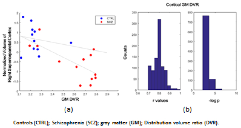
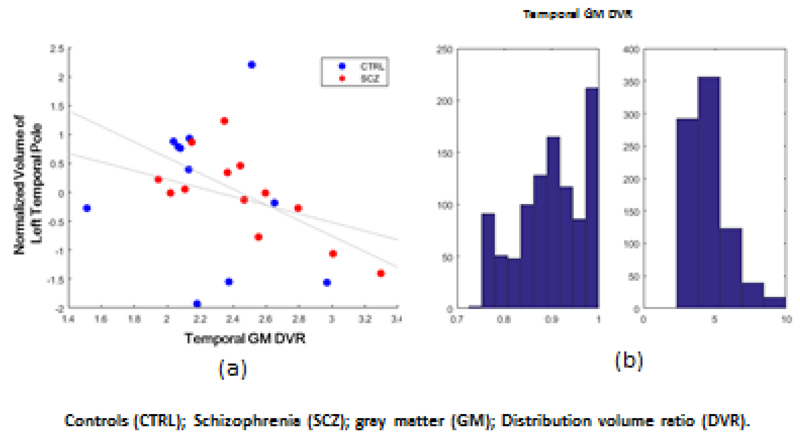
Methods: Fourteen patients with schizophrenia and 14 ultra high risk for psychosis (UHR) subjects alongside two groups of age and genotype matched healthy controls received [11C]PBR28 PET scans to index TSPO expression, a marker of microglial activation and a 3T MRI scan. We investigated the relationship between the volume changes of cortical regions and microglial activation in cortical GM (as indexed by [11C]PBR28 distribution volume ratio (DVR).
Results: The total cortical GM volume was significantly lower in SCZ than the controls [mean (SD)/cm3: SCZ=448.83 (39.2) and controls=499.6 (59.2) (p=0.02) but not in UHR (mean (SD)=503.06 (57.9) and controls=524.46 (45.3) p=0.3). Regression model fitted the total cortical GM DVR values with the cortical regional volumes in SCZ (r=0.81; p<0.001) and in UHR (r=0.63; p=0.02). We found a significant negative correlation between the TSPO signal and total cortical GM volume in SCZ with the highest absolute correlation coefficient in the right superior-parietal cortex (r=-0.72; p=0.006).
Conclusions: These findings suggest that microglial activity is related to the altered cortical volume seen in schizophrenia. Longitudinal investigations are required to determine whether microglial activation leads to cortical gray matter loss.
Keywords: Brain volume; Gray matter; Microglia; PET; Psychosis; Schizophrenia.
Copyright © 2017 Elsevier B.V. All rights reserved.
Conflict of interest statement
Dr. Howes has received investigator-initiated research funding from and/or participated in advisory/ speaker meetings organised by Astra-Zeneca, Autifony, BMS, Eli Lilly, Heptares, Jansenn, Lundbeck, Lyden-Delta, Otsuka, Servier, Sunovion, Rand and Roche. Neither Dr. Howes or his family have been employed by or have holdings/ a financial stake in any biomedical company. Sudhakar Selvaraj, Peter S Bloomfield, Cao Bo, Mattia Veronese and Federico Turkheimer have no conflicts of interest for this work. The authors would like to thank all the clinical imaging staff at Imanova for their help with this study.
Figures
Similar articles
-
Lower levels of the glial cell marker TSPO in drug-naive first-episode psychosis patients as measured using PET and [11C]PBR28.Mol Psychiatry. 2017 Jun;22(6):850-856. doi: 10.1038/mp.2016.247. Epub 2017 Feb 14. Mol Psychiatry. 2017. PMID: 28194003
-
The association of psychosocial risk factors for mental health with a brain marker altered by inflammation: A translocator protein (TSPO) PET imaging study.Brain Behav Immun. 2019 Aug;80:742-750. doi: 10.1016/j.bbi.2019.05.023. Epub 2019 May 18. Brain Behav Immun. 2019. PMID: 31112791
-
PET imaging of putative microglial activation in individuals at ultra-high risk for psychosis, recently diagnosed and chronically ill with schizophrenia.Transl Psychiatry. 2017 Aug 29;7(8):e1225. doi: 10.1038/tp.2017.193. Transl Psychiatry. 2017. PMID: 28850113 Free PMC article.
-
Brain structural abnormalities as potential markers for detecting individuals with ultra-high risk for psychosis: A systematic review and meta-analysis.Schizophr Res. 2019 Jul;209:22-31. doi: 10.1016/j.schres.2019.05.015. Epub 2019 May 16. Schizophr Res. 2019. PMID: 31104914
-
Structural brain changes in schizophrenia at different stages of the illness: A selective review of longitudinal magnetic resonance imaging studies.Aust N Z J Psychiatry. 2017 May;51(5):500-508. doi: 10.1177/0004867417699473. Epub 2017 Mar 21. Aust N Z J Psychiatry. 2017. PMID: 28415873 Review.
Cited by
-
Neurodevelopmental and Neuropsychiatric Disorders.Adv Neurobiol. 2024;37:457-495. doi: 10.1007/978-3-031-55529-9_26. Adv Neurobiol. 2024. PMID: 39207708 Review.
-
Selective Review of Neuroimaging Findings in Youth at Clinical High Risk for Psychosis: On the Path to Biomarkers for Conversion.Front Psychiatry. 2020 Sep 23;11:567534. doi: 10.3389/fpsyt.2020.567534. eCollection 2020. Front Psychiatry. 2020. PMID: 33173516 Free PMC article. Review.
-
The Inflamed Brain in Schizophrenia: The Convergence of Genetic and Environmental Risk Factors That Lead to Uncontrolled Neuroinflammation.Front Cell Neurosci. 2020 Aug 27;14:274. doi: 10.3389/fncel.2020.00274. eCollection 2020. Front Cell Neurosci. 2020. PMID: 33061891 Free PMC article. Review.
-
Predictive validity of conversion from the clinical high risk syndrome to frank psychosis.Schizophr Res. 2020 Feb;216:184-191. doi: 10.1016/j.schres.2019.12.002. Epub 2019 Dec 19. Schizophr Res. 2020. PMID: 31864837 Free PMC article.
-
Gliosis and Neurodegenerative Diseases: The Role of PET and MR Imaging.Front Cell Neurosci. 2020 Apr 2;14:75. doi: 10.3389/fncel.2020.00075. eCollection 2020. Front Cell Neurosci. 2020. PMID: 32327973 Free PMC article. Review.
References
-
- Abourbeh G, Thézé B, Maroy R, Dubois A, Brulon V, Fontyn Y, Dollé F, Tavitian B, Boisgard R. Imaging microglial/macrophage activation in spinal cords of experimental autoimmune encephalomyelitis rats by positron emission tomography using the mitochondrial 18 kDa translocator protein radioligand [18F]DPA-714. J Neurosci. 2012;32(17):5728–5736. - PMC - PubMed
-
- Arlicot N, Petit E, Katsifis A, Toutain J, Divoux D, Bodard S, Roussel S, Guilloteau D, Bernaudin M, Chalon S. Detection and quantification of remote microglial activation in rodent models of focal ischaemia using the TSPO radioligand CLINDE. Eur J Nucl Med Mol Imaging. 2010;37(12):2371–2380. - PubMed
-
- Banati RB, Cagnin A, Brooks DJ, Gunn RN, Myers R, Jones T, Birch R, Anand P. Long-term trans-synaptic glial responses in the human thalamus after peripheral nerve injury. Neuroreport. 2001;12(16):3439–3442. - PubMed
Publication types
MeSH terms
Substances
Grants and funding
LinkOut - more resources
Full Text Sources
Other Literature Sources
Medical
