. 2018 Jan:149:78-88.
doi: 10.1016/j.antiviral.2017.09.003. Epub 2017 Sep 8.

Raltegravir blocks the infectivity of red-fluorescent-protein (mCherry)-labeled HIV-1JR-FL in the setting of post-exposure prophylaxis in NOD/SCID/Jak3-/- mice transplanted with human PBMCs

Affiliations

Raltegravir blocks the infectivity of red-fluorescent-protein (mCherry)-labeled HIV-1JR-FL in the setting of post-exposure prophylaxis in NOD/SCID/Jak3-/- mice transplanted with human PBMCs

Hiromi Ogata-Aoki et al. Antiviral Res. 2018 Jan.

Abstract

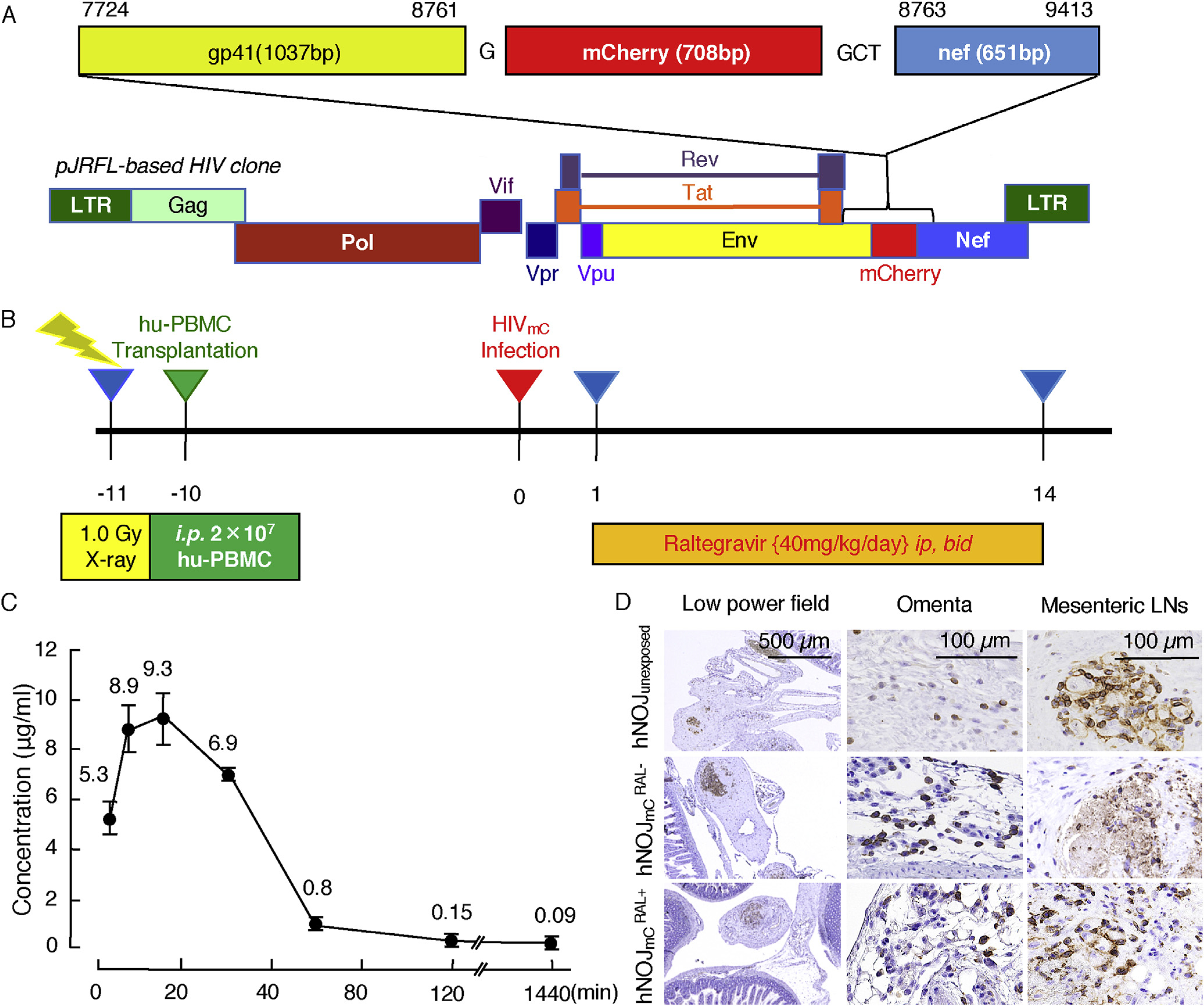
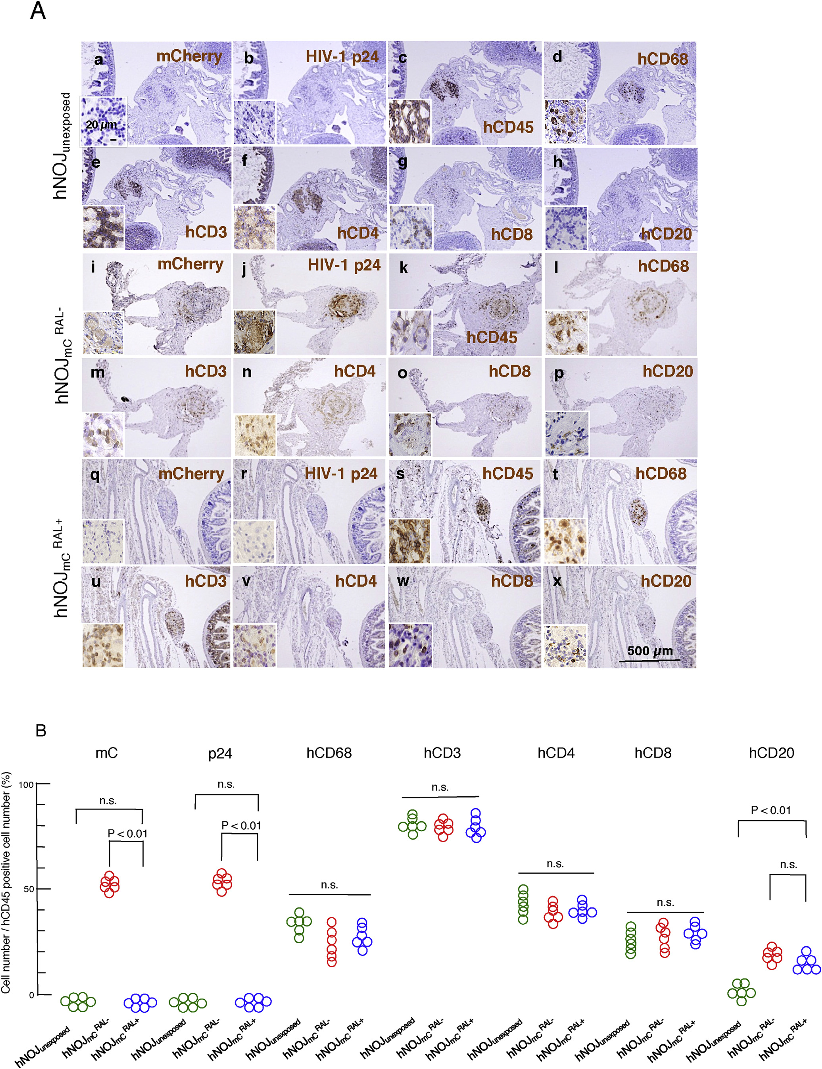
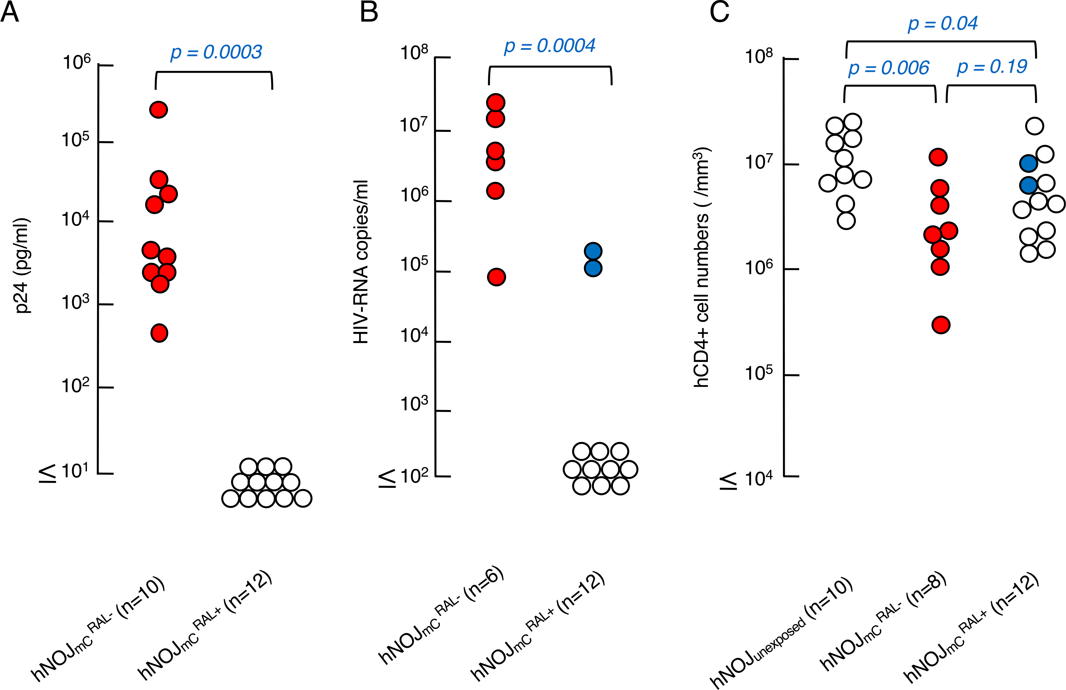
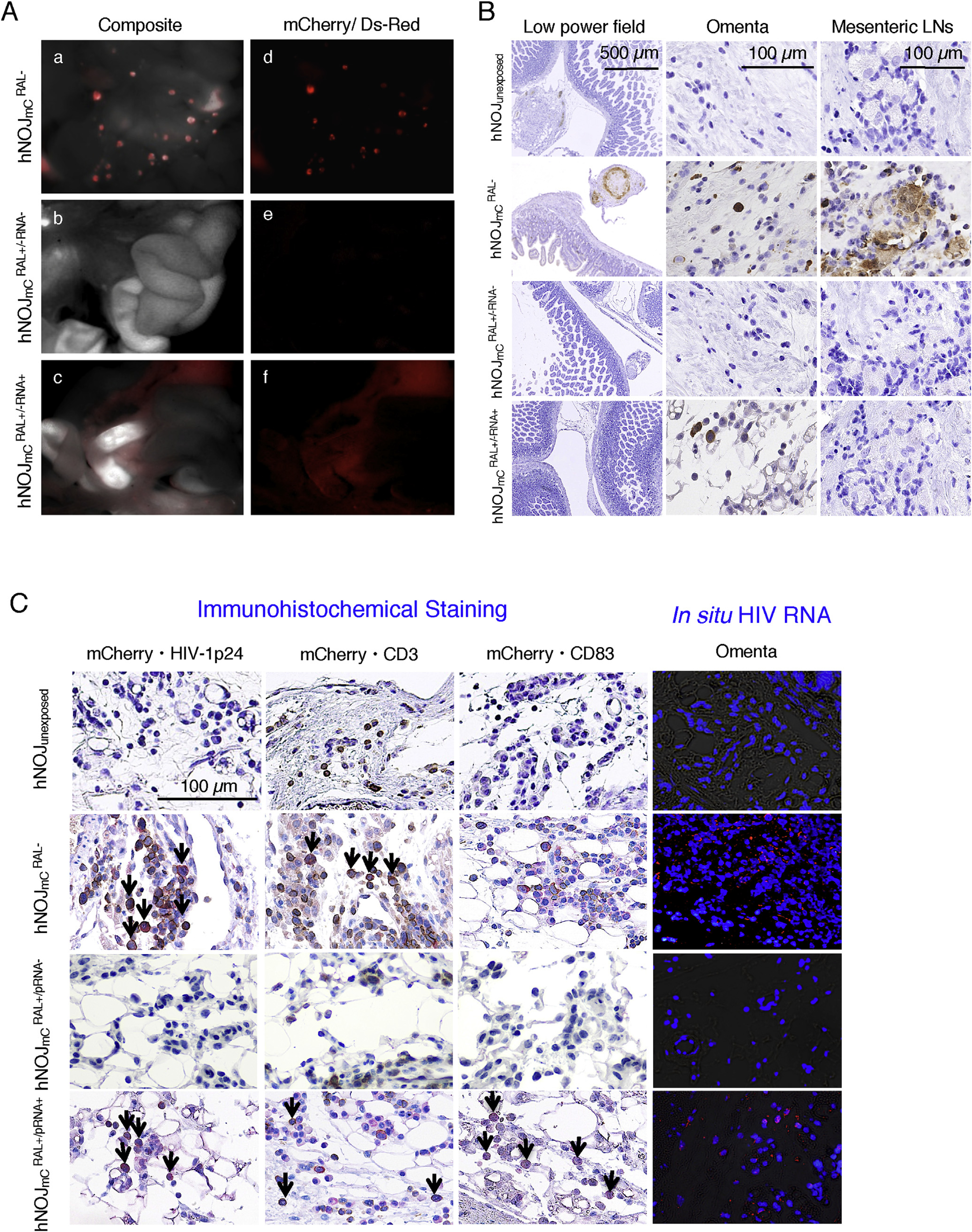
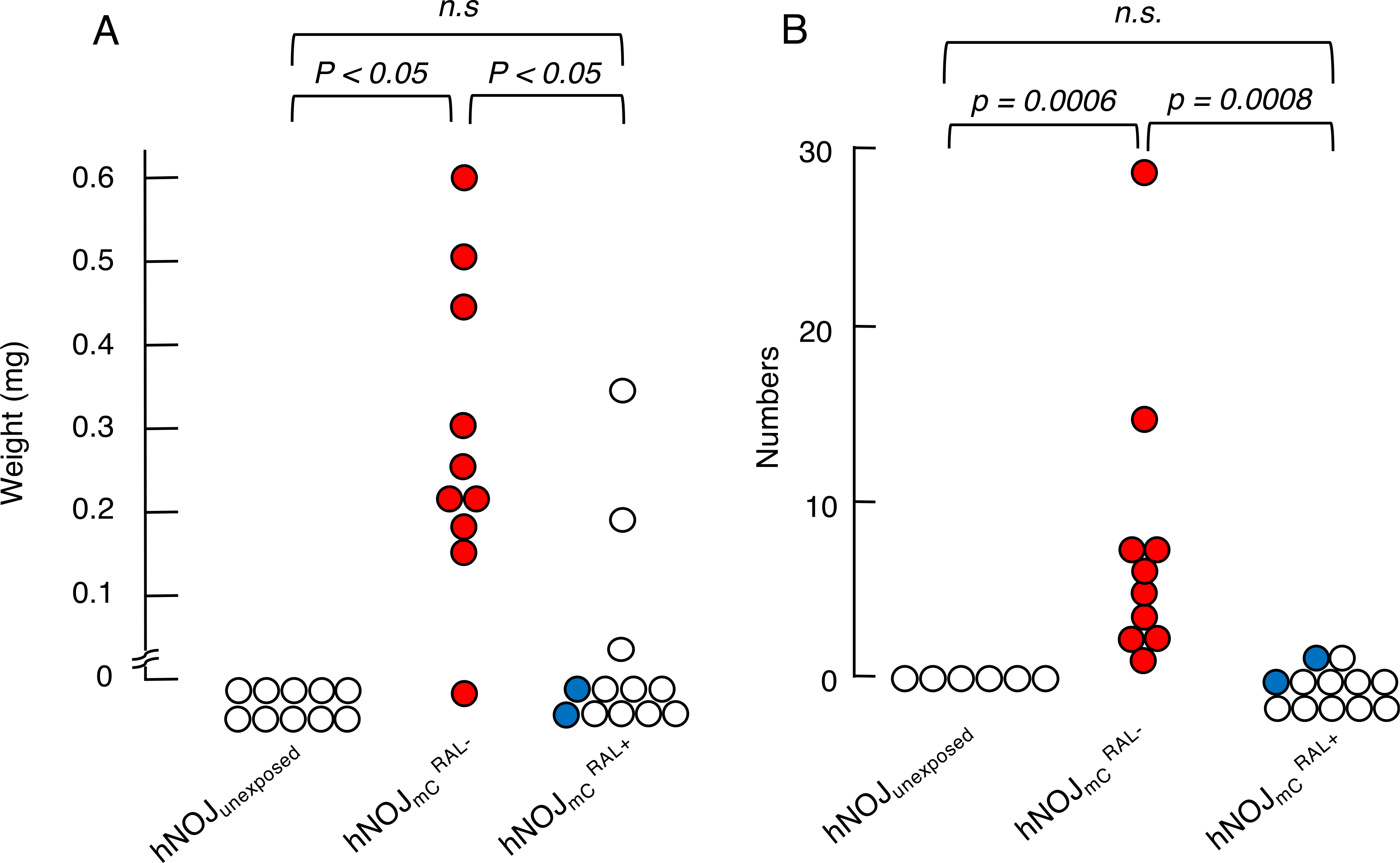
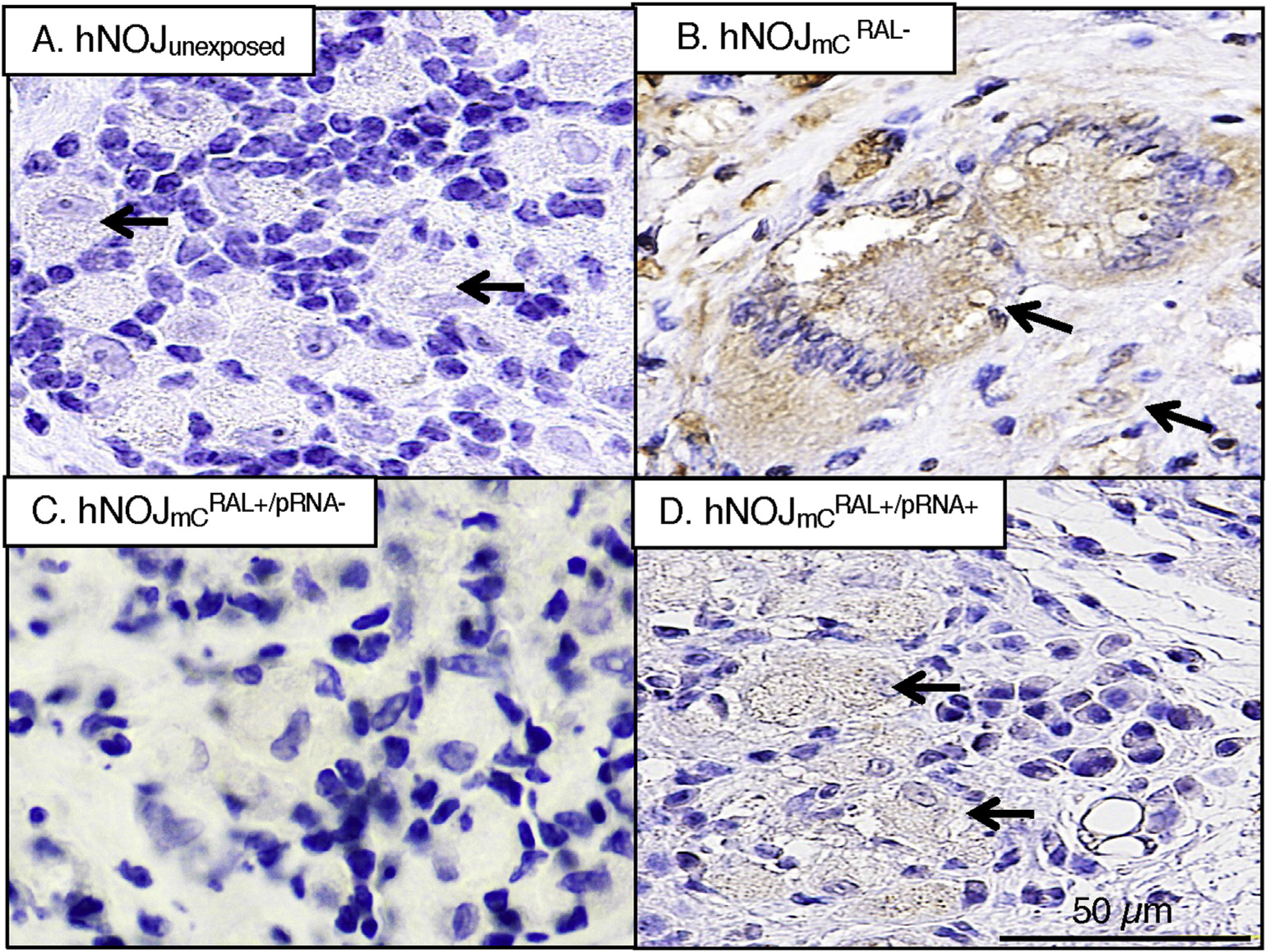
Employing NOD/SCID/Jak3-/- mice transplanted with human PBMCs (hNOJ mice) and replication-competent, red-fluorescent-protein (mCherry; mC)-labeled HIV-1JR-FL (HIVmC), we examined whether early antiretroviral treatment blocked the establishment of HIV-1 infection. The use of hNOJ mice and HIVmC enabled us to visually locate infection foci and to examine the early dynamics of HIVmC infection without using a large amount of antiretroviral unlike in non-human primate models. Although when raltegravir (RAL) administration was begun 1 day after intraperitoneal (ip) inoculation of HIVmC, no plasma p24 or plasma HIV-1-RNA (pRNA) were detected in 10 of 12 hNOJ (hNOJmCRAL+) mice as assessed on the last day of the 14-day continuous twice-daily RAL administration, all 10 untreated hNOJmC (hNOJmCRAL-) mice became positive for p24 and pRNA and had significantly swollen lymph nodes in peritoneal cavity and abundant p24+/mC+/CD3+/CD4+ T cells and p24+/mC+/CD68+ monocytes/macrophages were identified in their omenta and mesenteric lymphoid tissues/lymph nodes upon necropsy of the mice on day 14. In 12 hNOJmCRAL+ mice, no significantly swollen lymph nodes were seen compared to hNOJmCRAL- mice; however, in the omentum of the 2 hNOJmCRAL+ mice that were positive for pRNA and in site RNA, mC+/p24+/CD3+/CD83+ cells were identified, suggesting that viral breakthrough occurred later in the observation period. The present data suggest that the use of hNOJ mouse model and HIVmC may shed light on the study of early-phase dynamics of HIV-1 infection and cellular events in post-exposure/pre-exposure prophylaxis.

Keywords: HIV-1 breakthrough; In vivo imaging; NOD/SCID/Jak3−/− (NOJ) mice; Post-exposure prophylaxis; Raltegravir administration; mCherry-labeled HIV-1 (HIVmC).


Conflict of interest statement

Conflict of interest

All authors declare no conflict of interest.

Figures

Fig. 1.
Fig. 1.. Construction of pHIVmC and study design.
(A) Genetic map of HIV-1 containing the mC fluorescent protein-encoding gene. HIV-1Jr-Fl was constructed to contain mC fluorescent protein between gp41 and nef proteins. (B) Protocol for radiation, hPBMC transplantation, HIVmC inoculation, RAL treatment and monitoring the dynamics of HIVmC infection. Twenty million hPBMC were transplanted to each mouse 1 day after X-ray exposure. Ten days after the hPBMC transplantation, HIVmC was inoculated ip. Twenty-four hours following the HIVmC inoculation, RAL administration (20 mg/kg, twice a day) was implemented and continued over 14 days. Mice were sacrificed on day 14 and subjected to virological, histological, and immunological examinations. (C) Pharmacokinetics of RAL. RAL was administered to each mouse at a dose of 20 mg/kg. Blood samples were taken at 3, 7, 15, 30, 60, 120 and 1440 min and subjected to HPLC analysis (n = 4). The concentration of RAL reached the maximal concentration almost immediately after ip administration and subsequently decreased rapidly. The plasma half-life of RAL in this study was approximately 20 min. (D) Re-population of hPBMC in the mesenteric area of each mouse group. Fixed/paraffin-embedded tissue sections were immunohistologically stained with anti-hCD45 antibody (a pan-lymphocyte marker) and examined under light microscopy. Nuclei were counterstained with Mayer’s hematoxylin (blue). Note that hCD45+ cells (brown) had been re-populated within the mesenteric lymph nodes comparably among the three mouse groups: hNOJunexposed, hNOJmCRAL+, and hNOJmCRAL mice. (For interpretation of the references to colour in this figure legend, the reader is referred to the web version of this article.)
Fig. 2.
Fig. 2.. Representative immunohistochemical staining profiles of mesenteric lymph nodes.
(A) Re-population of human cells in the abdominal cavity. The paraffin sections of mesenteric lymph nodes in each group were immunostained with monoclonal antibodies against mC/DsRed, p24, hCD45, hCD68 (macrophage-specific antigen), hCD3, hCD4, hCD8 and hCD20 (brown). Nuclei were counterstained with Mayer’s hematoxylin (blue). Neither mCherry+ nor p24+ cells were seen in mesenteric (q and r) lymph nodes of hNOJmCRAL+ mice. Cells in mesenteric lymph nodes of hNOJmCRAL+ mice (s–w) showed similar features as seen in hNOJunexposed mice (c–g). (B) Quantitative analysis of each positive cell in the mesenteric lymph nodes. Numbers of mCherry+, p24+, hCD68+, hCD3+, hCD4+, hCD8+, and hCD20+ cells per unit area were divided by each corresponding hCD45+ cell number per unit area. No mC+ or p24+ cells were seen in hNOJunexposed and hNOJmCRAL+ mice and cell number/hCD45+ cell number was expressed as 0%. Note that the %hCD20+ was significantly greater in the mesenteric lymph node of hNOJmCRAL and hNOJmCRAL+ mice compared to that of hNOJunexposed mice (P < 0.01). No significant differences were observed in hCD68+, hCD3+, hCD4+, or hCD8+ cell numbers among the three mouse groups. (For interpretation of the references to colour in this figure legend, the reader is referred to the web version of this article.)
Fig. 3.
Fig. 3.. HIV-1 p24 amounts, pRNA copies and hCD4+ cell counts in each hNOJ mouse group with or without RAL treatment.
(A) Amounts of plasma p24 antigen. Plasma from the hNOJmCRAL and hNOJmCRAL+ mice were examined using a fully automated chemiluminescent enzyme immunoassay on day 14 after HIVmc inoculation. Note that the amounts of p24 in plasma were high in hNOJmCRAL mice while RAL significantly suppressed the plasma p24 antigen as examined on day 14 after HIVmc inoculation. (p = 0.0003 compared to hNOJmCRAL+ mice). (B) Amounts of pRNA copies. Blood samples were collected on day 14 and subjected to the determination of pRNA copy numbers. Note that the copy numbers in hNOJmCRAL mice were high on day 14. (P = 0.0004 compared to hNOJmCRAL+ mice). (C) Numbers of CD4+ cells in each mouse group. hPBMc recovered on day 14 after HIVmc inoculation were counted and subjected to flow cytometry.
Fig. 4.
Fig. 4.. Prevention of HIVmC infection with RAL treatment in hNOJ mice.
(A) Representative in vivo images. No mC signals were seen in abdominal area in all of hNOJmCRAL+ mice, except two hNOJmCRAL+/pRNA+ mice. Composite images (a–c) consisting of autoflurescence spectra and mC specific spectra demonstrate the location of mC positive organs. By day 14 after HIVmC inoculation, mC signal was mainly detected in mesenteric lymph nodes and the omentum of hNOJmCRAL mice (a and d). The mC signals were hardly recognized in hNOJmCRAL+ mice with undetectable pRNA levels (b and e). hNOJmCRAL+ mice with detectable pRNA copies showed diffuse mC signals in their omentum (c and f). In panels a and d of Figure 4-A, the multiple spotty red signals are mC+ mesenteric lymph nodes, whereas the dimly red areas are mC+ omenta. In panels c and f of Figure 4-A, although significant mC signals are seen in omenta, no mC+ lymph nodes are seen. (B) Representative p24 profiles among mouse groups. No p24 positive cells were observed in the abdominal area in all the hNOJmCRAL+ mice, except two hNOJmCRAL+/pRNA+ mice. The p24+ cells were observed in the omenta of those two hNOJmCRAL+/pRNA+ mice as well as all hNOJmCRAL mice. No p24+ cells were observed in the mesenteric lymph node of hNOJmCRAL+/pRNA+ mice, whereas a number of p24 positive cells were found in hNOJmCRAL mice. (C) Representative double-staining profiles and in situ hybridization data. HIV-RNA was not detected in the mesenteric lymph nodes of hNOJmCRAL+/pRNA, while no HIV-RNA was observed in hNOJunexposed (very right columns). HIV-RNA was detected in the omenta of hNOJmCRAL+/pRNA+, however, the number of such HIV-RNA+ cells was much smaller than in the hNOJmCRAL. Nuclei were stained with DAPI. Arrows denote mC+ plus p24+ cells, mC+ plus hCD3+ cells, and mC+ plus hCD83+ cells. Nuclei were stained with hematoxylin and seen in blue. mC + cells are stained in red, whereas HIV-1 p24+, CD3+, and CD83+ cells are stained in brown. (For interpretation of the references to colour in this figure legend, the reader is referred to the web version of this article.)
Fig. 5.
Fig. 5.. Comparison of the weights and numbers of mesenteric lymph nodes among three hNOJ mouse groups with or without RAL treatment.
For comparison, the largest lymph node in each mouse was examined. The largest lymph nodes in each mouse less than 0.01 mg were expressed as 0 mg in Panel A. For comparison of the numbers of lymph nodes, mC signal-emitting lymph nodes were counted using the Maestro™ system as shown in Panel B. Statistical analyses were performed using a nonparametric multiple comparison called Bonferroni method.
Fig. 6.
Fig. 6.. Morphologic profiles of mesenteric lymph nodes.
A number of multinuclear giant cells and cytolysis were seen in HIVmCRAL− mice. Only cytolysis was seen in one of the two hNOJmCRAL+/pRNA+ mice. Neither was seen in the other hNOJmCRAL+/pRNA+ mouse, hNOJunexposed mice, or hNOJmCRAL+/pRNA mice.

Similar articles

Cited by

References

    1. Alford R, Ogawa M, Choyke PL, Kobayashi H, 2009. Molecular probes for the in vivo imaging of cancer. Mol. Biosyst. 5,1279–1291. - PMC - PubMed
    1. Dargent JL, Lespagnard L, Kornreich A, Hermans P, Clumeck N, Verhest A, 2000. HIV-associated multinucleated giant cells in lymphoid tissue of the Waldeyer’s ring: a detailed study. Mod. Pathol. 13,1293–1299. - PubMed
    1. Denton PW, Krisko JF, Powell DA, Mathias M, Kwak YT, Martinez-Torres F, Zou W, Payne DA, Estes JD, Garcia JV, 2010. Systemic administration of antiretrovirals prior to exposure prevents rectal and intravenous HIV-1 transmission in humanized BLT mice. PLoS One 5, e8829. - PMC - PubMed
    1. Fang G, Weiser B, Visosky A, Moran T, Burger H, 1999. PCR-mediated recombination: a general method applied to construct chimeric infectious molecular clones of plasma-derived HIV-1 RNA. Nat. Med. 5, 239–242. - PubMed
    1. Garcia-Lerma JG, Otten RA, Qari SH, Jackson E, Cong ME, Masciotra S, Luo W, Kim C, Adams DR, Monsour M, Lipscomb J, johnson JA, Delinsky D, Schinazi RF, Janssen R, Folks TM, Heneine W, 2008. Prevention of rectal SHIV transmission in macaques by daily or intermittent prophylaxis with emtricitabine and tenofovir. PLoS Med. 5, e28. - PMC - PubMed

Publication types

MeSH terms