. 2017 Oct 20;292(42):17473-17481.
doi: 10.1074/jbc.M117.801860. Epub 2017 Sep 11.

Processive searching ability varies among members of the gap-filling DNA polymerase X family

Affiliations

Processive searching ability varies among members of the gap-filling DNA polymerase X family

Michael J Howard et al. J Biol Chem. .

Abstract

DNA repair proteins must locate rare damaged sites within the genome. DNA polymerase β (Pol β), a member of the DNA polymerase X family that is involved in base excision repair, uses a processive hopping search mechanism to locate substrates. This effectively enhances its search footprint on DNA, increasing the probability of locating damaged sites. Processive searching has been reported or proposed for many DNA-binding proteins, raising the question of how widespread or specific to certain enzymes the ability to perform this function is. To provide insight into this question, we compared the ability of three homologous DNA Pol X family members to perform a processive search for 1-nucleotide gaps in DNA using a previously developed biochemical assay. We found that at near-predicted physiological ionic strengths, the intramolecular searching ability of Pol β is at least 4-fold higher than that of Pol μ and ∼2-fold higher than that of Pol λ. Pol β also was able to perform intersegmental transfer with the intersegmental searching ability of Pol β being at least 6- and ∼2-fold higher than that of Pols μ and λ, respectively. Mutational analysis suggested that differences in the N-terminal domains of these polymerases are responsible for the varying degrees of searching competence. Of note, the differences in processive searching ability observed among the DNA Pol X family members correlated with their proposed biological functions in base excision repair and nonhomologous end joining.

Keywords: DNA binding protein; DNA polymerase; DNA repair; NHEJ; base excision repair (BER); enzyme kinetics; facilitated diffusion; intersegmental transfer; processive search; protein-DNA interaction.


Conflict of interest statement

The authors declare that they have no conflicts of interest with the contents of this article

Figures

Figure 1.
Figure 1.
Human DNA polymerase X family. A, domain organization of human DNA Pol X family members (except TdT). S/P, serine/proline-rich region. B, structural alignment of Pols β (red; Protein Data Bank code 3ISB), μ (amino acids 131–494) (gray; Protein Data Bank code 4LZG), and λ (amino acids 241–575) (blue; Protein Data Bank code 1XSL) bound to 1-nt-gapped DNA (binary complexes). C, measurement of single-turnover rate constants (kpol) for single nucleotide insertion (dCTP) for Pols β, μ, and λ on a 1-nt-gapped DNA substrate containing a G template. The concentration of dCTP was adjusted to saturation for each respective enzyme. Data from two independent experiments were fit to a single-exponential resulting in kpol values of 2.9 ± 0.2, 1.4 ± 0.2, and 0.17 ± 0.01 s−1, respectively. The error is from the fit.
Figure 2.
Figure 2.
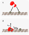
Processive searching model. The processive intramolecular substrate contains two 1-nt gaps separated by 20 bp. There is equal probability of a polymerase reacting with either site (panel 1). After catalyzing nucleotide insertion at the first encountered site (forming a nick product), the polymerase can dissociate (kdiss) into bulk solution or search to the adjacent 1-nt gap (ksearch) (panel 2).
Figure 3.
Figure 3.
Intramolecular searching abilities of DNA Pols β, μ, and λ. A, representative gels of processive assays performed with 500 nm P20 substrate, 1 nm Pol β, and 3 nm Pols μ and λ at 100 mm ionic strength. Time points are indicated at the top of the gels. S indicates substrate bands, P indicates processive product, and D represents distributive products. B, representative time course for Pol μ-catalyzed nucleotide insertion on P20 substrate, resulting in Fp and kcat values of 0.1 and 0.01 s−1, respectively. C, bar graph representing the fraction processive values for full-length and truncated forms of Pol β, μ, and λ enzymes (supplemental Table S1). The error bars represent the S.D. from at least two experiments.
Figure 4.
Figure 4.
Pol β can perform intersegmental transfer. A, the intersegmental substrate is composed of two DNA duplexes, each containing a 1-nt gap (indicated by circle), attached through varying PEG-6 unit lengths, termed IS4 and IS8 for four and eight PEG-6 units, respectively. B, representative gel of a processive assay performed with Pol β and the intersegmental transfer substrate (IS8). P indicates processive product, and D represents distributive products. C, representative plot (quantification of B) of intersegmental transfer processive assay (Fp = 0.45).
Figure 5.
Figure 5.
Pol β intersegmental transfer ionic strength and PEG linker length dependence. A, the ionic strength dependence of intersegmental transfer (IS4) is indistinguishable from the hopping substrate (P20) (supplemental Table S2). The ionic strength data for P20 is taken from Ref. . B, variation of the PEG linker length with the intersegmental transfer substrate has no effect on the fraction processive (supplemental Table S3). The KΔ3A mutant reduces intersegmental transfer ability of Pol β. The error bars represent the S.D. from at least two assays.
Figure 6.
Figure 6.
Comparison of intersegmental transfer abilities of Pol X family members. A, gels representing processive assays performed with Δ131Pol μ and Δ241Pol λ truncated enzymes and the intersegmental transfer substrate (IS8). Assays were performed as described in Fig. 4 at 100 mm ionic strength. S indicates substrate bands, P indicates processive product, and D represents distributive products. B, bar graph representing the Fp values obtained with the indicated enzyme and the IS8 substrate (supplemental Table S4). The error bars represent the mean and S.D. from at least two assays except for the values reported for Pol μ.
Figure 7.
Figure 7.
Measurement of nucleotide insertion efficiency for Pols β, μ, and λ. A, partition assay design schematic. Once a Pol dissociates from the FAM-DNA (DNA*) substrate it will be trapped, leading to a decrease in the amplitude of the fraction product. An equal volume of dCTP (100 μm dCTP and 2 μm dGPcPP) or dCTP and trap (100 μm dCTP, 2 μm dGPcPP, and 4 μm trap) is mixed with a binary complex with the indicated Pol (400 nm Pol and 200 nm DNA). B–D, Pol β (B), Pol μ (C), and Pol λ (D) partition assays. The black circles represent the amount of product formed when the binary complex is mixed with an equal volume of a solution containing 100 μm dCTP and 2 μm dGPcPP. These reactions are fit to a single-exponential equation (Pols μ and λ) or a line (Pol β). The open red circles represent the amount of product formed in the presence of trap, and these data are fit to a burst equation (Pols μ and λ) or a line (Pol β). The blue squares represent a trap control reaction where the indicated binary complex was incubated with trap DNA and 2 μm dGPcPP for 5 min at 37 °C before the reaction was initiated by addition of dCTP to a final concentration of 50 μm. Data from two independent reactions are shown for Pol λ. Efficiency values are 0.78, 0.39, and 0.93 for Pols β, μ, and λ, respectively (supplemental Table S5).
Figure 8.
Figure 8.
Proposed models of DNA Pol X localization to respective substrates. A, processive searching model for Pol β. After catalyzing product (forming a nick), a series of steps occurs for productive processive searching: 1, microdissociation from product; 2, reassociation to undamaged DNA or direct transfer to an adjacent proximal DNA; 3, dissociation from undamaged DNA (steps 2 and 3 repeated give rise to a hopping mechanism); and 4, association to substrate. B, proposed model for Pol μ and λ localization to a double strand (Ds) break during NHEJ. Pols μ and λ are recruited to double strand breaks through the Ku70/80 complex by their BRCT domains.

References

    1. Berg O. G., Winter R. B., and von Hippel P. H. (1981) Diffusion-driven mechanisms of protein translocation on nucleic acids. 1. Models and theory. Biochemistry 20, 6929–6948 - PubMed
    1. Halford S. E., and Marko J. F. (2004) How do site-specific DNA-binding proteins find their targets? Nucleic Acids Res. 32, 3040–3052 - PMC - PubMed
    1. Hedglin M., Zhang Y., and O'Brien P. J. (2013) Isolating contributions from intersegmental transfer to DNA searching by alkyladenine DNA glycosylase. J. Biol. Chem. 288, 24550–24559 - PMC - PubMed
    1. Carey D. C., and Strauss P. R. (1999) Human apurinic/apyrimidinic endonuclease is processive. Biochemistry 38, 16553–16560 - PubMed
    1. Hedglin M., and O'Brien P. J. (2008) Human alkyladenine DNA glycosylase employs a processive search for DNA damage. Biochemistry 47, 11434–11445 - PMC - PubMed

MeSH terms