. 2017 Nov;92(5):588-600.
doi: 10.1124/mol.117.108555. Epub 2017 Sep 11.

Agonist-Dependent and -Independent κ Opioid Receptor Phosphorylation: Distinct Phosphorylation Patterns and Different Cellular Outcomes

Affiliations

Agonist-Dependent and -Independent κ Opioid Receptor Phosphorylation: Distinct Phosphorylation Patterns and Different Cellular Outcomes

Yi-Ting Chiu et al. Mol Pharmacol. 2017 Nov.

Erratum in

Abstract

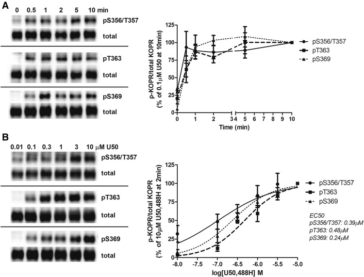
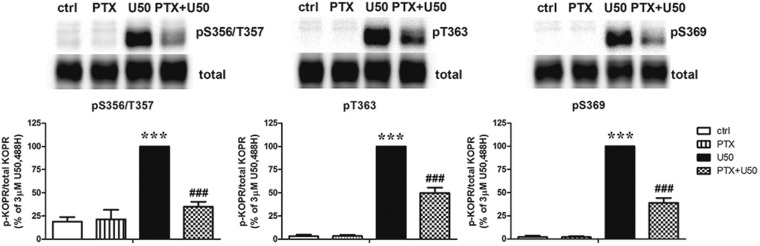
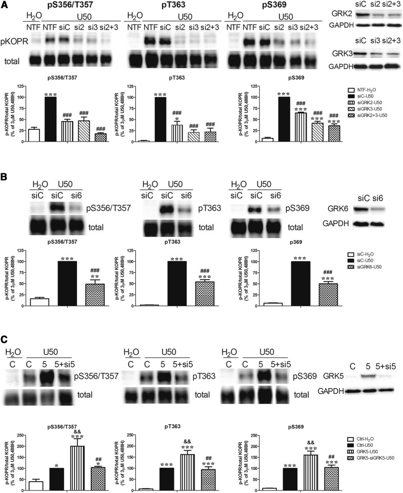
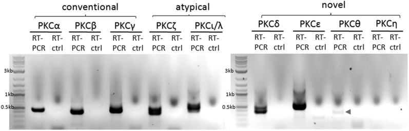
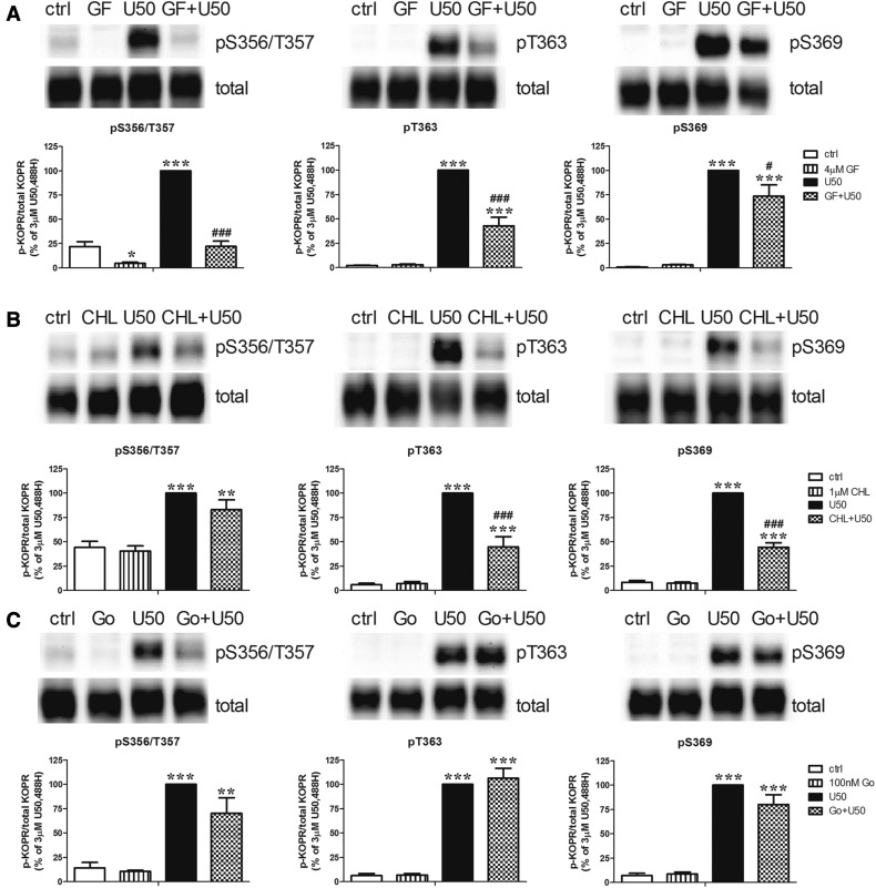
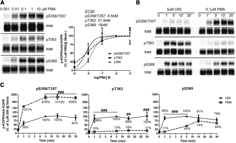
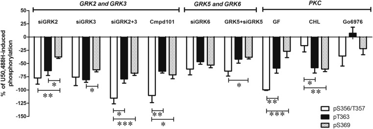
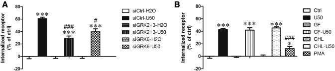
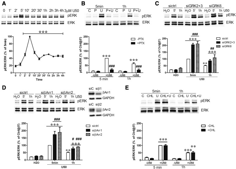
We reported previously that the selective agonist U50,488H promoted phosphorylation of the mouse κ opioid receptor (KOPR) at residues S356, T357, T363, and S369. Here, we found that agonist (U50,488H)-dependent KOPR phosphorylation at all the residues was mediated by Gi/o α proteins and multiple protein kinases [GRK2, GRK3, GRK5, GRK6 and protein kinase C (PKC)]. In addition, PKC activation by phorbol ester induced agonist-independent KOPR phosphorylation. Compared with U50,488H, PKC activation promoted much higher S356/T357 phosphorylation, much lower T363 phosphorylation, and similar levels of S369 phosphorylation. After U50,488H treatment, GRKs, but not PKC, were involved in agonist-induced KOPR internalization. In contrast, PKC activation caused a lower level of agonist-independent KOPR internalization, compared with U50,488H. U50,488H-induced activation of extracellular signal-regulated kinase 1/2 (ERK1/2) was G protein-, but not β-arrestin-, dependent. After U50,488H treatment, GRK-mediated, but not PKC-mediated, KOPR phosphorylation followed by β-arrestin recruitment desensitized U50,488H-induced ERK1/2 response. Therefore, agonist-dependent (GRK- and PKC-mediated) and agonist-independent (PKC-promoted) KOPR phosphorylations show distinct phosphorylation patterns, leading to diverse cellular outcomes.


Figures

Fig. 1.
Fig. 1.
Time-course and dose-response relationship of U50,488H-induced KOPR phosphorylation. N2A-FmK6H cells were treated with 0.1 μM U50,488H for different durations (0, 0.5, 1, 2, 5, and 10 minutes) (A) or at different concentrations of U50,488H (0.01, 0.1, 0.3, 1, 3, and 10 μM) for 2 minutes (B) at 37°C. Cells were lysed with lysis buffer, and then receptors were affinity purified using Ni-NTA agarose beads. Eluted samples were subjected to Western blot using the three phospho-KOPR–specific antibodies. Membranes were stripped and blotted again for total KOPR. See Materials and Methods for details. One representative blot of each phospho-KOPR antibody is shown. Staining intensity was quantified and normalized against that of 10-minute treatment (A) or treatment with 10 μM U50,488H (B). Each value is the mean ± S.E.M. (n = 3–7).
Fig. 2.
Fig. 2.
Effects of PTX on U50,488H-induced KOPR phosphorylation. Cells were pretreated with 200 ng/ml PTX for 2 hours followed by a 2-minute incubation with 3 µM U50,488H. Experiments were performed as described in the Fig. 1 legend. The data shown are reported as the mean ± S.E.M. (n = 3) and were analyzed by two-way ANOVA followed by Bonferroni post hoc test (***P < 0.001, compared with control (ctrl) group; ###P < 0.001, compared with U50,488H-treated group).
Fig. 3.
Fig. 3.
Involvement of GRKs in U50,488-induced KOPR phosphorylation. (A, B) Effects of knockdown of GRK2, GRK3, and GRK6 on U50,488H-induced KOPR phosphorylation. Cells were left NTF or transfected with siCtrl (siC) or siRNAs targeting GRK2 (si2), GRK3 (si3), GRKs2+3 (si2+3), or GRK6 (si6). Two days later, cells were treated with vehicle or 3 μM U50,488H for 2 minutes. Data were normalized against that of the U50,488H-treated siRNA control group (siC-U50), and values are reported as the mean ± S.E.M. (n = 3–6). Knockdown of GRK2, GRK3, and GRK6 was verified by Western blot with specific antibodies. (C) Effects of GRK5 expression and expression plus knockdown on U50,488H-induced KOPR phosphorylation. Cells were cotransfected with the vector pcDNA3 and siCtrl (C), GRK5 cDNA and siCtrl (5), and GRK5 cDNA and GRK5 siRNA (5+si5) with Lipofectamine 2000. Two days later, cells were treated with vehicle or 3 µM U50,488H for 2 minutes. The results were normalized against that of the U50,488H-treated vector control group (Ctrl-U50), and values are reported as the mean ± S.E.M. (n = 6–8). The expression of GRK5 and the knockdown of GRK5 were validated by Western blot. Data were analyzed with one-way ANOVA followed by Tukey post hoc test [*P < 0.05, **P < 0.01, ***P < 0.001, compared with vehicle control group (siC-H2O) or H2O-treated control group (Ctrl-H2O); ##P < 0.01, ###P < 0.001, compared with U50,488H-treated siRNA control group (siC-U50) or U50,488H-treated GRK5 overexpression group (GRK5-U50); &&P < 0.01, compared with U50,488H-treated vector control group (Ctrl-U50)].
Fig. 4.
Fig. 4.
Endogenous PKC isoforms in N2A cells detected with RT-PCR. N2A cells were solubilized with TRIzol reagent and then RNA was extracted. RNA was used for RT-PCR for the detection of different PKC isoforms (conventional PKCs: PKCα, PKCβ, and PKCγ; atypical PKCs: PKCζ and PKCι/λ; novel PKCs: PKCδ, PKCε, PKCθ, and PKCη). For the control, no PCR primers were added (RT-ctrl). The following PKC isoforms (predicted PCR product sizes) were detected: PKCα (496 bp), PKCβ (428 bp), PKCγ (552 bp), PKCζ (428 bp), PKCι/λ (456 bp), PKCδ (428 bp), PKCε (547 bp), and PKCθ (494 bp). PKCθ PCR product was barely detectable (arrowhead). No PKCη PCR product was detected. The experiment was carried out twice with similar results.
Fig. 5.
Fig. 5.
Effect of PKC inhibitors on U50,488H-induced KOPR phosphorylation at S356/T357, T363, and S369. Cells were pretreated with the nonselective PKC inhibitor GF109203X (GF) (4 µM) (A), CHL (1 µM) (B), or the selective PKC α and β1 inhibitor Go6976 (100 nM) (C) for 30 minutes followed by 3 µM U50,488H or vehicle for 2 minutes. Each point is shown as the mean ± S.E.M. (n = 3–6). Data were analyzed by two-way ANOVA followed by Tukey post hoc test (*P < 0.05, **P < 0.01, ***P < 0.001, compared with control (ctrl) group; #P < 0.05, ###P < 0.001, compared with U50,488H-treated group).
Fig. 6.
Fig. 6.
Effect of PKC activation on KOPR phosphorylation. (A) Dose-response relationship of PMA-induced KOPR phosphorylation: cells were treated with different concentrations of PMA (0.001, 0.01, 0.1, 1, and 10 µM) for 30 minutes. (B and C) Time course of PMA-induced KOPR phosphorylation and comparison with that of U50,488H: cells were incubated with 3 µM U50,488H or 0.1 µM PMA for 0, 1, 5, 15, and 30 minutes. In (A), staining intensity was normalized against that of 1 μM PMA, and each value is reported as the mean ± S.E.M. (n = 3–4). The mean EC50 values of PMA in promoting phosphorylation at S356/T357, T363 and S369 are shown. In (B), there was an empty lane between U50,488H-treated and PMA-treated samples. In (C), the staining intensity in (B) was normalized against that of U50,488H at 5 minutes. Data are shown as the mean ± S.E.M. (n = 3–4) and were analyzed by two-way ANOVA followed by Bonferroni post hoc test (##P < 0.01, ###P < 0.001, compared with U50,488H-treated group).
Fig. 7.
Fig. 7.
Regulation of U50,488H-induced KOPR phosphorylation at S356/T357, T363, and S369 by protein kinases. This figure summarizes and compares the percentage of reduction by inhibitors and GRK siRNAs of U50,488H-induced KOPR phosphorylation at all the four phosphosites. The value was calculated according to the equation: % reduction = {[(inhibitor or siRNA + U50,488H) − U50,488H]/U50,488H} × 100%. Data were analyzed by two-way ANOVA followed by Tukey-Kramer post hoc test (*P < 0.05, **P < 0.01, ***P < 0.001).
Fig. 8.
Fig. 8.
Roles of GRKs and PKC in KOPR internalization. (A) GRKs mediated U50,488H-induced KOPR internalization. Cells were transfected with siCtrl or siRNAs targeting GRKs2+3 (siGRKs2+3) or GRK6 (siGRK6). Two days later, cells were treated with vehicle or 3 μM U50,488H for 30 minutes. Cells were collected with 1 mM EDTA in PBS and then washed three times with 0.1% bovine serum albumin in PBS for 10 minutes at 4°C. Receptor internalization was determined using [3H]diprenorphine binding to total and cell surface KOPR in cells as described in the Materials and Methods. Internalized receptors are presented as the percentage of the U50,488H-treated group over that of the water-treated group. The data were shown the mean ± S.E.M. (n = 3) and analyzed with two-way ANOVA followed by Bonferroni post hoc test [***P < 0.001, compared with vehicle control group (siCtrl-H2O); #P < 0.05, ###P < 0.001, compared with U50,488H-treated siRNA control group (siCtrl-U50)]. (B) PKC promoted agonist-independent KOPR internalization but was not involved in U50,488H-dependent KOPR internalization. Cells were treated with 4 µM GF109203X (GF) or 1 µM CHL for 30 minutes and then treated with water or 3 µM U50,488H for 30 minutes. Some cells were treated with 0.1 µM PMA for 30 minutes. Cells were treated as described above. Results are shown as the mean ± S.E.M. (n = 3). Data for PKC inhibitor-treated groups were analyzed by two-way ANOVA followed by Bonferroni post hoc test (***P < 0.001, compared with control-treated group). The data for PMA were analyzed with one-way ANOVA followed by Tukey post hoc test [*P < 0.05; ***P < 0.001, compared with control group (Ctrl); ###P < 0.001, compared with U50,488H-treated group (U50)].
Fig. 9.
Fig. 9.
U50,488H-induced ERK1/2 phosphorylation: roles of G proteins, GRKs, and β-arrestins. (A) Cells were treated with 3 μM U50,488H for different periods of time (0, 1, 2, 5, 10, 20, and 30 minutes and 1, 2, 3, and 4 hours). (B) Cells were pretreated with 200 ng/ml PTX (P) for 2 hours followed by 3 μM U50,488H (U) for 5 minutes or 1 hour. (C and D) Cells were transfected with siCtrl (siC) or siRNAs targeting GRK2 and GRK3 (si2+3), GRK6 (si6), β-arrestin 1 (siβ1 or siβArr1), or β-arrestin 2 (siβ2 or siβArr2). Two days later, cells were treated with H2O or 3 μM U50,488H for 5 minutes or 1 hour. (E) Cells were pretreated with 1 µM CHL for 30 minutes and then incubated with H2O or 3 μM U50,488H for 5 minutes or 1 hour. ERK1/2 phosphorylation was determined as described in Materials and Methods. Data shown are the mean ± S.E.M. (n = 3–4). (A) Data were analyzed by one-way ANOVA followed by Tukey post hoc test (***P < 0.001, compared with time zero group). (B) Data were analyzed by two-way ANOVA followed by Bonferroni post hoc test (***P < 0.001, compared with vehicle-treated group; ###P < 0.001, compared with U50,488H-treated group). (C) Data were analyzed by two-way ANOVA followed by Bonferroni post hoc test (**P < 0.01, ***P < 0.001, compared with water-treated group; ##P < 0.01, ###P < 0.001, compared with siCtrl-U50,488H-treated group). (D) Data were analyzed by two-way ANOVA followed by Bonferroni post hoc test (**P < 0.01, ***P < 0.001, compared with water-treated group; ###P < 0.001, compared with sictrl-U50,488H-treated group at 5 minutes) and by t test for 1-hour U50,488H treatment between sictrl and siβ-arrestin (#P < 0.05, ###P < 0.001). (E) Data were analyzed by two-way ANOVA followed by Bonferroni post hoc test (**P < 0.01, ***P < 0.001, compared with vehicle-treated group).

References

    1. Al-Hasani R, Bruchas MR. (2011) Molecular mechanisms of opioid receptor-dependent signaling and behavior. Anesthesiology 115:1363–1381. - PMC - PubMed
    1. Ansonoff MA, Zhang J, Czyzyk T, Rothman RB, Stewart J, Xu H, Zjwiony J, Siebert DJ, Yang F, Roth BL, et al. (2006) Antinociceptive and hypothermic effects of Salvinorin A are abolished in a novel strain of kappa-opioid receptor-1 knockout mice. J Pharmacol Exp Ther 318:641–648. - PubMed
    1. Appleyard SM, Celver J, Pineda V, Kovoor A, Wayman GA, Chavkin C. (1999) Agonist-dependent desensitization of the kappa opioid receptor by G protein receptor kinase and beta-arrestin. J Biol Chem 274:23802–23807. - PubMed
    1. Belcheva MM, Vogel Z, Ignatova E, Avidor-Reiss T, Zippel R, Levy R, Young EC, Barg J, Coscia CJ. (1998) Opioid modulation of extracellular signal-regulated protein kinase activity is ras-dependent and involves Gbetagamma subunits. J Neurochem 70:635–645. - PMC - PubMed
    1. Belcheva MM, Clark AL, Haas PD, Serna JS, Hahn JW, Kiss A, Coscia CJ. (2005) Mu and kappa opioid receptors activate ERK/MAPK via different protein kinase C isoforms and secondary messengers in astrocytes. J Biol Chem 280:27662–27669. - PMC - PubMed

Publication types

MeSH terms

Substances

LinkOut - more resources