. 2017 Sep 11;7(1):11218.
doi: 10.1038/s41598-017-10125-3.

Cinaciguat ameliorates glomerular damage by reducing ERK1/2 activity and TGF-ß expression in type-1 diabetic rats

Affiliations

Cinaciguat ameliorates glomerular damage by reducing ERK1/2 activity and TGF-ß expression in type-1 diabetic rats

Szabina Czirok et al. Sci Rep. .

Abstract

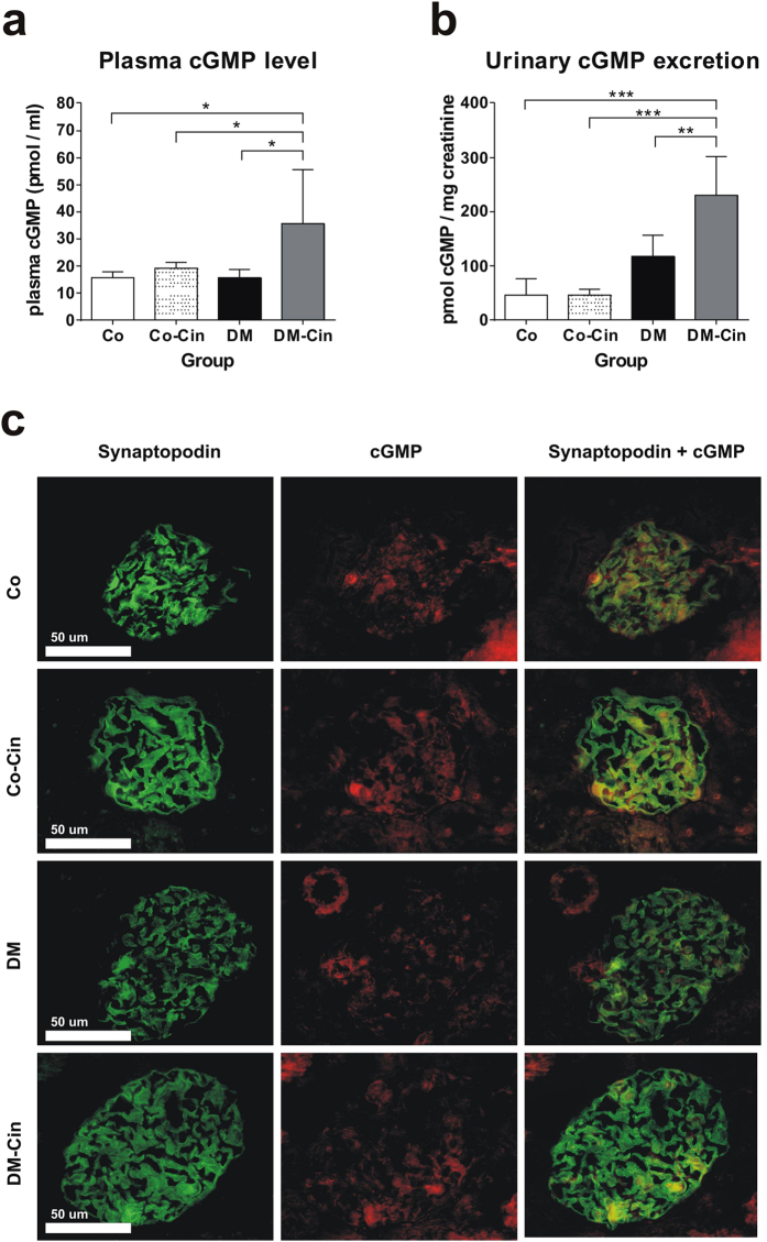
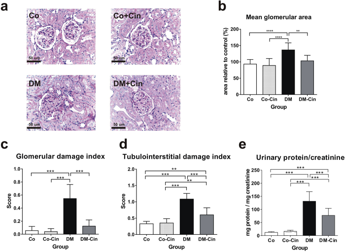
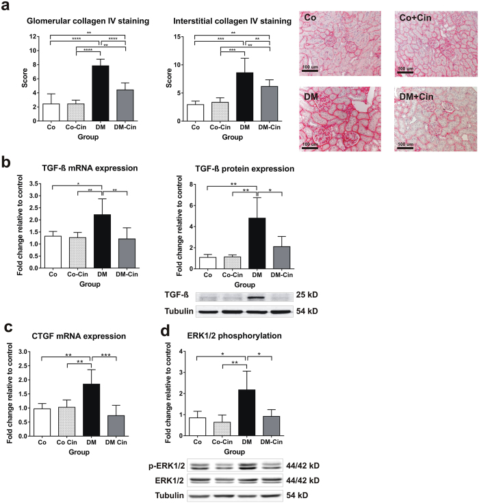
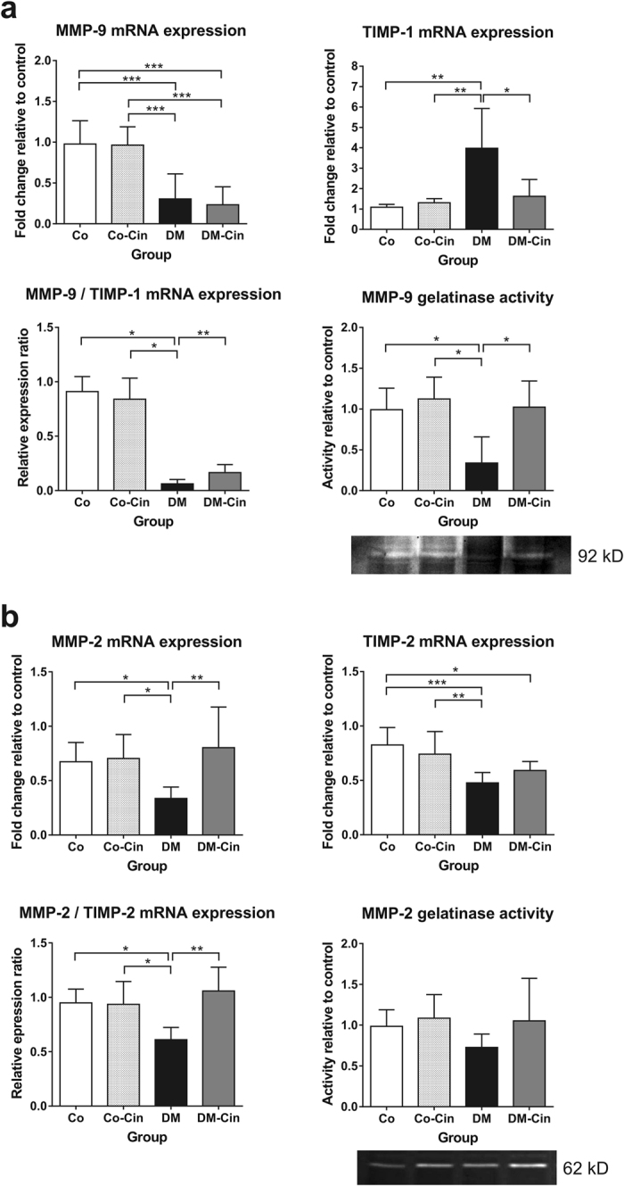
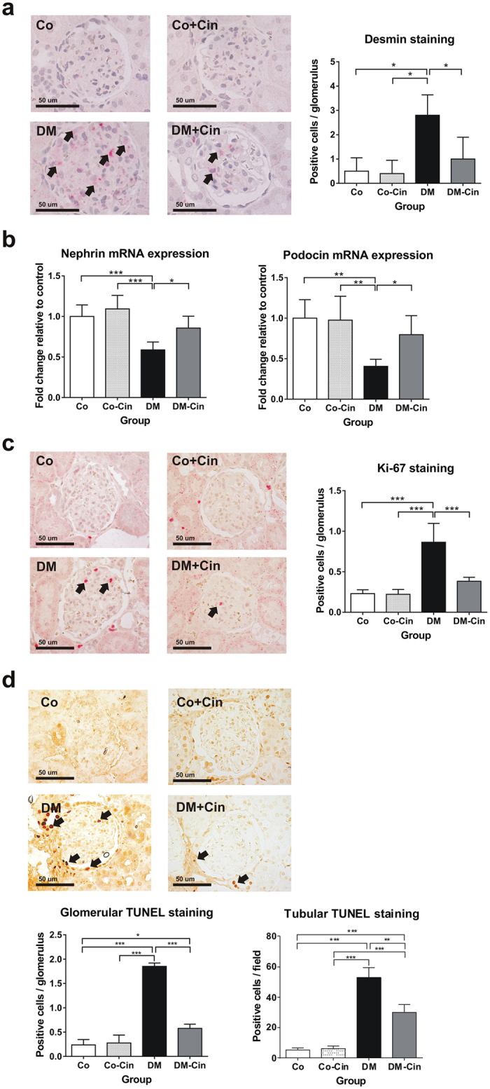
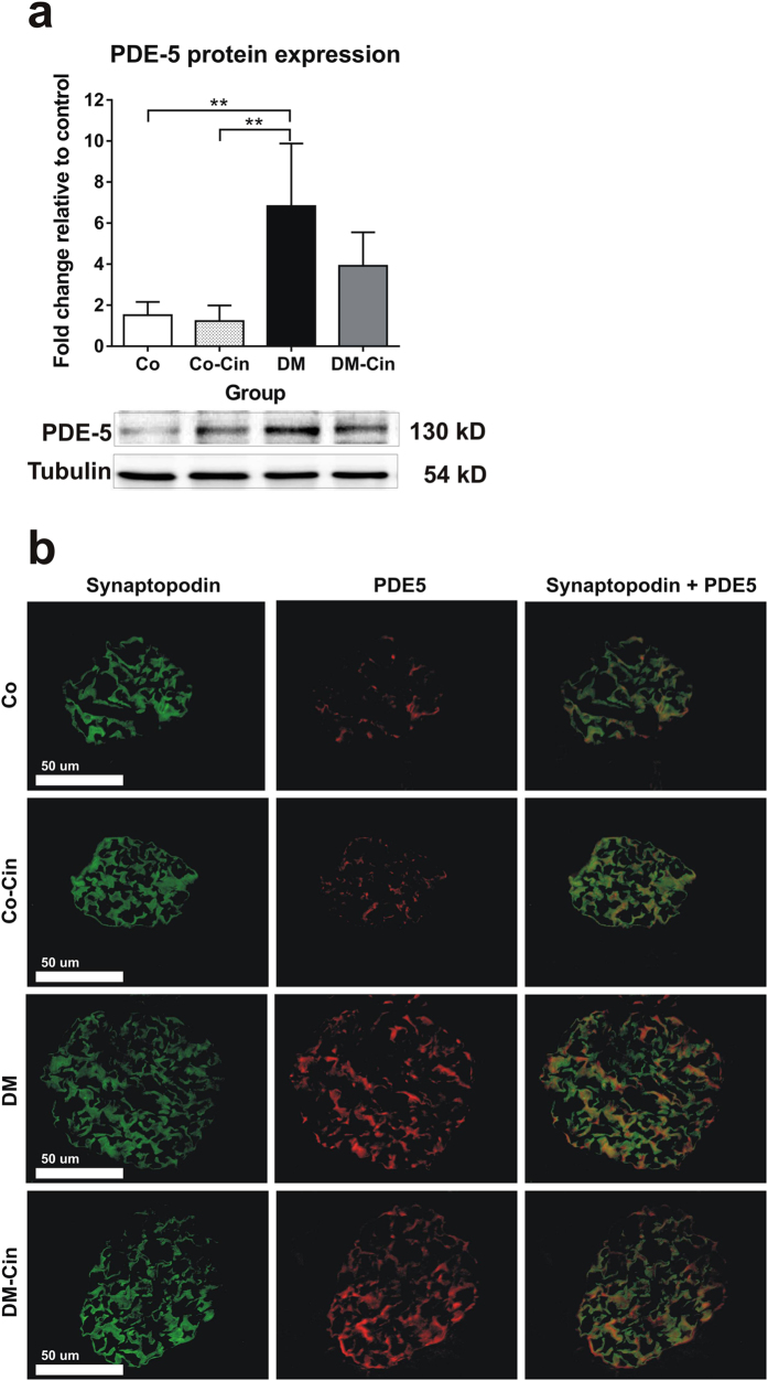
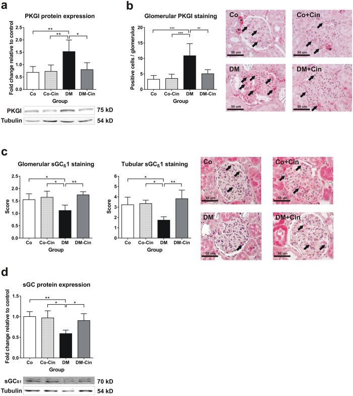
Decreased soluble guanylate cyclase activity and cGMP levels in diabetic kidneys were shown to influence the progression of nephropathy. The regulatory effects of soluble guanylate cyclase activators on renal signaling pathways are still unknown, we therefore investigated the renal molecular effects of the soluble guanylate cyclase activator cinaciguat in type-1 diabetic (T1DM) rats. Male adult Sprague-Dawley rats were divided into 2 groups after induction of T1DM with 60 mg/kg streptozotocin: DM, untreated (DM, n = 8) and 2) DM + cinaciguat (10 mg/kg per os daily, DM-Cin, n = 8). Non-diabetic untreated and cinaciguat treated rats served as controls (Co (n = 10) and Co-Cin (n = 10), respectively). Rats were treated for eight weeks, when renal functional and molecular analyses were performed. Cinaciguat attenuated the diabetes induced proteinuria, glomerulosclerosis and renal collagen-IV expression accompanied by 50% reduction of TIMP-1 expression. Cinaciguat treatment restored the glomerular cGMP content and soluble guanylate cyclase expression, and ameliorated the glomerular apoptosis (TUNEL positive cell number) and podocyte injury. These effects were accompanied by significantly reduced TGF-ß overexpression and ERK1/2 phosphorylation in cinaciguat treated diabetic kidneys. We conclude that the soluble guanylate cyclase activator cinaciguat ameliorated diabetes induced glomerular damage, apoptosis, podocyte injury and TIMP-1 overexpression by suppressing TGF-ß and ERK1/2 signaling.


Conflict of interest statement

The authors declare that they have no competing interests.

Figures

Figure 1
Figure 1
Effect of diabetes and cinaciguat treatment on cyclic guanosine monophosphate (cGMP) levels in the plasma and urine, and glomerular cGMP content. (a) Plasma cGMP levels of DM rats were similar to Co and Co-Cin controls, but DM-Cin rats had significantly higher circulating cGMP levels. (b) According to plasma cGMP, urinary cGMP excretion increased significantly in DM-Cin rats, as compared to DM or to both controls. (c) Double immunostaining for synaptopodin (green) and cGMP (red) depicted visible glomerular cGMP content in both Co and Co-Cin kidneys, mainly co-localized with synaptopodin (yellow). Immunoreactivity for cGMP was practically absent in glomeruli of DM rats, but it was restored in DM-Cin kidneys. Data are presented as mean ± SD (n = 8–10/group). *p < 0.05, **p < 0.01, ***p < 0.001 (two-way ANOVA with Sidak’s multiple comparison test).
Figure 2
Figure 2
Effect of cinaciguat on kidney histology and proteinuria in diabetic rats. (a,b) Representative photomicrographs of PAS stained kidneys (400x magnification) show normal glomerular structure in both Co and Co-Cin controls, mesangial expansion and glomerular hypertrophy in DM and almost normal structure in DM-Cin groups (scale bar represents 50 μm). (c,d) Glomerular and tubulointerstitial damage index scores of each group are shown. (e) The extent of proteinuria was determined by urinary protein/creatinine ratio (mg protein/mg creatinine), showing significant proteinuria in DM that was reduced in DM-Cin rats. Data are presented as mean ± SD (n = 8–10/group). **p < 0.01, ***p < 0.001 (two-way ANOVA with Sidak’s multiple comparison test).
Figure 3
Figure 3
Effect of diabetes and cinaciguat on renal fibrosis. (a) Immunostaining for collagen IV was performed on paraffin embedded kidney sections. Representative photomicrographs (200x magnification) show moderate tubulointerstitial and glomerular staining in controls, strong tubulointerstitial and thickened glomerular expression in DM but only mild collagen IV expression in DM-Cin groups (scale bar represents 50 μm). (b,c) Compared to controls, the renal mRNA and protein expression analysis of profibrotic TGF-ß as well as CTGF mRNA expression showed a marked overexpression in DM kidneys, which was significantly attenuated in DM-Cin group. TGF-ß protein expression of each sample was normalized for tubulin expression and given as fold change relative to a calibrator. The mRNA expression were normalized for GAPDH expression using the formula 2 −ΔΔCt. (d) DM kidneys showed significantly increased phosphorylation of the p44/p42 MAPK (ERK1/2) as compared to non-diabetic controls, which was inhibited by cinaciguat treatment, as seen in DM-Cin kidneys. Representative immunoblots are shown. Phospho-ERK1/2 expression was normalized for total ERK1/2 expression of each sample and given as fold change. Data are presented as mean ± SD (n = 8–10/group). **p < 0.01, ***p < 0.001 (two-way ANOVA with Sidak’s multiple comparison test).
Figure 4
Figure 4
Effect of cinaciguat on the expression of renal extracellular matrix turnover components. (a) Compared to the non-diabetic controls, MMP-9 mRNA expression was markedly reduced in DM kidneys, accompanied by 4-fold increased TIMP-1 expression. Although cinaciguat did not alter MMP-9 expression, it attenuated TIMP-1 overexpression, which resulted in slightly better MMP-9/TIMP-1 ratio in DM-Cin kidneys, as compared to DM, supported by the MMP-9 zymography results. (b) The renal mRNA expression of MMP-2 dropped in DM rats, but was normalized in DM-Cin group. TIMP-2 mRNA expression reduced in both DM and DM-Cin groups, but the MMP-2/TIMP-2 imbalance was attenuated in DM-Cin kidneys. Zymography did not show, in contrast, significant changes in MMP-2 activity. Representative zymogram bands are shown. The mRNA expression was normalized for GAPDH expression using the formula 2 −ΔΔCt. Data are presented as mean ± SD (n = 8–10/group). *p < 0.05, **p < 0.01, ***p < 0.001 (two-way ANOVA with Sidak’s multiple comparison test).
Figure 5
Figure 5
Evaluation of podocyte damage, glomerular cell proliferation and apoptosis. (a) Immunostaining for desmin, as marker of podocyte damage, was performed on paraffin embedded kidney sections. Representative photomicrographs (400x magnification) show no staining in non-diabetic controls, but several positive podocytes (arrows) in DM with significantly reduced staining intensity in DM-Cin kidneys (scale bar represents 50 μm). (b) Compared to controls, the mRNA expression of nephrin and podocin was reduced by at least 40% in DM kidneys, showing diabetic podocyte damage. Both nephrin and podocin expression was significantly ameliorated in DM-Cin kidneys. The mRNA expression was normalized for GAPDH expression using the formula 2 −ΔΔCt. (c) Representative photomicrographs for Ki-67 are shown (400x magnification), as a marker of cell proliferation. We detected significantly more Ki-67 positive proliferating glomerular cells in DM rats than in non-diabetic controls, which difference was abolished by cinaciguat treatment. (d) TUNEL assay demonstrated a marked increase of apoptotic glomerular and tubular cell count in DM rats vs. controls, which was significantly reduced in DM-Cin kidneys. Scale bar represents 50 μm. Data are presented as mean ± SD (n = 8–10/group). *p < 0.05, **p < 0.01, ***p < 0.001 (two-way ANOVA with Sidak’s multiple comparison test).
Figure 6
Figure 6
Analyses of renal PDE-5 protein expression and glomerular immunoreactivity. (a) As compared to non-diabetic controls, DM kidneys showed 6-fold increased PDE-5 expression. Cinaciguat treatment only tended to lower PDE-5 expression in DM-Cin kidneys. (b) Double immunostaining with the podocyte marker synaptopodin (green) revealed minimal basal PDE-5 expression (red) in controls but strong glomerular co-localization in DM and DM-Cin kidneys (yellow color). Bar represents 50 μm (400x magnification). Representative PDE-5 immunoblot is shown. Protein expressions were normalized to tubulin and expressed as fold change relative to a calibrator control sample. Data are presented as mean ± SD (n = 8–10/group). **p < 0.01 (two-way ANOVA with Sidak’s multiple comparison test).
Figure 7
Figure 7
Immunoblot analysis of the renal NO-cGMP pathway components. (a,b) Glomerular and tubular guanylate cyclase (sGCβ1) immunohistochemistry depicted strong glomerular and tubular staining in non-diabetic controls (arrows), with 40% reduction of expression both in total kidney homogenates (a) and in glomeruli and tubuli of DM rats (b). Both glomerular and tubular sGC staining was significantly stronger in DM-Cin kidneys, as compared to DM. (c,d) As compared to non-diabetic controls, DM kidneys showed significant PKG overexpression (c) and increased number of positive glomerular cells (d). Cinaciguat treatment ameliorated PKG overexpression in DM-Cin as shown by immunblot (c) and immunostaining (d). Bar represents 50 μm (400x magnification). Representative immunoblots are shown. All protein expressions were normalized to tubulin and expressed as fold change relative to a calibrator control sample. Data are presented as mean ± SD (n = 8–10/group). *p < 0.05, **p < 0.01, ***p < 0.001 (two-way ANOVA with Sidak’s multiple comparison test).

Similar articles

Cited by

References

    1. Jha V, et al. Chronic kidney disease: global dimension and perspectives. Lancet. 2013;382:260–272. doi: 10.1016/S0140-6736(13)60687-X. - DOI - PubMed
    1. Brewster UC, Setaro JF, Perazella MA. The renin-angiotensin-aldosterone system: cardiorenal effects and implications for renal and cardiovascular disease states. The American journal of the medical sciences. 2003;326:15–24. doi: 10.1097/00000441-200307000-00003. - DOI - PubMed
    1. Ito D, et al. Effects of Ipragliflozin on Diabetic Nephropathy and Blood Pressure in Patients With Type 2 Diabetes: An Open-Label Study. Journal of clinical medicine research. 2017;9:154–162. doi: 10.14740/jocmr2875w. - DOI - PMC - PubMed
    1. Suzuki D, et al. Immunohistochemical evidence for an increased oxidative stress and carbonyl modification of proteins in diabetic glomerular lesions. Journal of the American Society of Nephrology: JASN. 1999;10:822–832. - PubMed
    1. Wolf G, Chen S, Ziyadeh FN. From the periphery of the glomerular capillary wall toward the center of disease: podocyte injury comes of age in diabetic nephropathy. Diabetes. 2005;54:1626–1634. doi: 10.2337/diabetes.54.6.1626. - DOI - PubMed

Publication types

MeSH terms