. 2017 Sep 11;7(1):11242.
doi: 10.1038/s41598-017-11687-y.

A pH-dependent Antibacterial Peptide Release Nano-system Blocks Tumor Growth in vivo without Toxicity

Affiliations

A pH-dependent Antibacterial Peptide Release Nano-system Blocks Tumor Growth in vivo without Toxicity

Jing Cao et al. Sci Rep. .

Abstract

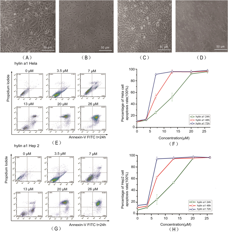
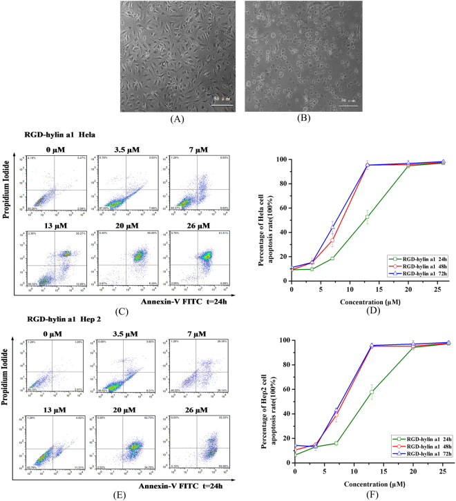
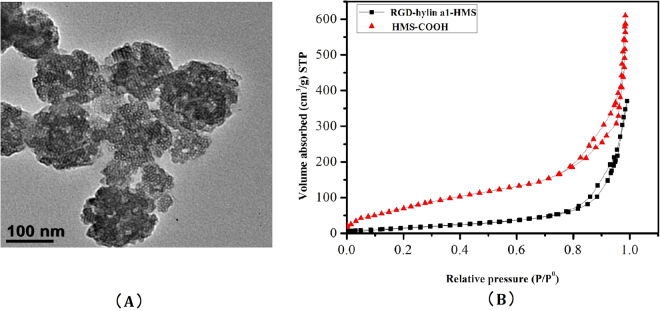
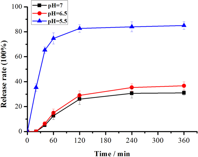
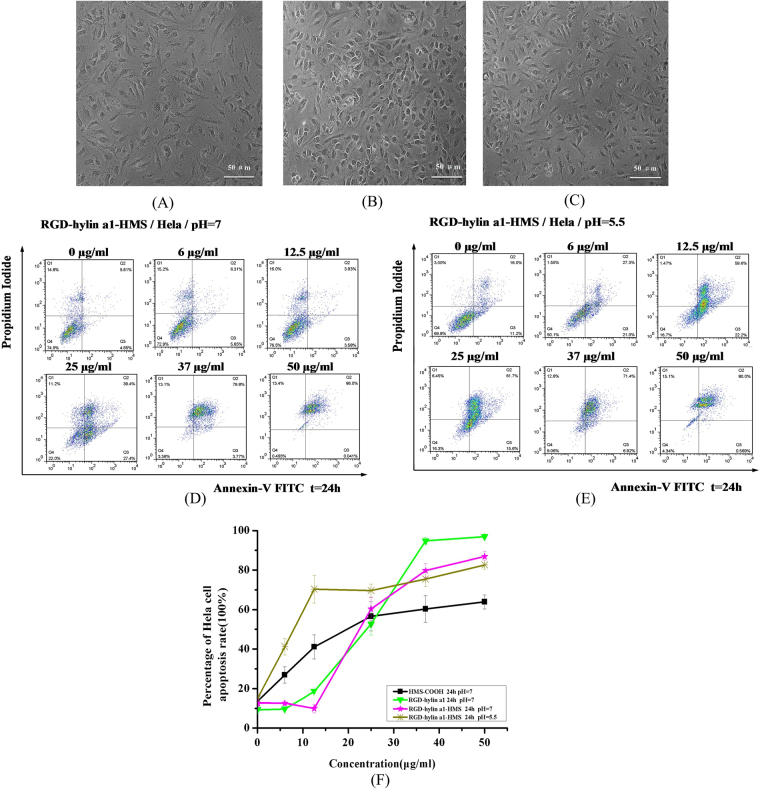
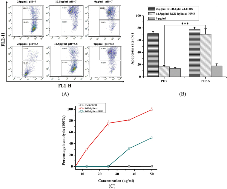
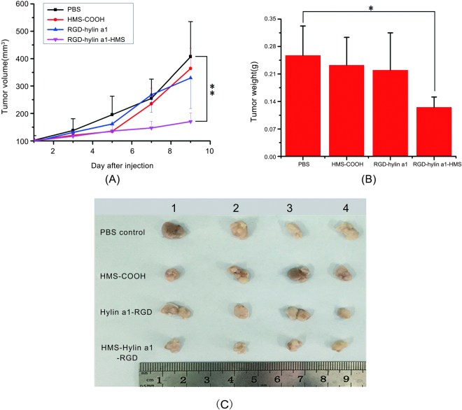
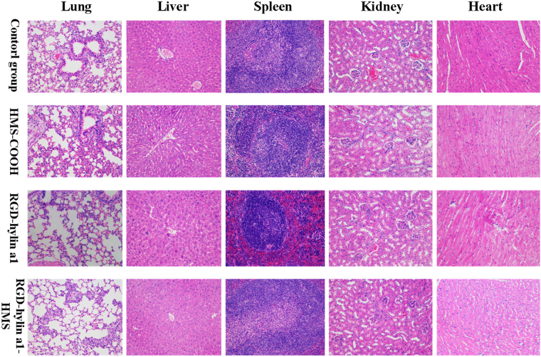
In this study, we designed a nano-system where a novel antibacterial peptide RGD-hylin a1 with reduced hemolysis than the commonly studied melittin was loaded onto mesoporous silica (HMS). We found out that the designed nano-system, RGD-hylin a1-HMS, released RGD-hylin a1 in a pH-dependent manner. It caused apoptosis of cancer cells at low dosage of the antibacterial peptide at pH = 5.5, but was safe to the cells at pH = 7. The hemolytic activity of RGD-hylin a1 itself was reduced by 50~100% by the nano-system depending on the dosage. When this nano-system was administered to tumor-bearing mice at low dosage via intravenous injection, the growth of the solid tumor was blocked by the RGD-hylin a1-HMS nano-system with a 50-60% inhibition rate relative to the PBS-treated control group in terms of tumor volume and weight. Further, the hemolytic activity of RGD-hylin a1 was completely eliminated within the delivery system with no other side effects observed. This study demonstrates that this smart pH-dependent antibacterial peptide release nano-system has superior potential for solid tumor treatments through intravenous administration. This smart-releasing system has great potential in further clinical applications.


Conflict of interest statement

The authors declare that they have no competing interests.

Figures

Figure 1
Figure 1
Hylin a1 cytotoxicity assays in Hela and Hep2 cells in vitro. Cellular morphology was observed by 10 × inverted microscope. All data was presented as the means ± SD, n = 3. (A) Hela cells grew for 24 h. (B) Hela cells were treated with 20 μM hylin a1 for 24 h. (C) Hep2 cells grew for 24 h. (D) Hep2 cells were treated with 20 μM hylin a1 for 24 h. (E) and (F) Apoptosis assays evaluating the cytotoxicity of hylin a1 in Hela cells. (G) and (H) Apoptosis assays evaluating the cytotoxicity of hylin a1 in Hep2 cells.
Figure 2
Figure 2
RGD-hylin a1 cytotoxicity assays in Hela and Hep2 cells in vitro. Cellular morphology was observed by 10× inverted microscope. All data was presented as the means ± SD, n = 3. (A) Hela cells were treated with 20 μM RGD-hylin a1 for 24 h. (B) Hep2 cells were treated with 20 μM RGD-hylin a1 for 24 h. (C) and (D) Apoptosis assays evaluating the cytotoxicity of RGD-hylin a1 in Hela cells. (E) and (F) Apoptosis assays evaluating the cytotoxicity of RGD-hylin a1 in Hep2 cells.
Figure 3
Figure 3
(A) Transmission Electron Microscopy (TEM) image of HMS-COOH, the black bar indicates 100 nm. (B) Nitrogen adsorption/desorption isotherms of HMS-COOH and RGD-hylina1-HMS samples.
Figure 4
Figure 4
Release rates of RGD-hylina1 from RGD-hylina1-HMS at pH values of 7, 6.5, and 5.5 with different times.
Figure 5
Figure 5
Cytotoxicity assays of HMS-COOH and RGD-hylin a1-HMS (pH = 7 and pH = 5.5) in Hela cells. Cellular morphology was observed by 10× inverted microscope. (A) Hela cells were treated with 25 μg/ml HMS-COOH for 24 h; (B) Hela cells were treated with 25 μg/ml RGD-hylin a1-HMS (pH = 7) for 24 h; (C) Hela cells were treated with 25 μg/ml RGD-hylin a1-HMS (pH = 5.5) for 24 h; (D) RGD-hylin a1-HMS induced apoptosis of Hela cells. Hela cells were treated with various concentrations of RGD-hylin a1-HMS (pH = 7) for 24 h. (E) RGD-hylin a1-HMS induced apoptosis of Hela cells. Hela cells were treated with various concentrations of RGD-hylin a1-HMS (pH = 5.5) for 24 h. (F) Quantification of apoptosis rates of HMS-COOH, RGD-hylin a1, and RGD-hylin a1-HMS (pH = 7 and pH = 5.5) in Hela cells.
Figure 6
Figure 6
Mitochondrial membrane potential assays and hemolysis assays in Hela cells. All data was presented as the means ± SD, n = 3. (A) and (B) Hela cells were treated with RGD-hylin a1-HMS for 24 h, stained with JC-1, and analyzed by FACS. Results were presented as percentage of apoptosis. (C) Hemolytic assays for HMS-COOH, RGD-hylin a1 and RGD-hylin a1-HMS (pH = 7) in RBC.
Figure 7
Figure 7
In vivo evaluation of the effect of RGD-hylin a1-HMS on the inhibition of tumor growth. All data was presented as the means ± SD, n = 4, **p < 0.01, *p < 0.05. (A) and (B) Tumor volume and weight in each group with increasing days. (C) Photographs of the tumors from each group on the 9th day.
Figure 8
Figure 8
Evaluation of the side effects of RGD-hylin a1-HMS in vivo. All data was presented as the means + SD, n = 3, *p < 0.05, **p < 0.01, ***p < 0.001. (A,B) Biochemical analyses of glutamate pyruvate transaminase (ALT) and glutamic-oxalacetic transaminease (AST). (C,D) Blood hemanalysis of red blood cells (RBC) and hemoglobin (HGB).
Figure 9
Figure 9
Histopathologic analyses of H&E-stained tissue sections from the hearts, livers, spleens, lungs, and kidneys of tumor-bearing mice.

References

    1. Patrzykat A, Douglas SE. Gone gene fishing: how to catch novel marine antimicrobials. Trends in biotechnology. 2003;21:362–369. doi: 10.1016/S0167-7799(03)00145-8. - DOI - PubMed
    1. Papo N, Shai Y. Host defense peptides as new weapons in cancer treatment. Cellular and molecular life sciences: CMLS. 2005;62:784–790. doi: 10.1007/s00018-005-4560-2. - DOI - PMC - PubMed
    1. Hoskin DW, Ramamoorthy A. Studies on anticancer activities of antimicrobial peptides. Biochimica et biophysica acta. 2008;1778:357–375. doi: 10.1016/j.bbamem.2007.11.008. - DOI - PMC - PubMed
    1. Suttmann H, et al. Antimicrobial peptides of the Cecropin-family show potent antitumor activity against bladder cancer cells. BMC urology. 2008;8:5. doi: 10.1186/1471-2490-8-5. - DOI - PMC - PubMed
    1. Liu S, et al. Melittin prevents liver cancer cell metastasis through inhibition of the Rac1-dependent pathway. Hepatology. 2008;47:1964–1973. doi: 10.1002/hep.22240. - DOI - PubMed

Publication types

Substances

LinkOut - more resources