. 2017 Dec;22(12):1673-1679.
doi: 10.1038/mp.2017.176. Epub 2017 Sep 12.

Gβγ subunit activation promotes dopamine efflux through the dopamine transporter

Affiliations

Gβγ subunit activation promotes dopamine efflux through the dopamine transporter

J Garcia-Olivares et al. Mol Psychiatry. 2017 Dec.

Abstract

The dopamine transporter (DAT) is an important regulator of brain dopamine (DA) homeostasis, controlling the intensity and duration of DA signaling. DAT is the target for psychostimulants-like cocaine and amphetamine-and plays an important role in neuropsychiatric disorders, including attention-deficit hyperactivity disorder and drug addiction. Thus, a thorough understanding of the mechanisms that regulate DAT function is necessary for the development of clinical interventions to treat DA-related brain disorders. Previous studies have revealed a plethora of protein-protein interactions influencing DAT cellular localization and activity, suggesting that the fine-tuning of DA homeostasis involves multiple mechanisms. We recently reported that G-protein beta-gamma (Gβγ) subunits bind directly to DAT and decrease DA clearance. Here we show that Gβγ induces the release of DA through DAT. Specifically, a Gβγ-binding/activating peptide, mSIRK, increases DA efflux through DAT in heterologous cells and primary dopaminergic neurons in culture. Addition of the Gβγ inhibitor gallein or DAT inhibitors prevents this effect. Residues 582 to 596 in the DAT carboxy terminus were identified as the primary binding site of Gβγ. A TAT peptide containing the Gβγ-interacting domain of DAT blocked the ability of mSIRK to induce DA efflux, consistent with a direct interaction of Gβγ with the transporter. Finally, activation of a G-protein-coupled receptor, the muscarinic M5R, results in DAT-mediated DA efflux through a Gβγ-dependent mechanism. Collectively, our data show that Gβγ interacts with DAT to promote DA efflux. This novel mechanism may have important implications in the regulation of brain DA homeostasis.


Conflict of interest statement

CONFLICT OF INTEREST

The authors declare no conflict of interest.

Figures
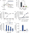
Figure 1
Figure 1
Activation of Gβγ subunits by mSIRK promotes DA efflux in heterologous cells. (a) Amperometric currents obtained from DA-preloaded CHO and CHO-DAT cells after bath application of mSIRK (10 μM) and scb-mSIRK (10 μM). DAT was blocked with cocaine (20 μM) for 5 min followed by the application of mSIRK (10 μM). (b) Maximum DA current induced in CHO-DAT cells by mSIRK (n = 9), scb-SIRK (n = 3) and mSIRK after cocaine (n = 3). (cf) Efflux assay in CHO-DAT cells preloaded with 0.02 μM [3H]-DA. (c) Time-course of [3H]-DA efflux in CHO-DAT cells with and without (CTL) mSIRK (20 μM), and scb-mSIRK (20 μM) (n = 3). (d) mSIRK dose response in the presence of cocaine (20 μM), mazindol (50 μM) or GBR12935 (10 μM) (n = 6). (e) Effect of Gβγ inhibition on the mSIRK-induced DA efflux. CHO-DAT cells were incubated with mSIRK (10 μv) in the absence or presence of gallein (10, 25, 50 μv) (n = 3). (f) Effect of the Gβγ scavenger peptide TAT-Kpept (20 μM) on mSIRK-induced DA efflux (n = 3). In d, e and f, released [3H]-DA was measured 10 min after mSIRK. *P<0.05, **P<0.01
Figure 2
Figure 2
Activation of Gβγ subunits by mSIRK promotes DA efflux in dopaminergic neurons. (a) Dose-dependent effect of mSIRK on DA efflux in primary cultures of DA neurons. [3H]-DA efflux was measured 15 min after application of mSIRK (10 μM) (n = 3). (b) Effect of mSIRK (10 μM) and control peptide (scb-mSIRK, 10 μM) on [3H]-DA efflux. mSIRK-induced efflux was tested in presence of the DAT inhibitor GBR12935 (0.5 μM) or the Gβγ inhibitor gallein (10 μM) (n = 4). **P<0.01.
Figure 3
Figure 3
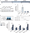
Characterization of the Gβγ binding motif within the DAT carboxy-terminus. (a) Schematic representation of peptides corresponding to the carboxy terminus of hDAT. (b) Representative (n = 4) in vitro binding assay (I.B.). Biotinylated peptides were immobilized with Neutravinagarose resin, followed by incubation with purified Gβγ. Bound Gβγ was detected by SDS-PAGE followed by Gβ-immunoblotting. In lower panel, flow-through from binding assay represents the unbound Gβγ present in all samples as loading control. (c) Densitometric analysis of the in vitro binding assays (n = 4). Relative binding of Gβγ to biotinylated peptides was calculated relative to control group (no peptide bound) (d) Depiction of in vitro binding assay and sequences of DATct1 peptides. (e) Purified Gβγ was added to peptides immobilized on an avidin-coated microplate. For detection of bound Gβγ, a Gβ pan-antibody, Protein A-HRP-conjugated antibody and QuantaBlu kit was used. All results are shown as relative binding to group without Gβγ (n = 3). *P<0.05, **P<0.01.
Figure 4
Figure 4
A TAT-DATct1 peptide containing the DAT-Gβγ binding motif inhibits mSIRK-stimulated DA efflux. (a) Efflux of [3H]-DA in CHO-DAT cells after application of mSIRK (10 μM) and increasing concentrations of TAT-DTct1 peptide (n = 3). (b) Preloaded dopaminergic neurons with [3H]-DA were incubated for 15 min with different combinations of TAT-DATct1 peptide (20 μM) or TAT-scbDATct1 control peptide (20 μM) followed by mSIRK (20 μM) (n = 5). **P<0.01.
Figure 5
Figure 5
Muscarinic receptor activation induces Gβγ-dependent DAT-mediated efflux. (a, b) [3H]-MPP+ efflux assay in CHO-DAT and CHO-DAT cells expressing hM5R. (a) [3H]-MPP+ efflux after the addition of increasing concentrations of carbachol. (n = 4). (b) Effect of gallein (20 μM) and TAT-DATct1 peptide (20 μM) on [3H]-MPP+ efflux induced by carbachol (Cchol, 100 μM) (n = 3, **P<0.01).

References

    1. Torres GE, Gainetdinov RR, Caron MG. Plasma membrane monoamine transporters: structure, regulation and function. Nat Rev Neurosci. 2003;4:13–25. - PubMed
    1. Cragg SJ, Rice ME. DAncing past the DAT at a DA synapse. Trends Neurosci. 2004;27:270–277. - PubMed
    1. Giros B, Caron MG. Molecular characterization of the dopamine transporter. Trends Pharmacol Sci. 1993;14:43–49. - PubMed
    1. Sulzer D, Sonders MS, Poulsen NW, Galli A. Mechanisms of neurotransmitter release by amphetamines: a review. Prog Neurobiol. 2005;75:406–433. - PubMed
    1. Robbins TW, Everitt BJ. Drug addiction: bad habits add up. Nature. 1999;398:567–570. - PubMed

MeSH terms

Substances