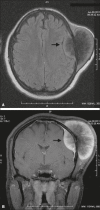
Primary osteosarcoma of the cranial vault
- PMID: 28894335
- PMCID: PMC5586518
- DOI: 10.1590/0100-3984.1914-2014
Primary osteosarcoma of the cranial vault
Abstract
Only 5-10% of osteosarcomas arise from the craniofacial bones. We report the case of a 14-year-old female patient who presented with headache and a mass that had been growing in the left frontoparietal region for six months. We describe the findings on conventional radiography, computed tomography, and magnetic resonance imaging.
Osteossarcomas que se originam dos ossos craniofaciais correspondem a apenas 5-10% dos casos. Neste artigo relatamos caso de uma paciente de 14 anos de idade com quadro de cefaleia e crescimento de massa tumoral na região frontoparietal esquerda com evolução de seis meses. São descritos os achados na radiografia simples, tomografia computadorizada e ressonância magnética.
Keywords: Neoplasms; Osteosarcoma; Skull.
Figures


References
-
- Greenspan A. Radiologia ortopédica - uma abordagem prática. 5ª ed. Rio de Janeiro: Guanabara Koogan; 2012.
-
- Mascarenhas L, Peteiro A, Ribeiro CA, et al. Skull osteosarcoma: illustrated review. Acta Neurochir (Wien) 2004;146:1235–1239. - PubMed
-
- Fukunaga M. Low-grade central osteosarcoma of the skull. Pathol Res Pract. 2005;201:131–135. - PubMed
-
- Chander B, Ralte AM, Dahiya S, et al. Primary osteosarcoma of the skull. A report of 3 cases. J Neurosurg Sci. 2003;47:177–181. - PubMed
-
- Patel AJ, Rao VY, Fox BD, et al. Radiation-induced osteosarcomas of the calvarium and skull base. Cancer. 2011;117:2120–2126. - PubMed
Publication types
LinkOut - more resources
Full Text Sources
Other Literature Sources
