

Corneal Cell Morphology in Keratoconus: A Confocal Microscopic Observation
- PMID: 28894403
- PMCID: PMC5566061
- DOI: 10.21315/mjms2017.24.2.6
Corneal Cell Morphology in Keratoconus: A Confocal Microscopic Observation
Abstract
Purpose: To evaluate corneal cell morphology in patients with keratoconus using an in vivo slit scanning confocal microscope.
Methods: A cross-sectional study was conducted to evaluate the corneal cell morphology of 47 keratoconus patients and 32 healthy eyes without any ocular disease. New keratoconus patients with different disease severities and without any other ocular co-morbidity were recruited from the ophthalmology department of a public hospital in Malaysia from June 2013 to May 2014. Corneal cell morphology was evaluated using an in vivo slit-scanning confocal microscope. Qualitative and quantitative data were analysed using a grading scale and the Nidek Advanced Visual Information System software, respectively.
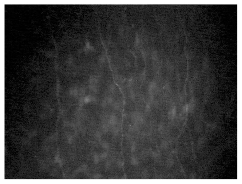
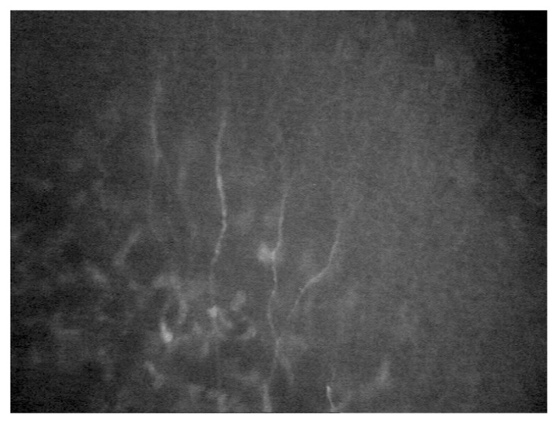
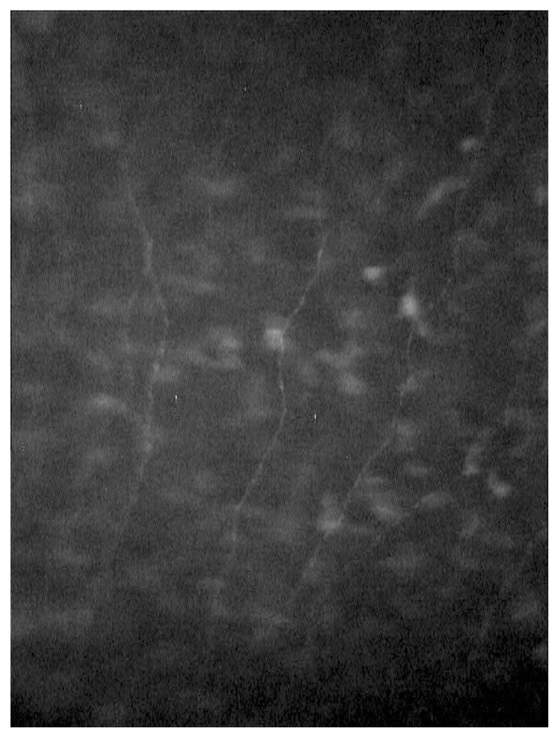
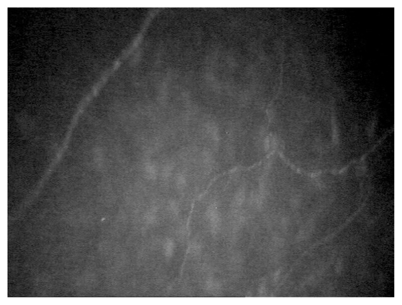
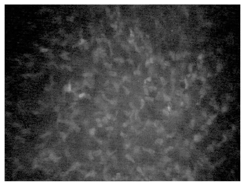
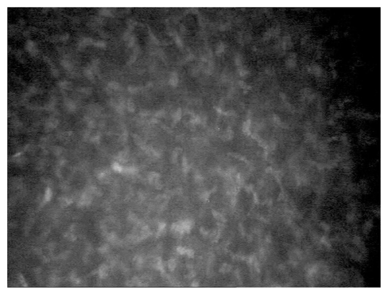
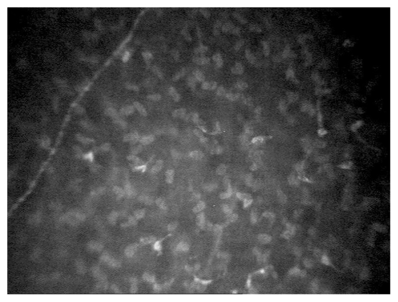
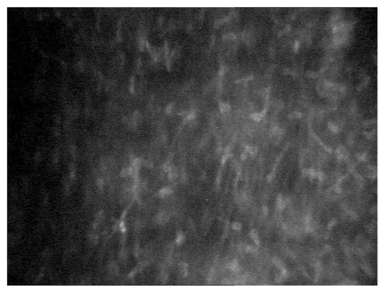
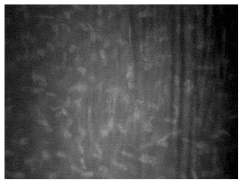
Results: The corneal cell morphology of patients with keratoconus was significantly different from that of healthy eyes except in endothelial cell density (P = 0.072). In the keratoconus group, increased level of stromal haze, alterations such as the elongation of keratocyte nuclei and clustering of cells at the anterior stroma, and dark bands in the posterior stroma were observed with increased severity of the disease. The mean anterior and posterior stromal keratocyte densities and cell areas among the different stages of keratoconus were significantly different (P < 0.001 and P = 0.044, respectively). However, the changes observed in the endothelium were not significantly different (P > 0.05) among the three stages of keratoconus.
Conclusion: Confocal microscopy observation showed significant changes in corneal cell morphology in keratoconic cornea from normal healthy cornea. Analysis also showed significant changes in different severities of keratoconus. Understanding the corneal cell morphology changes in keratoconus may help in the long-term monitoring and management of keratoconus.
Keywords: corneal cell morphology; in vivo confocal microscopy; keratoconus.
Conflict of interest statement
Conflict of Interest The authors have no potential conflict of interests to declare.
Figures
References
-
- Rabinowitz Y. Keratoconus. Surv Ophthalmol. 1998;42(4):297–319. https://doi.org/10.1016/S0039-6257(97)00119-127. - DOI - PubMed
-
- Romero-Jiménez M, Santodomingo-Rubido J, Wolffsohn JS. Keratoconus: a review. Cont Lens Anterior Eye. 2010;33(4):157–166. https://doi.org/10.1016/j.clae.2010.04.006. - DOI - PubMed
-
- Erie JC, Patel SV, McLaren JW, Nau CB, Hodge DO, Bourne WM. Keratocyte density in keratoconus. A confocal microscopy studya. Am J Ophthalmol. 2002;13(5):689–695. https://doi.org/10.1016/S0002-9394(02)01698-7. - DOI - PubMed
-
- Uçakhan OO, Kanpolat A, Ylmaz N, Ozkan M. In vivo confocal microscopy findings in keratoconus. Eye Contact Lens. 2006;32(4):183–191. https://doi.org/10.1097/01.icl.0000189038.74139. - DOI - PubMed
-
- Weed KH, MacEwen CJ, Cox A, McGhee CNJ. Quantitative analysis of corneal microstructure in keratoconus utilising in vivo confocal microscopy. Eye (Lond) 2007;21(5):614–623. https://doi.org/10.1038/sj.eye.6702286. - DOI - PubMed
LinkOut - more resources
Full Text Sources
Other Literature Sources