Case Reports
. 2017 Mar;24(2):106-115.
doi: 10.21315/mjms2017.24.2.14. Epub 2017 Apr 14.

Bilateral Hydroureteronephrosis with a Hypertrophied, Trabeculated Urinary Bladder

Affiliations
Case Reports

Bilateral Hydroureteronephrosis with a Hypertrophied, Trabeculated Urinary Bladder

Showkathali Iqbal et al. Malays J Med Sci. 2017 Mar.

Abstract

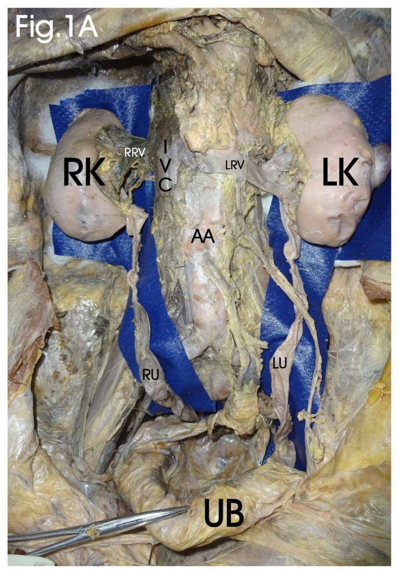
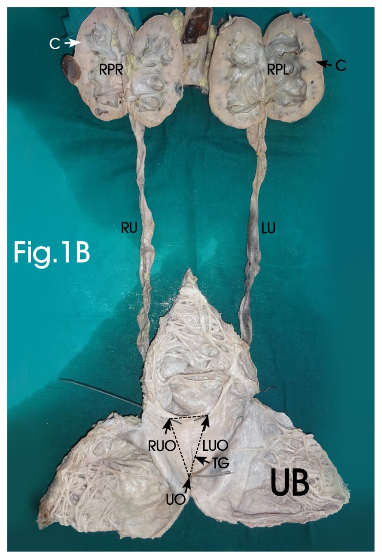
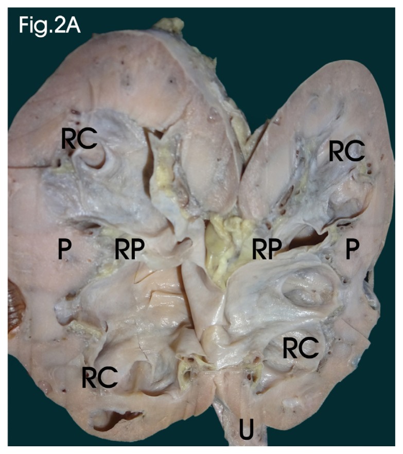
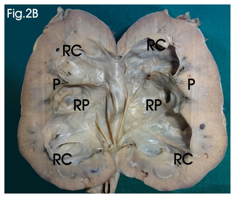
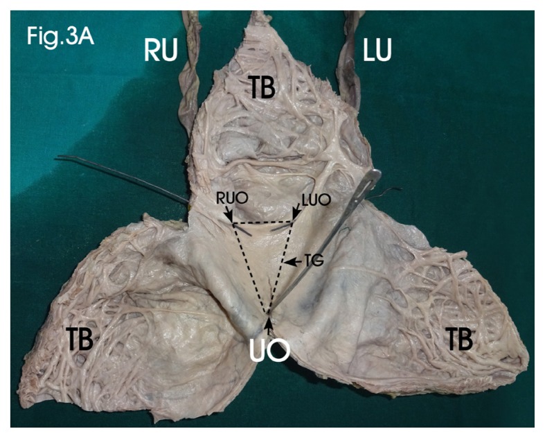
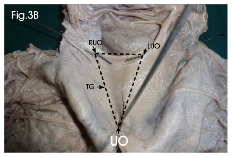
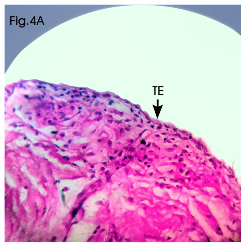
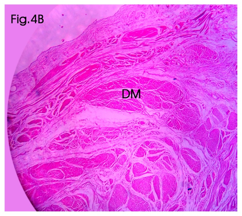
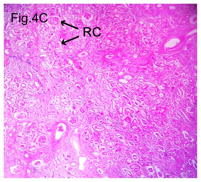
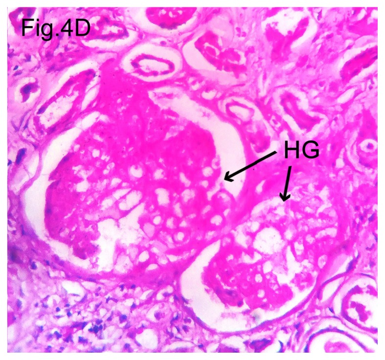
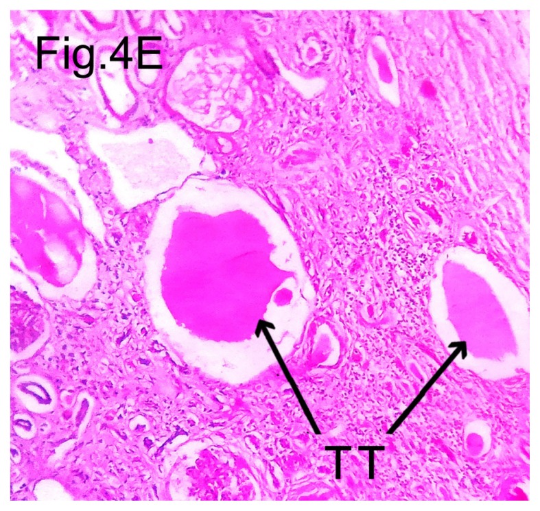
Bilateral hydroureteronephrosis involves the dilatation of the renal pelvis, calyces and ureter; it develops secondary to urinary tract obstruction and leads to a build-up of back pressure in the urinary tract, and it may lead to impairment of renal function and ultimately culminate in renal failure. Although clinically silent in most cases, it can be diagnosed as an incidental finding during evaluation of an unrelated cause. In a minority of patients, it presents with signs and symptoms. Renal calculus is the most common cause, but there are multiple non-calculus aetiologies, and they depend on age and sex. Pelviureteric junction obstruction, benign prostatic hypertrophy, urethral stricture, neurogenic bladder, retroperitoneal mass and bladder outlet obstruction are some of the frequent causes of hydroureteronephrosis in adults. The incidence of non-calculus hydronephrosis is more common in males than in females. Ultrasonography is the most important baseline investigation in the evaluation of patients with hydronephrosis. Here, we report a rarely seen case of bilateral hydroureteronephrosis associated with a hypertrophied, trabeculated bladder in an adult male cadaver, suspected to be due to a primary bladder neck obstruction, and analyse its various other causes, clinical presentations and outcomes.

Keywords: hydronephrosis; prostatic hyperplasia; ultrasonography; urinary bladder neck obstruction; urodynamics.


Conflict of interest statement

Conflicts of Interest None

Figures

Figure 1A
Figure 1A
Bilateral hydronephrotic kidneys, hydroureters and hypertrophied, trabeculated urinary bladder. Kidney shows bilateral, multiple communicating cysts; ureters are dilated bilaterally. RK: Right kidney; LK: left kidney; RRV: Right renal vein; LRV: Left renal vein; AA: Abdominal aorta; RU: Right ureter; LU: Left ureter.
Figure 1B
Figure 1B
Sagittal sections of hydronephrotic kidneys, hydroureters and interior of the hypertrophied bladder showing trigone. Renal pelvis and calyces are markedly dilated; pyramids and medulla are attenuated; bladder shows trabeculated anterior wall; both ureterovesical junctions are patent and probed; urethral orifice was patent and probed RPR: Dilated right renal pelvis; RPL: Dilated left renal pelvis; RU: Right hydroureter; LU: Left hydroureter; TG: Trigone; RUO: Right ureteric orifice; LUO: Left ureteric orifice; UO: Urethral orifice.
Figure 2A
Figure 2A
Sagittal section of right kidney Dimensions: Kidney: 88.3 mm × 41.9 mm × 46.1 mm; cortical thickness: 15.05 mm (range: 14.7 mm to 15.4 mm); renal pelvis: 57.9 mm × x 28.6 mm × 30.9 mm; Ureter length: 260 mm; width max: 19.5 mm; min: 4.7 mm; intramural length: 26.8 mm. RP: Dilated renal pelvis; RC: Dilated renal calyces; P: Attenuated renal pyramids; U: Dilated ureter
Figure 2B
Figure 2B
Sagittal section of left kidney Dimensions: Kidney: 84.1 mm × 29.7 mm × 34.8 mm; cortical thickness: 12.8 mm (range: 11.5 mm to 14.1 mm); renal pelvis: 58.7 mm × 24.9 mm × 22.1 mm; Ureter: length: 278 mm; width max: 14.2 mm; min: 3.9 mm; intramural length: 27.1 mm. RP: Dilated renal pelvis; RC: Dilated renal calyces; P: Attenuated renal pyramids; U: Dilated ureter
Figure 3A
Figure 3A
Interior of hypertrophied urinary bladder Enlarged bladder shows thickened and trabeculated anterior wall; both ureterovesical junctions are probed and ureteric orifices are made out bilaterally; urethra was patent and probed Dimensions: 142 mm × 135 mm × 6.3 mm; Trigone interureteric distance: 36.7 mm; uretero-urethral distance: 57.6 mm; diameter of ureteric orifice left: 1.5 mm; right: 1.2 mm; urethral orifice: 3.3 mm × 1.9 mm. TB: Trabeculated wall of the bladder; TG: Smooth trigone; RUO: Right ureteric orifice; LUO: Left ureteric orifice; UO: Urethral orifice
Figure 3B
Figure 3B
Enlarged view of trigone of the bladder TG: Smooth trigone; RUO: Right ureteric orifice; LUO: Left ureteric orifice; UO: Urethral orifice
Figure 4A
Figure 4A
Cut section of urinary bladder showing transitional epithelium (H & E stain, 10X magnification) Sections of the bladder wall showing the attenuated transitional epithelium with hypertrophied detrusor muscular layer TE: Transitional epithelium
Figure 4B
Figure 4B
Cut section of urinary bladder showing hypertrophied detrusor muscle fibres (H & E stain, 10X magnification) DM: Hypertrophied detrusor muscle fibres
Figure 4C
Figure 4C
Cut section of hydronephrotic kidney showing attenuated renal parenchyma (H & E stain, 4X magnification) Sections of the kidney showing interstitial fibrosis with chronic inflammatory infiltrate in interstitium; many of the glomeruli show periglomerular fibrosis and some of them were completely hyalinised; renal tubules show dilation with amorphous eosinophilic material deposition in the lumen (thyroidisation); blood vessels show thick hyalinised vessel wall and the transitional epithelium of the pelvis was attenuated. RC: Renal corpuscles
Figure 4D
Figure 4D
Cut section of hydronephrotic kidney showing hyalinised glomerulus (H & E stain, 10X magnification) HG: Hyalinised glomerulus
Figure 4E
Figure 4E
Cut section of hydronephrotic kidney showing thyroidsation of renal tubules (H & E stain, 10X magnification) TT: Thyroidsation of renal tubules

References

    1. Kumar V, Abbas AK, Fausto N, Aster JC. Robbins and Cotran pathologic basis of disease. 8th ed. Philadelphia PA: Elsevier Saunders; 2010. pp. 960–962.
    1. Mergener K, Weinerth JL, Baillie J. Dietl’s crisis: a syndrome of episodic abdominal pain of urologic origin that may present to a gastroenterologist. Am J Gastroenterol. 1997;92(12):2289–2291. - PubMed
    1. Merck Manuals Home Health Handbook. Hydronephrosis. (cited 2010 Nov 5) Available from: http://www.merckmanuals.com/home/index.html.
    1. Tazi MF, Riyach O, Ahallal Y, Mellas S, Khallouk A, El Fassi MJ, et al. Giant urinary bladder and bilateral giant hydronephrosis due to bladder neck obstruction: one case report and literature review. Case Rep Urol. 2012;2012:817519. doi: 10.1155/2012/817519. - DOI - PMC - PubMed
    1. Lameire N, Van Biesen W, Vanholder R. Acute renal failure. Lancet. 2005;365:417–430. - PubMed

Publication types

LinkOut - more resources