Review
. 2018 May;10(3):e1496.
doi: 10.1002/wnan.1496. Epub 2017 Sep 11.

Two decades of dendrimers as versatile MRI agents: a tale with and without metals

Affiliations
Review

Two decades of dendrimers as versatile MRI agents: a tale with and without metals

Michael T McMahon et al. Wiley Interdiscip Rev Nanomed Nanobiotechnol. 2018 May.

Abstract

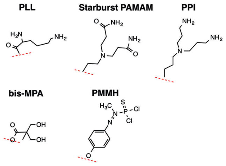
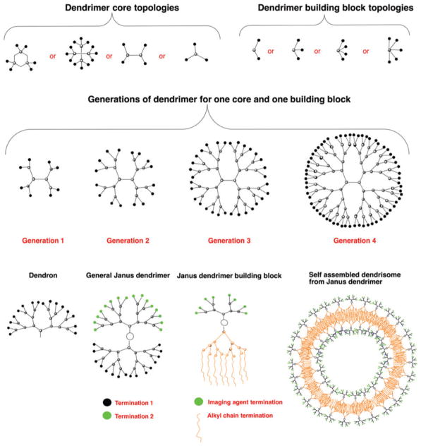
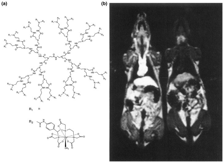
Dendrimers or dendritic polymers are a class of compounds with great potential for nanomedical use. Some of their properties, including their rigidity, low polydispersity and the ease with which their surfaces can be modified make them particularly well suited for use as MRI diagnostic or theranostic agents. For the past 20 years, researchers have recognized this potential and refined dendrimer formulations to optimize these nanocarriers for a host of MRI applications, including blood pool imaging agents, lymph node imaging agents, tumor-targeted theranostic agents and cell tracking agents. This review summarizes the various types of dendrimers according to the type of MR contrast they can provide. This includes the metallic T1 , T2 and paraCEST imaging agents, and the non-metallic diaCEST and fluorinated (19 F) heteronuclear imaging agents. This article is categorized under: Diagnostic Tools > In Vivo Nanodiagnostics and Imaging Implantable Materials and Surgical Technologies > Nanomaterials and Implants.


Figures

FIGURE 1
FIGURE 1
Building blocks that have been used for production of commercially available dendrimers. These include poly-L-lysine (PLL), polyaminoamine (PAMAM), polypropylimine [PPI, also known as diaminobutane (DAB)], 2,2-bis-methylolpropionic acid (bis-MPA), and phenoxymethyl(methylhydrazone) (PMMH).
FIGURE 2
FIGURE 2
Available topologies for synthesizing dendrimers. Shown are representative core and building block topologies (top row), the topology for Generations 1–4 for one core and one building block as present in polyaminoamine (PAMAM) and poly-L-lysine (PLL) dendrimers (middle row), and a topology for a dendron, a generalized Janus dendrimer with two dendrons presenting different terminal groups which are grafted to a single core, a Janus dendrimer suitable for self-assembly into MR dendrisomes, and an MR dendrisome (bottom row).
FIGURE 3
FIGURE 3
Chronological representation of milestone publications on dendrimer development and their (green) use as MRI agents (red).
FIGURE 4
FIGURE 4
The first dendrimer platform used as MR contrast agent. (a) General structure of PAMAM-Gd-DOTA dendrimers showing a G = 3PAMAM dendrimer and Gd-DOTA with linker. (b) The first image acquired with PAMAM Gd-DOTA dendrimers as MR contrast agent. A Gd-DOTA-conjugated G = 6 PAMAM dendrimer, having a half-life of approximately 200 min, was injected in the left rat at 0.005 mmol Gd kg−1. A clear vascular enhancement can be seen compared with the uninjected rat on the right. (Reprinted with permission from Ref Copyright 1994 John Wiley & Sons, Inc.)
FIGURE 5
FIGURE 5
Variable field relaxometry of a Dy-DOTA-conjugated G = 5 PAMAM dendrimer and single Dy(III) chelates. Shown are the T2 relaxivities of (a) Dy-DOTA-PAMAM G = 5 dendrimers, (b) Dy-DOTA single chelates, and (c) Dy-DTPA single chelates as a function of magnetic field strength. Data are shown at 3 (▲), 10 (▼), 20 (●), and 37°C (+). Solid lines represent quadratic fits to the equation 1/T2 = a + bB0,2 with B0 being the external magnetic field strength. For comparison, the T1 relaxivities are negligible, shown as dashed lines at 3 (■) and 37 C (·). (Reprinted with permission form Ref Copyright 1998 Wolters Kluwer Health)
FIGURE 6
FIGURE 6
Structure of magnetodendrimers. Shown is a schematic representation of the stabilization of maghemite nanoparticles by G = 4.5 carboxyl-terminated PAMAM dendrimers. (Reprinted with permission from Ref Copyright 2001 American Chemical Society)
FIGURE 7
FIGURE 7
(a) General structure of PAMAM-SA-Ac dendrimers showing a G = 3PAMAM dendrimer, salicylic acid (SA) with linker, and acetyl termination. (B–E) In vivo images of salicylic acid methyl ester (SAME) G = 5 PAMAM dendrimer conjugates infused into a mouse carrying a glioblastoma xenograft. Shown are (b) T2w (arrow highlights tumor) and CEST MR images obtained (c) pre- and (d) 30 and (e) 60 min postinjection of a 500 μM solution of diaCEST dendrimer. (Reprinted with permission from Ref Copyright 2016 American Chemical Society)
FIGURE 8
FIGURE 8
Two decades of (a) publications and (b) citations on dendrimer MRI agents (Source: ISI Web of Science).

References

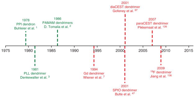
    1. Buhleier E, Wehner W, Vogtle F. Cascade-chain-like and nonskid-chain-like syntheses of molecular cavity topologies. Synthesis (Stuttg) 1978;2:155–158.
    1. Tomalia DA, Baker H, Dewald J, Hall M, Kallos G, Martin S, Roeck J, Ryder J, Smith P. A new class of polymers –starburst-dendritic macromolecules. Polym J. 1985;17:117–132.
    1. Denkewalter RG, Kolc J, Lukasavage WJ. Macromolecular highly branched homogeneous compound based on lysine units. 1981 Available at: http://www.google.com.au/patents/US4289872.
    1. Newkome GR, Yao ZQ, Baker GR, Gupta VK. Micelles. 1. Cascade molecules: a new approach to micelles – A 27-arborol. J Org Chem. 1985;50:2003–2004.
    1. Duncan R, Izzo L. Dendrimer biocompatibility and toxicity. Adv Drug Deliv Rev. 2005;57:2215–2237. - PubMed

LinkOut - more resources