. 2017 Sep 20;9(37):31645-31656.
doi: 10.1021/acsami.7b11438. Epub 2017 Sep 12.

Injectable Carbon Nanotube-Functionalized Reverse Thermal Gel Promotes Cardiomyocytes Survival and Maturation

Affiliations

Injectable Carbon Nanotube-Functionalized Reverse Thermal Gel Promotes Cardiomyocytes Survival and Maturation

Brisa Peña et al. ACS Appl Mater Interfaces. .

Abstract

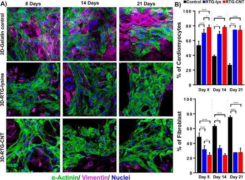
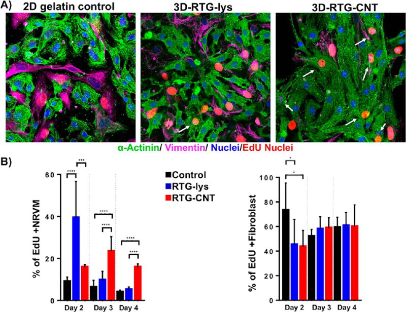
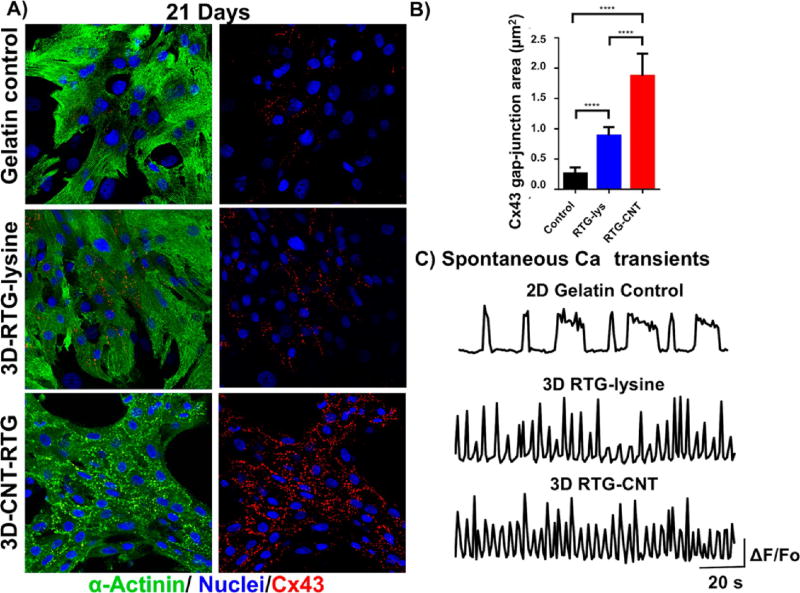
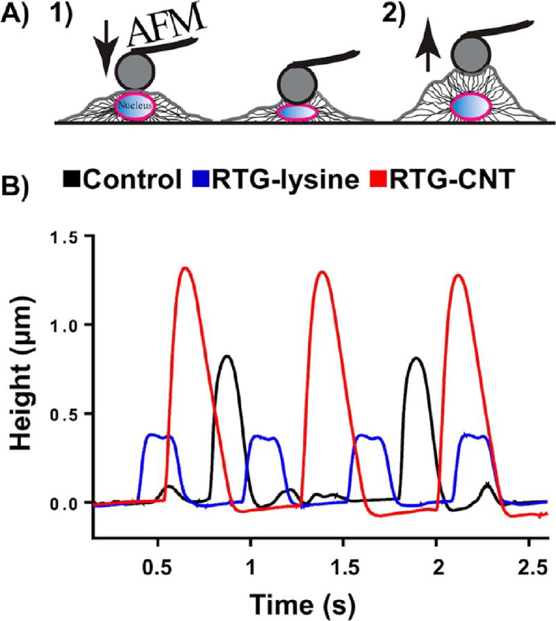
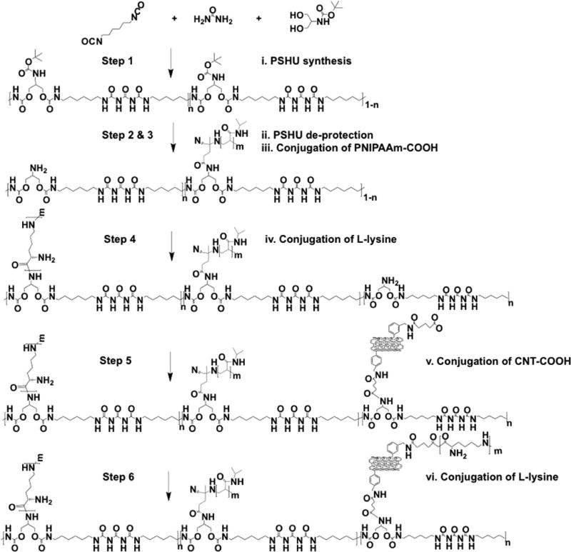
The ability of the adult heart to regenerate cardiomyocytes (CMs) lost after injury is limited, generating interest in developing efficient cell-based transplantation therapies. Rigid carbon nanotubes (CNTs) scaffolds have been used to improve CMs viability, proliferation, and maturation, but they require undesirable invasive surgeries for implantation. To overcome this limitation, we developed an injectable reverse thermal gel (RTG) functionalized with CNTs (RTG-CNT) that transitions from a solution at room temperature to a three-dimensional (3D) gel-based matrix shortly after reaching body temperature. Here we show experimental evidence that this 3D RTG-CNT system supports long-term CMs survival, promotes CMs alignment and proliferation, and improves CMs function when compared with traditional two-dimensional gelatin controls and 3D plain RTG system without CNTs. Therefore, our injectable RTG-CNT system could potentially be used as a minimally invasive tool for cardiac tissue engineering efforts.

Keywords: carbon nanotube; cardiac tissue engineering; hybrid-biomimetic hydrogel; injectable polymer; reverse thermal gel.


Figures

Figure 1
Figure 1
MWCNT-COOH. (A) Schematic representation of MWCNT-COOH synthesis. (B) TGA of MWCNT-NH2 (blue) and MWCNT-COOH (green). (C) TEM shows MWCNT-COOH dispersion in DMF. Scale bar 2000 nm.
Figure 2
Figure 2
(A) Cross-section micrographs of the RTG systems at different magnifications. (top) RTG-lysine and (bottom) RTG-CNT. A fibrous CNT mesh network can be observed in the RTG-CNT system (red arrows). (B) Storage and loss moduli of both RTG-lysine (red) and RTG-CNT (blue) and temperature-dependent phase transition: the chemical conjugation of CNT improves the mechanical properties of the RTG-lysine system. (bottom, 1) An aqueous solution of RTG-CNT at room temperature turns to (bottom, 2) physical gel at 37 °C. (C) Resistance measurements: the hybrid CNT-RTG presented a resistance between both the RTG polymer and the CNT-COOH. (ANOVA-Dunn’s test) (CNT-COOH “vs” RTG-lysine ****p value: <0.0001; n = 20; RTG-lysine “vs” RTG-CNT ***p value: 0.0002; n = 20; RTG-CNT “vs” CNT-COOH ***p value: 0.0002, n = 20). (D) Both RTG systems present lower viscosity than media. (ANOVA-Bonferroni’s test) (media “vs” RTG-lysine **p value: 0.0013; n = 5; RTG-lysine “vs” RTG-CNT *p value: 0.0238; n = 5; RTG-CNT “vs” media ****p value: <0.0001, n = 5).
Figure 3
Figure 3
Fluorescence staining of NRVMs and fibroblasts cultured in different substrates. Sarcomeric α-actinin (green), vimentin (pink), and DAPI (blue). (A, top) NRVMs cultured on 2D gelatin control. (A, middle) NRVMs cultured in 3D RTG-lysine. (A, bottom) NRVMs cultured in 3D RTG-CNT. (B) Quantification of the percentage of NRVMs and fibroblast growing in gelatin-control and the RTG systems. Significant differences on the % of NRVMs can be observed at all the time points between the gelatin control group and RTG systems. (ANOVA-Bonferroni’s test). Day 8: RTG-lysine “vs” gelatin control ****p value: <0.0001; n = 8; RTG-CNT “vs” gelatin control ****p value: <0.0001; n = 8; RTG-CNT “vs” RTG-lysine *p value: <0.018; n = 8. (ANOVA-Dunn’s test) Day 14: RTG-lysine “vs” gelatin control *p value: 0.0487, n = 8; RTG-CNT “vs” gelatin control ****p value: <0.0001, n = 8. (ANOVA-Bonferroni’s) Day 21: RTG-lysine “vs” gelatin control ****p value: <0.0001, n = 8; RTG-CNT “vs” gelatin control ****p value: <0.0001, n = 8. Significant differences on the % of fibroblast were also observed at all time points between the gelatin control group and RTG systems. (ANOVA-Bonferroni’s test) Day 8: RTG-lysine “vs” gelatin control ****p value: <0.0001, n = 8; RTG-CNT “vs” gelatin control ****p value: <0.0001 n = 8. RTG-CNT “vs” RTG-lysine *p value: 0.018 n = 8. (ANOVA-Dunn’s test) Day 14: RTG-lysine “vs” gelatin control *p value: 0.0487, n = 8; RTG-CNT “vs” gelatin control ****p value: <0.0001 n = 8. (ANOVA-Bonferroni’s test) Day 21: RTG-lysine “vs” gelatin control ****p value: <0.0001; n = 8; RTG-CNT “vs” gelatin control ****p value: <0.0001; n = 8. Data are presented as mean ± standard deviation (SD).
Figure 4
Figure 4
Proliferation assay of NRVMs and fibroblasts. (A, left to right) NRVMs and fibroblast cultured on 2D gelatin control, 3D RTG-lysine and 3D RTG-CNT at day 4. (B) Significant differences of dividing NRVMs on the RTGlysine system were observed at day 2 when compared with the gelatin controls (****p value: <0.0001, n = 8) and with the RTG-CNT scaffold (***p value: 0.0004, n = 8). Significant differences of dividing NRVMs on the RTG-CNT system were observed at days 3 and 4 when compared with the gelatin control and the RTG-lysine system. Day 3: RTG-CNT “vs” gelatin control ****p value: <0.0001, n = 8; RTG-CNT “vs” RTG-lysine ****p value: <0.0001, n = 8. Day 4: RTG-CNT “vs” gelatin control ****p value: <0.0001, n = 8; RTG-CNT “vs” RTG-lysine ****p value: <0.0001, n = 8; significant differences of dividing fibroblast were observed at day 2 between the gelatin controls and the RTG polymers. Day 2: gelatin control “vs” RTG-lysine *p value: 0.0241, n = 8; gelatin control “vs” RTG-CNT *p value: 0.0166, n = 8. Data are presented as mean ± SD.
Figure 5
Figure 5
Intercellular communication of NRVMs growing in different substrates after 21 d of culture. (A) Fluorescence images of connexin 43 (red dots), sarcomeric α-actinin (green), and DAPI (blue) staining of NRVMs (top) NRVMs cultured on 2D gelatin control; (middle) NRVMs cultured in 3D RTG-lysine; (bottom) NRVMs cultured in 3D RTG-CNT. (B) Quantification of Cx43 gap junction area: Significant differences on Cx43 gap-junction were observed between the gelatin control groups and the RTG systems. RTG-lysine “vs” gelatin control ****p value: <0.0001, n = 8; RTG-CNT “vs” gelatin control ****p value: <0.0001, n = 8; RTG-CNT “vs” RTG-lysine ****p value: <0.0001, n = 5. Data are presented as mean ± SD (n = 5). (ANOVA-Bonferroni’s test). Data are presented as mean ± SD (n = 5). (C) Spontaneous calcium transients of NRVMs growing on 2D gelatin control and in 3D RTG systems (n = 5).
Figure 6
Figure 6
Spontaneous beating of NRVMs cultured for 21 d in different substrates by AFM. (A) Schematic representation of the beating activity of NRVMs measurements by the deflection of the AFM cantilever. (left to right) AFM cantilever gently pushing the nuclear region the CM, the beating activity of CM pushed away the AFM cantilever, providing information about beating height and frequency. (B) NRVMs cultured in the 3D RTG-CNT presented higher contraction, while beating suggesting that the CNT-RTG niche supports a stronger cardiomyocyte contraction.
Scheme 1
Scheme 1
RTG-CNT Synthesis

References

    1. Cahill TJ, Ashrafian H, Watkins H. Genetic Cardiomyopathies Causing Heart Failure. Circ. Res. 2013;113(6):660–675. - PubMed
    1. Mestroni L, Rocco C, Gregori D, Sinagra G, Di Lenarda A, Miocic S, Vatta M, Pinamonti B, Muntoni F, Caforio ALP, McKenna WJ, Falaschi A, Giacca M, et al. Familial Dilated Cardiomyopathy: Evidence for Genetic and Phenotypic Heterogeneity. J. Am. Coll. Cardiol. 1999;34(1):181–190. - PubMed
    1. Eulalio A, Mano M, Dal Ferro M, Zentilin L, Sinagra G, Zacchigna S, Giacca M. Functional Screening Identifies miRNAs Inducing Cardiac Regeneration. Nature. 2012;492(7429):376–381. - PubMed
    1. Van Amerongen MHJ, Engel FB. Features of Cardiomyocyte Proliferation and Its Potential for Cardiac Regeneration: Stem Cells Review Series. J. Cell. Mol. Med. 2008;12(6A):2233–2244. - PMC - PubMed
    1. Alraies MC, Eckman P. Adult Heart Transplant: Indications and Outcomes. J. Thorac. Dis. 2014;6(8):1120–1128. - PMC - PubMed