. 2017 Sep 12;7(3):67.
doi: 10.3390/biom7030067.

Functional Amyloid Protection in the Eye Lens: Retention of α-Crystallin Molecular Chaperone Activity after Modification into Amyloid Fibrils

Affiliations

Functional Amyloid Protection in the Eye Lens: Retention of α-Crystallin Molecular Chaperone Activity after Modification into Amyloid Fibrils

Megan Garvey et al. Biomolecules. .

Abstract

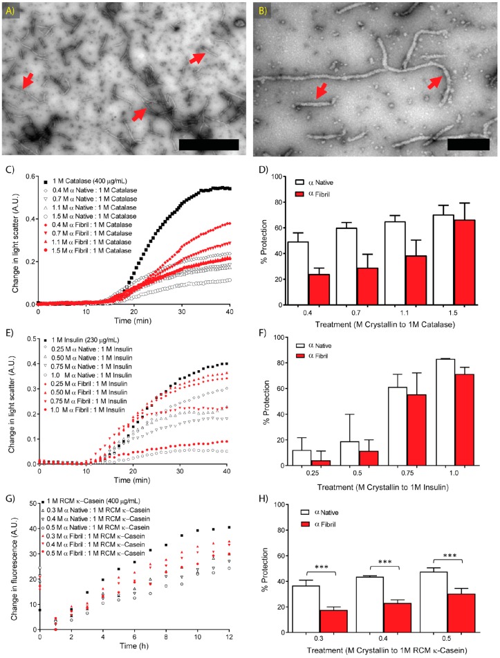
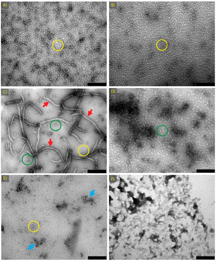
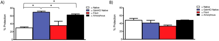
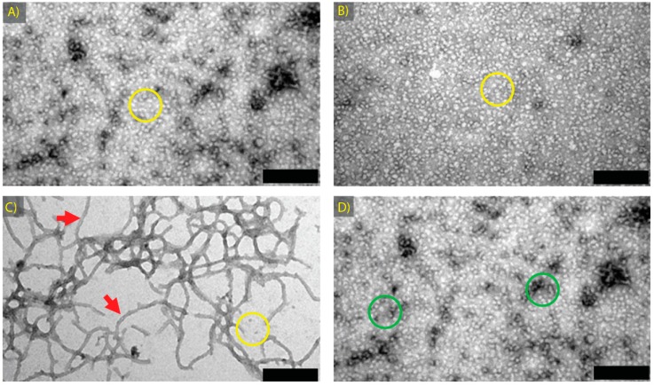
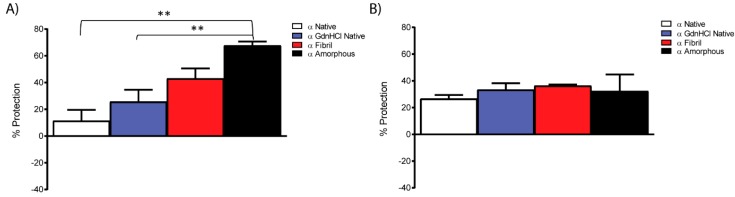
Amyloid fibril formation occurs from a wide range of peptides and proteins and is typically associated with a loss of protein function and/or a gain of toxic function, as the native structure of the protein undergoes major alteration to form a cross β-sheet array. It is now well recognised that some amyloid fibrils have a biological function, which has led to increased interest in the potential that these so-called functional amyloids may either retain the function of the native protein, or gain function upon adopting a fibrillar structure. Herein, we investigate the molecular chaperone ability of α-crystallin, the predominant eye lens protein which is composed of two related subunits αA- and αB-crystallin, and its capacity to retain and even enhance its chaperone activity after forming aggregate structures under conditions of thermal and chemical stress. We demonstrate that both eye lens α-crystallin and αB-crystallin (which is also found extensively outside the lens) retain, to a significant degree, their molecular chaperone activity under conditions of structural change, including after formation into amyloid fibrils and amorphous aggregates. The results can be related directly to the effects of aging on the structure and chaperone function of α-crystallin in the eye lens, particularly its ability to prevent crystallin protein aggregation and hence lens opacification associated with cataract formation.

Keywords: amyloid fibril; molecular chaperone; protein aggregation; protein unfolding; small heat-shock protein.


Conflict of interest statement

The authors declare no conflict of interest. The funding sponsors had no role in the design of the study; in the collection, analyses, or interpretation of data; in the writing of the manuscript, and in the decision to publish the results.

Figures

Figure 1
Figure 1
Fibril formation and chaperone activity of native and fibrillar α-crystallin. (A,B) Transmission electron micrographs of α-crystallin fibrils formed in 1 M guanidine hydrochloride (GdnHCl), 0.1 M sodium phosphate, pH 7.4 at 60 °C for 2 h, with red arrows indicating fibril structures. Scale bars are: (A) 1 μm and (B) 200 nm; (CH) Native and fibrillar α-crystallin chaperone protection of (C,D) catalase at 400 μg/mL in 0.1 M sodium phosphate buffer at pH 7.4, undergoing thermal destabilisation at 60 °C; (E,F) reduced insulin, 230 μg/mL in 0.1 M sodium phosphate buffer pH 7.4, at 37 °C in the presence of 10 mM 1,4-dithiothreitol (DTT); and (G,H) reduced and carboxymethylated (RCM) κ-casein, 400 μg/mL incubated at 37 °C in 0.1 M sodium phosphate, 10 μM thioflavin T (ThT) for 12 h; (C,E,G) show representative profiles of light scattering; (D,F,H) show the percentage of protection provided by each chaperone, calculated from the difference between the maximum light scattering or fluorescence of the target protein alone and the target protein in the presence of the stated concentrations of α-crystallin. Data shown are means ± standard error (SE) of (D,F) three or (H) of six separate experiments; p-values indicating significant difference from two-way ANOVA (Šidák post-hoc test) are: *** p < 0.001.
Figure 2
Figure 2
Transmission electron microscopy (TEM) images of samples formed to assess the effects of structural variation on the chaperone activity of α-crystallin: (A) α Native; (B) α GdnHCl Native; (C) α Fibril; (D) α Amorphous; samples of α-crystallin. (E) βH Fibril; and (F) aldehyde dehydrogenase (ADH) Amorphous samples. All samples were prepared as described in Table 1. Scale bars are 200 nm. Features of importance are highlighted, including long amyloid fibrils (red arrows), curvilinear amyloid fibrils (blue arrows) and spherical aggregates of ~15 nm in diameter (yellow circles) or larger spherical aggregates (green circles).
Figure 3
Figure 3
Chaperone protection provided by native, fibrillar and amorphous α-crystallin species against the (A) amorphous aggregation of reduced insulin, 250 μg/mL in 0.1 M sodium phosphate, pH 7.4 and 20 mM DTT at 37 °C (0.9 chaperone: 1.0 insulin on a molar basis); and (B) fibrillar aggregation of RCM-κ-casein, 400 μg/mL incubated at 37 °C for 22 h in the presence of various native, amorphous and fibrillar chaperone species (0.5 chaperone: 1.0 RCM κ-casein on a molar basis) and monitored via ThT fluorescence. The percentage of protection provided by each chaperone is calculated from the difference between the maximal light scattering or fluorescence of the target protein alone and the target protein in the presence of the stated concentrations of α-crystallin. Results are mean ± SE of the percentage protection given by chaperones for three experiments; p-values, derived by one-way ANOVA with Tukey post-test, are * p < 0.05.
Figure 4
Figure 4
TEM images of αB-crystallin species formed to assess the effects of structural variation on chaperone activity: (A) αB Native; (B) αB GdnHCl Native; (C) αB Fibril; and (D) αB Amorphous. All samples were prepared as described in Table 1. Scale bars are 200 nm. Features of importance are highlighted, including long amyloid fibrils (red arrows) and spherical aggregates of ~15 nm in diameter (yellow circles) or larger spherical aggregates (green circles).
Figure 5
Figure 5
Chaperone protection provided by native, fibrillar and amorphous αB-crystallin species against the (A) amorphous aggregation of insulin, 250 μg/mL at 37 °C in 0.1 M sodium phosphate, 20 mM DTT, pH 7.4 (0.9 chaperone: 1.0 insulin on a molar basis); and (B) fibrillar aggregation of RCM κ-casein 400 μg/mL, 10 μM ThT at 37 °C, pH 7.4 (0.5 chaperone: 1.0 RCM κ-casein on a molar basis). The percentage of protection provided by each chaperone is calculated from the difference between the maximal light scattering or fluorescence of the target protein alone and the target protein in the presence of the stated concentrations of αB-crystallin. Results are mean ± SE of the percentage protection given by chaperones for three experiments; p-values, derived by one-way ANOVA with Tukey post-test, are ** p < 0.01.

Similar articles

Cited by

References

    1. Bloemendal H., de Jong W., Jaenicke R., Lubsen N.H., Slingsby C., Tardieu A. Ageing and vision: Structure, stability and function of lens crystallins. Prog. Biophys. Mol. Biol. 2004;86:407–485. doi: 10.1016/j.pbiomolbio.2003.11.012. - DOI - PubMed
    1. Ray N.J. Biophysical chemistry of the ageing eye lens. Biophys. Rev. 2015;7:353–368. doi: 10.1007/s12551-015-0176-4. - DOI - PMC - PubMed
    1. Augusteyn R.C. On the growth and internal structure of the human lens. Exp. Eye Res. 2010;90:643–654. doi: 10.1016/j.exer.2010.01.013. - DOI - PMC - PubMed
    1. Meehan S., Berry Y., Luisi B., Dobson C.M., Carver J.A., MacPhee C.E. Amyloid fibril formation by lens crystallin proteins and its implications for cataract formation. J. Biol. Chem. 2004;279:3413–3419. doi: 10.1074/jbc.M308203200. - DOI - PubMed
    1. Bassnett S. Lens organelle degradation. Exp. Eye Res. 2002;74:1–6. doi: 10.1006/exer.2001.1111. - DOI - PubMed

Publication types

LinkOut - more resources