. 2017 Oct 18;16(20):1954-1964.
doi: 10.1080/15384101.2017.1367071. Epub 2017 Sep 12.

MiR-26b reverses temozolomide resistance via targeting Wee1 in glioma cells

Affiliations

MiR-26b reverses temozolomide resistance via targeting Wee1 in glioma cells

Lixia Wang et al. Cell Cycle. .

Abstract

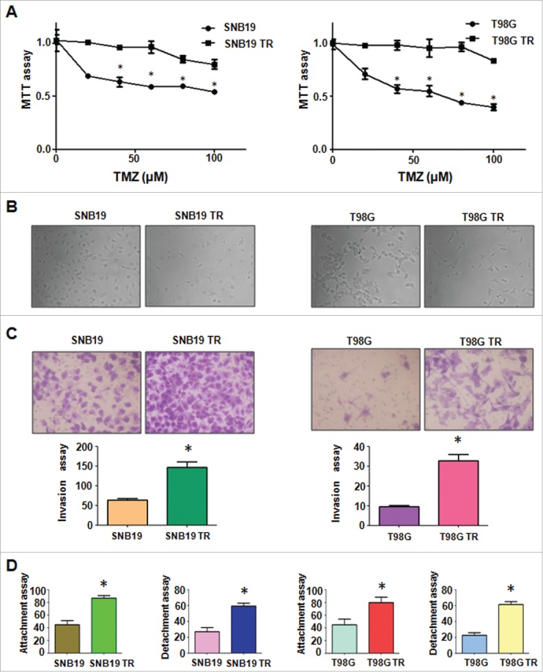
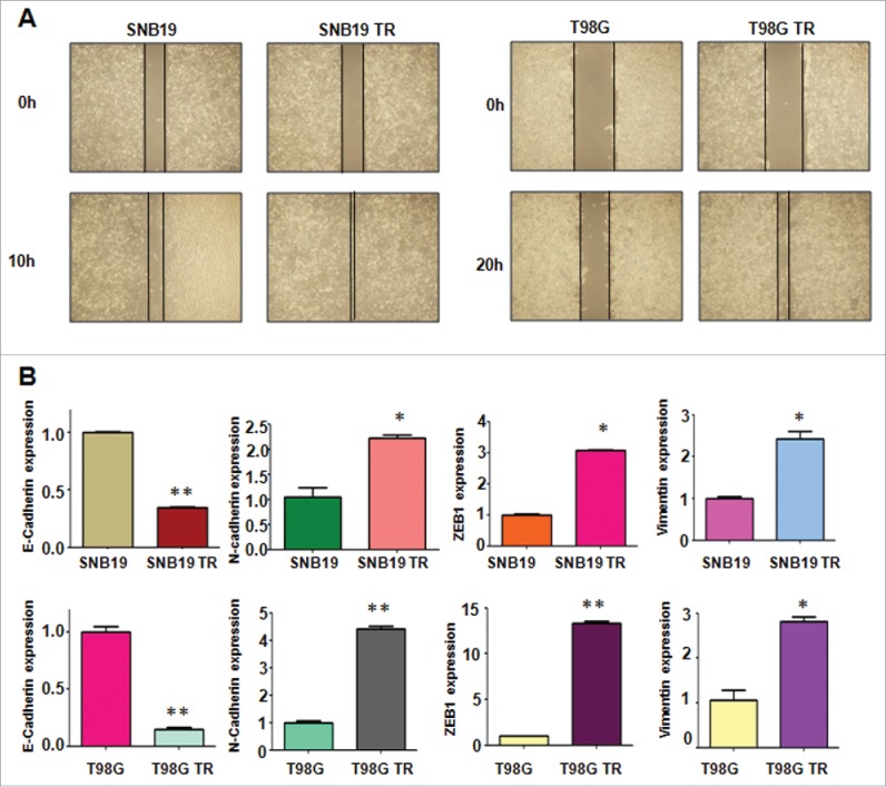
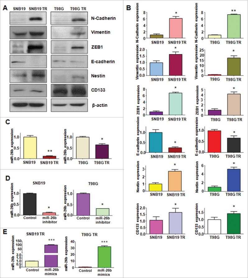
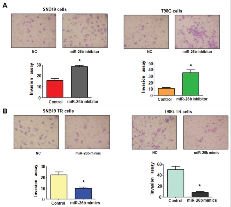
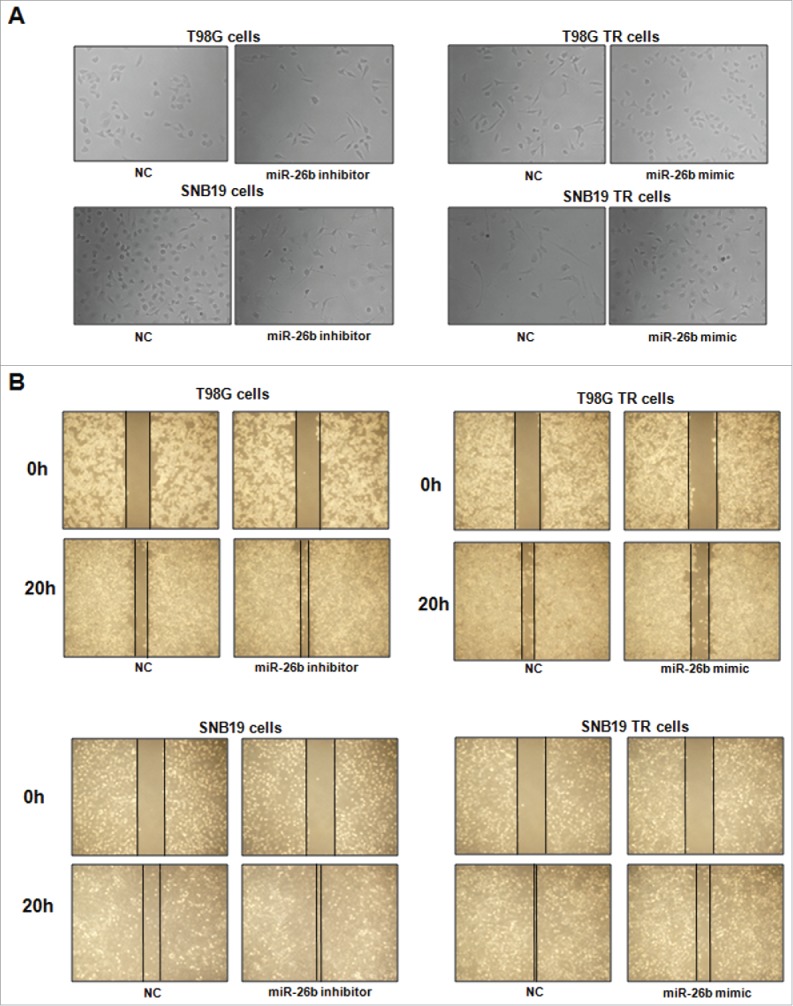
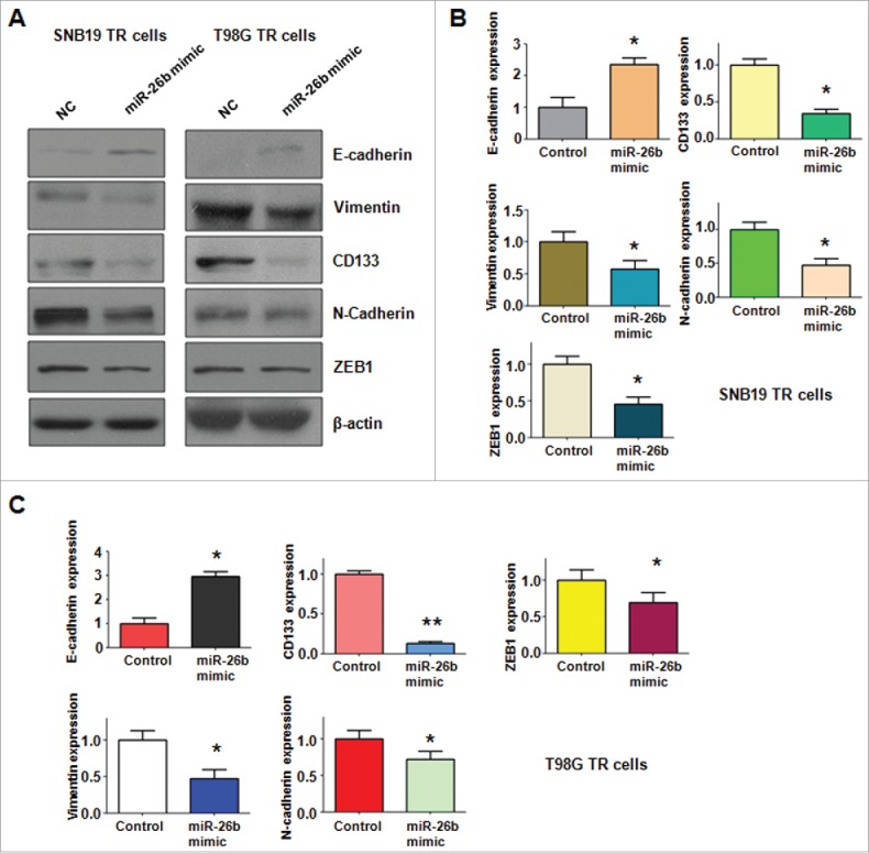
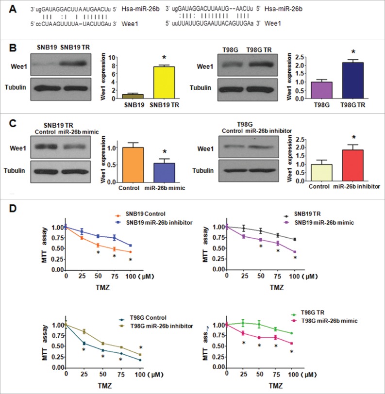
Emerging evidence has demonstrated that microRNAs (miRNA) play a critical role in chemotherapy-induced epithelial-mesenchymal transition (EMT) in glioma. However, the underlying mechanism of chemotherapy-triggered EMT has not been fully understood. In the current study, we determined the role of miR-26b in regulation of EMT in stable temozolomide (TMZ)-resistant (TR) glioma cells, which have displayed mesenchymal features. Our results illustrated that miR-26b was significantly downregulated in TR cells. Moreover, ectopic expression of miR-26b by its mimics reversed the phenotype of EMT in TR cells. Furthermore, we found that miR-26b governed TR-mediate EMT partly due to governing its target Wee1. Notably, overexpression of miR-26b sensitized TR cells to TMZ. These findings suggest that upregulation of miR-26b or targeting Wee1 could serve as novel approaches to reverse chemotherapy resistance in glioma.

Keywords: Glioma; Wee1; growth; miR-26b; temozolomide.


Figures

Figure 1.
Figure 1.
TR cells exhibited EMT phenotype. (A) MTT assay was conducted in parental and TR glioma cells. * P < 0.05 vs their parental cells. (B) Cell morphology was observed by microscope in parental and TR glioma cells. (C) Invasion assay was performed to measure the invasive capacity in parental and TR glioma cells. * P< 0.05 vs their parental cells. (D) Cell attachment and detachment assays were assessed in parental and TR glioma cells. * P < 0.05 vs their parental cells.
Figure 2.
Figure 2.
TR cells have enhanced motility activity. (A) Wound assays were performed to measure the motility activity in parental and TR glioma cells. (B) Real-time RT-PCR assay was conducted to detect the expression of EMT markers in parental and TR glioma cells. * P < 0.05; ** P < 0.01 vs their parental cells.
Figure 3.
Figure 3.
TR cells have EMT marker changes. (A) Western blotting analysis was used to detect the expression of E-cadherin, vimentin, ZEB1, N-cadherin, and Cancer Stem Cell marker, such as CD133 and Nestin in parental and TR glioma cells. (B) Quantitative results are illustrated for panel A. * P < 0.05; ** P < 0.01 vs their parental cells. (C) Real-time RT-PCR assay was conducted to detect the expression of miR-26b in parental and TR cells. * P < 0.05; ** P < 0.01 vs their parental cells. (D-E) Real-time RT-PCR was performed to detect the efficacy of miR-26b inhibitor and mimics transfection. * P < 0.05; *** P < 0.001 vs Control.
Figure 4.
Figure 4.
miR-26b mimics inhibited cell invasion in TR glioma cells. (A) Top panel: Invasion assays were conducted in glioma cells transfected with miR-26b inhibitor. Bottom panel: Quantitative results are illustrated for top panel. * P < 0.05 vs Control. (B) Top panel: Invasion assays were performed in TR glioma cells transfected with miR-26b mimics. Bottom panel: Quantitative results are illustrated for top panel. * P < N0.05 vs Control.
Figure 5.
Figure 5.
miR-26b inhibitor enhanced motility and invasion in glioma cells.(A) Cell morphology was taken by microscopy in parental cells treated with miR-26b inhibitor or miR-26b mimics. (B) Wound healing assays were used to detect the motility in SNB19 and T98G cells transfected with miR-26b inhibitor or miR-26b mimics.
Figure 6.
Figure 6.
miR-26b mimic regulated the expression of EMT markers in TR glioma cells. (A) Western blotting analysis was performed to detect the expression of EMT markers in TR glioma cells. (B-C) Quantitative results are illustrated for panel A. * P < 0.05; ** P < 0.01 vs Control.
Figure 7.
Figure 7.
miR-26b targeted Wee1 expression. (A) Sequences of target sites for miR-26b in Wee1 are shown. (B) Western blotting analysis was conducted to measure the expression of Wee1 in TR and their parental cells. (C) Western blotting analysis was performed to detect the expression of Wee1 in cells treated with miR-26b inhibitor or mimics. (D) The effect of miR-26b inhibitor and mimics on the cell viability by MTT assay. * P < 0.05 vs Control.

References

    1. Jordan JT, Gerstner ER, Batchelor TT, Cahill DP, Plotkin SR. Glioblastoma care in the elderly. Cancer. 2016;122:189-97. doi: 10.1002/cncr.29742. PMID:26618888 - DOI - PubMed
    1. Lathia JD, Mack SC, Mulkearns-Hubert EE, Valentim CL, Rich JN. Cancer stem cells in glioblastoma. Genes Dev. 2015;29:1203-17. doi: 10.1101/gad.261982.115. PMID:26109046 - DOI - PMC - PubMed
    1. Wait SD, Prabhu RS, Burri SH, Atkins TG, Asher AL. Polymeric drug delivery for the treatment of glioblastoma. Neuro-Oncol. 2015;17 Suppl 2:ii9-ii23. doi: 10.1093/neuonc/nou360. PMID:25746091 - DOI - PMC - PubMed
    1. Han J, Chen Q. MiR-16 modulate temozolomide resistance by regulating BCL-2 in human glioma cells. Int J Clin Exp Pathol. 2015;8:12698-707. PMID:26722459 - PMC - PubMed
    1. Huang D, Duan H, Huang H, Tong X, Han Y, Ru G, Qu L, Shou C, Zhao Z. Cisplatin resistance in gastric cancer cells is associated with HER2 upregulation-induced epithelial-mesenchymal transition. Scientific Reports. 2016;6:20502. doi: 10.1038/srep20502. PMID:26846307 - DOI - PMC - PubMed

MeSH terms

LinkOut - more resources