

Slow wave sleep disruption increases cerebrospinal fluid amyloid-β levels
- PMID: 28899014
- PMCID: PMC5790144
- DOI: 10.1093/brain/awx148
Slow wave sleep disruption increases cerebrospinal fluid amyloid-β levels
Abstract
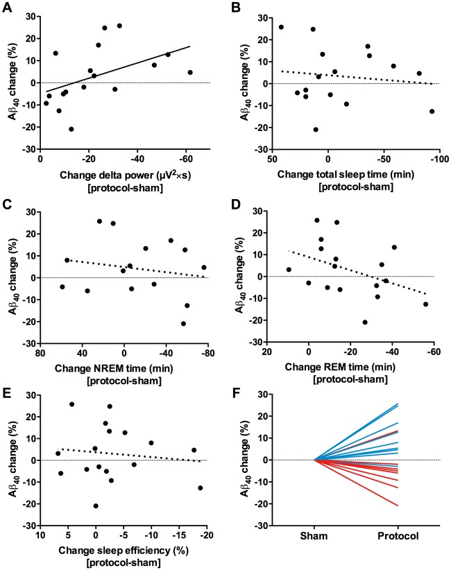
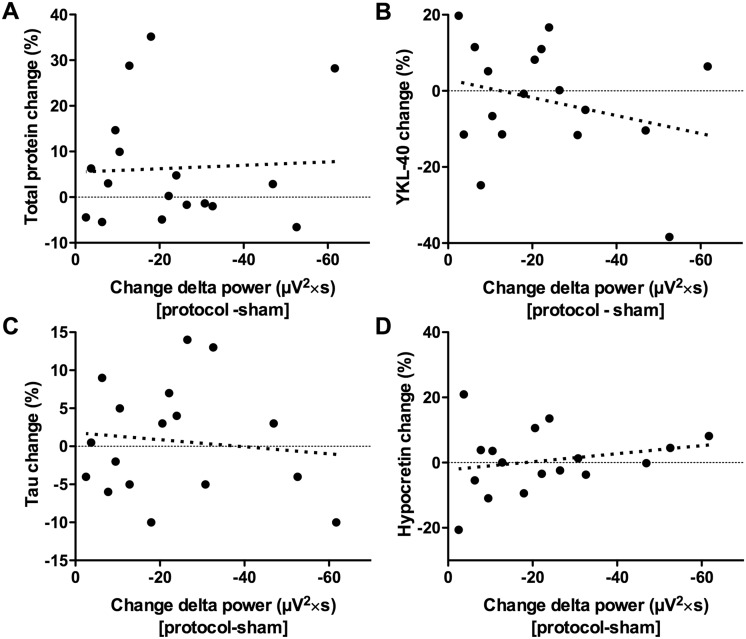
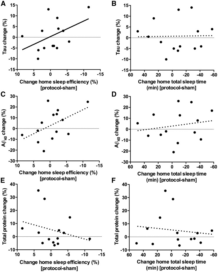
See Mander et al. (doi:10.1093/awx174) for a scientific commentary on this article.Sleep deprivation increases amyloid-β, suggesting that chronically disrupted sleep may promote amyloid plaques and other downstream Alzheimer's disease pathologies including tauopathy or inflammation. To date, studies have not examined which aspect of sleep modulates amyloid-β or other Alzheimer's disease biomarkers. Seventeen healthy adults (age 35-65 years) without sleep disorders underwent 5-14 days of actigraphy, followed by slow wave activity disruption during polysomnogram, and cerebrospinal fluid collection the following morning for measurement of amyloid-β, tau, total protein, YKL-40, and hypocretin. Data were compared to an identical protocol, with a sham condition during polysomnogram. Specific disruption of slow wave activity correlated with an increase in amyloid-β40 (r = 0.610, P = 0.009). This effect was specific for slow wave activity, and not for sleep duration or efficiency. This effect was also specific to amyloid-β, and not total protein, tau, YKL-40, or hypocretin. Additionally, worse home sleep quality, as measured by sleep efficiency by actigraphy in the six nights preceding lumbar punctures, was associated with higher tau (r = 0.543, P = 0.045). Slow wave activity disruption increases amyloid-β levels acutely, and poorer sleep quality over several days increases tau. These effects are specific to neuronally-derived proteins, which suggests they are likely driven by changes in neuronal activity during disrupted sleep.
Keywords: EEG; beta-amyloid; sleep; slow wave activity; tau.
© The Author (2017). Published by Oxford University Press on behalf of the Guarantors of Brain. All rights reserved. For Permissions, please email: journals.permissions@oup.com.
Figures
Comment in
-
A restless night makes for a rising tide of amyloid.Brain. 2017 Aug 1;140(8):2066-2069. doi: 10.1093/brain/awx174. Brain. 2017. PMID: 28899024 No abstract available.
References
-
- Buysse DJ, Reynolds CF, Monk TH, Berman SR, Kupfer DJ. The Pittsburgh Sleep Quality Index: a new instrument for psychiatric practice and research. Psychiatry Res 1989; 28: 193–213. - PubMed
-
- Cirrito JR, Yamada KA, Finn MB, Sloviter RS, Bales KR, May PC, et al. Synaptic activity regulates interstitial fluid amyloid-beta levels in vivo. Neuron 2005; 48:913–22. - PubMed
-
- Fagan AM, Mintun MA, Mach RH, Lee S-Y, Dence CS, Shah AR, et al. Inverse relation between in vivo amyloid imaging load and CSF Aβ42 in humans. Ann Neurol 2006; 59: 512–9. - PubMed
MeSH terms
Substances
Grants and funding
LinkOut - more resources
Full Text Sources
Other Literature Sources
Medical
