

The impact of preload on 3-dimensional deformation parameters: principal strain, twist and torsion
- PMID: 28899401
- PMCID: PMC5596939
- DOI: 10.1186/s12947-017-0111-x
The impact of preload on 3-dimensional deformation parameters: principal strain, twist and torsion
Abstract
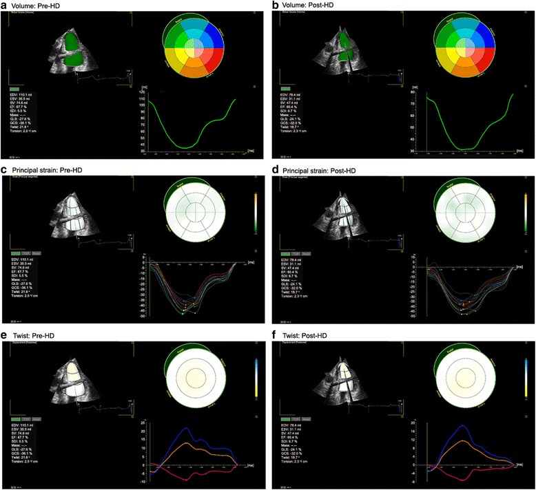
Background: Strain analysis is feasible using three-dimensional (3D) echocardiography. This approach provides various parameters based on speckle tracking analysis from one full-volume image of the left ventricle; however, evidence for its volume independence is still lacking.
Methods: Fifty-eight subjects who were examined by transthoracic echocardiography immediately before and after hemodialysis (HD) were enrolled. Real-time full-volume 3D echocardiographic images were acquired and analyzed using dedicated software. Two-dimensional (2D) longitudinal strain (LS) was also measured for comparison with 3D strain values.
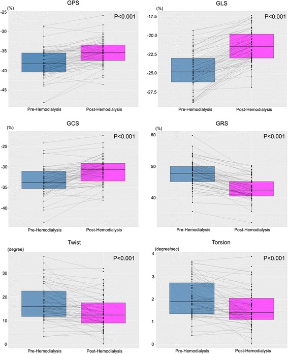
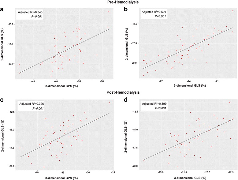
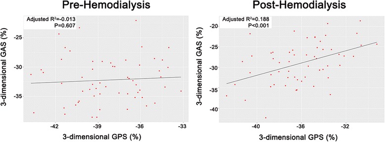
Results: Longitudinal (pre-HD: -24.57 ± 2.51, post-HD: -21.42 ± 2.15, P < 0.001); circumferential (pre-HD: -33.35 ± 3.50, post-HD: -30.90 ± 3.22, P < 0.001); and radial strain (pre-HD: 46.47 ± 4.27, post-HD: 42.90 ± 3.61, P < 0.001) values were significantly decreased after HD. The values of 3D principal strain (PS), a unique parameter of 3D images, were affected by acute preload changes (pre-HD: -38.10 ± 3.71, post-HD: -35.33 ± 3.22, P < 0.001). Twist and torsion values were decreased after HD (pre-HD: 17.69 ± 7.80, post-HD: 13.34 ± 6.92, P < 0.001; and pre-HD: 2.04 ± 0.86, post-HD:1.59 ± 0.80, respectively, P < 0.001). The 2D LS values correlated with the 3D LS and PS values.
Conclusion: Various parameters representing left ventricular mechanics were easily acquired from 3D echocardiographic images; however, like conventional parameters, they were affected by acute preload changes. Therefore, strain values from 3D echocardiography should be interpreted with caution while considering the preload conditions of the patients.
Keywords: Hemodialysis; Myocardial strain; Three-dimensional echocardiography.
Conflict of interest statement
Ethics approval and consent to participate
This study was approved by the Institutional Review Board of Bucheon St. Mary’s Hospital and was in compliance with the Declaration of Helsinki. Written consent was obtained from the subjects before performing the screening echocardiography.
Consent for publication
Not applicable.
Competing interests
The authors declare that they have no competing interests.
Publisher’s Note
Springer Nature remains neutral with regard to jurisdictional claims in published maps and institutional affiliations.
Figures
References
-
- Shiota T, McCarthy PM, White RD, Qin JX, Greenberg NL, Flamm SD, Wong J, Thomas JD. Initial clinical experience of real-time three-dimensional echocardiography in patients with ischemic and idiopathic dilated cardiomyopathy. Am J Cardiol. 1999;84(9):1068–1073. doi: 10.1016/S0002-9149(99)00500-7. - DOI - PubMed
-
- Qin JX, Jones M, Shiota T, Greenberg NL, Tsujino H, Firstenberg MS, Gupta PC, Zetts AD, Xu Y, Ping Sun J, et al. Validation of real-time three-dimensional echocardiography for quantifying left ventricular volumes in the presence of a left ventricular aneurysm: in vitro and in vivo studies. J Am Coll Cardiol. 2000;36(3):900–907. doi: 10.1016/S0735-1097(00)00793-2. - DOI - PubMed
Publication types
MeSH terms
LinkOut - more resources
Full Text Sources
Other Literature Sources
Medical
