. 2017 Nov 6;214(11):3207-3217.
doi: 10.1084/jem.20170580. Epub 2017 Sep 12.

B cell-derived IL-6 initiates spontaneous germinal center formation during systemic autoimmunity

Affiliations

B cell-derived IL-6 initiates spontaneous germinal center formation during systemic autoimmunity

Tanvi Arkatkar et al. J Exp Med. .

Abstract

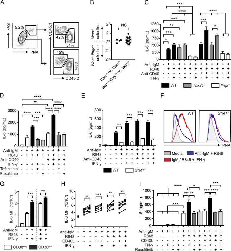
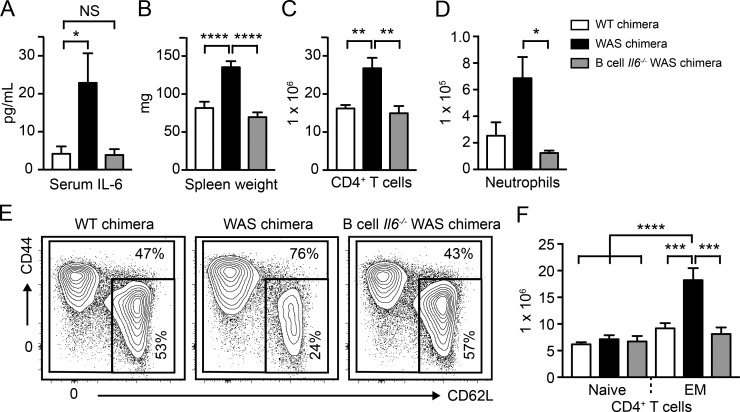
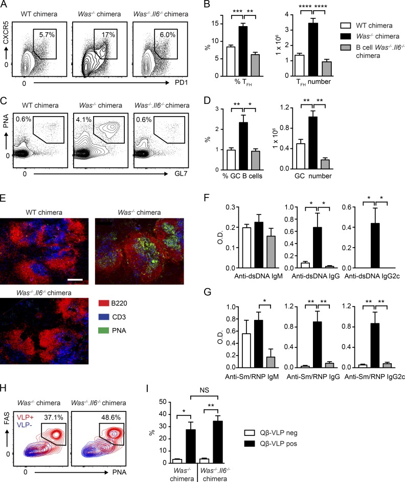
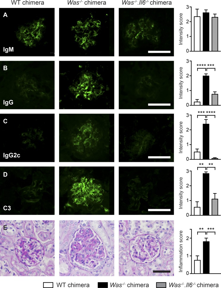
Recent studies have identified critical roles for B cells in triggering autoimmune germinal centers (GCs) in systemic lupus erythematosus (SLE) and other disorders. The mechanisms whereby B cells facilitate loss of T cell tolerance, however, remain incompletely defined. Activated B cells produce interleukin 6 (IL-6), a proinflammatory cytokine that promotes T follicular helper (TFH) cell differentiation. Although B cell IL-6 production correlates with disease severity in humoral autoimmunity, whether B cell-derived IL-6 is required to trigger autoimmune GCs has not, to our knowledge, been addressed. Here, we report the unexpected finding that a lack of B cell-derived IL-6 abrogates spontaneous GC formation in mouse SLE, resulting in loss of class-switched autoantibodies and protection from systemic autoimmunity. Mechanistically, B cell IL-6 production was enhanced by IFN-γ, consistent with the critical roles for B cell-intrinsic IFN-γ receptor signals in driving autoimmune GC formation. Together, these findings identify a key mechanism whereby B cells drive autoimmunity via local IL-6 production required for TFH differentiation and autoimmune GC formation.


Figures

Figure 1.
Figure 1.
IFN-γ synergizes with B cell activation signals to promote IL-6 production by mouse and human B cells. (A) Representative FACS plots showing CD45.1+ Was−/− versus CD45.2+ Was−/−Ifngr−/− B cells in GC and non-GC B cell compartments. (A, left) Gated on CD19+ B cells. (A, right) Gated on PNA+FAS+ GC B cells (top) and PNAFAS non-GC B cells (bottom). Number equals percentage within gate. (B) Selection of CD45.1+ Was−/− versus CD45.2+ Was−/−Ifngr−/− B cells into the GC compartment. NS, not significant. (C) IL-6 from splenic WT (black), Tbx21−/− (gray), and Ifngr−/− (white) B cells cultured for 48 h with anti–IgM, R848, anti–CD40 and/or IFN-γ. (D) IL-6 production by stimulated mouse B cells with or without ruxolitinib or tofacitinib (500 nM). (E) IL-6 in WT (black) and Stat1−/− (white) B cells stimulated as indicated. (F) Surface PNA binding in WT (left) and Stat1−/− (right) splenic B cells stimulated as indicated. (G) IL-6 intracellular staining at 72 h in cultured human B cells, gated as CD38CD27 “naïve” (white) or CD38+CD27 “GC” (black) B cells. (H) IL-6 mean fluorescence intensity by intracellular staining in human CD38+CD27 B cells stimulated for 24 h. (I) IL-6 from human B cells stimulated for 72 h with or without ruxolitinib (500 nM). (B–E and G–I) Error bars indicate means ± SEM. *, P < 0.05; **, P < 0.01; ***, P < 0.001; ****, P < 0.0001, by two-tailed Student’s t test (B); by one-way ANOVA and Tukey's multiple comparison test (C–E and I); or by paired two-tailed Student’s t test (G and H). (H) Data shown as paired analysis of different stimulation conditions from individual human donors. (A and B) Data representative of three independent Was−/− versus Was−/−Ifngr−/− competitive chimeras. (C-H) Data representative of at least two independent experiments.
Figure 2.
Figure 2.
B cell–derived IL-6 promotes systemic inflammation in SLE. (A) Serum IL-6 levels in WT, Was−/−, and B cell–intrinsic Was−/−Il6−/− chimeras. (B–D) Spleen weight (B), number of splenic CD4+ T cells (C), and CD11b+GR1+ neutrophils (D) in indicated chimeras. (E) Representative FACS plots (gated on CD4+ T cells) showing naive (CD44loCD62Lhi) and effector/memory (CD44hiCD62Llo/hi) CD4+ T cells. The percentages are those within the gate. (F) Number of naive and effector/memory CD4+ T cells. (A–F) Error bars indicate means ± SEM. *, P < 0.05; **, P < 0.01; ***, P < 0.001; ****, P < 0.0001, by one-way ANOVA and Tukey's multiple comparison test. Data are representative of four WT (white, n = 11), four Was−/− (black, n = 13), and two B cell–intrinsic Was−/−Il6−/− (gray, n = 6) independent chimeras.
Figure 3.
Figure 3.
B cell IL-6 production initiates spontaneous, autoimmune GCs. (A and B) Representative FACS plots (gated on splenic CD4+ T cells) showing PD1+CXCR5+ TFH cell percentage (left) and total number (right) of splenic TFH cells in indicated chimeras. (C) Representative FACS plots (gated on splenic CD19+ B cells) showing loss of PNA+GL7+ GC B cells after B cell–intrinsic Il6−/− deletion. (D) Percentage (left) and total number (right) of splenic GC B cells in indicated chimeras. (E) Representative splenic sections stained with B220 (red), PNA (green), and CD3 (blue). Bar, 150 µm. (F and G) Anti–dsDNA (F) and anti–Sm/RNP (G) IgM, IgG, and IgG2c autoAb at 12 (F) and 24 (G) wk after transplant. (H) Representative overlaid FACS plots showing PNA+FAS+ GC phenotype in Qβ-VLP+ (red) versus Qβ-VLP (blue) B cells in Was−/− (left) and B cell–intrinsic Was−/−Il6−/− (right) chimeras, 12 d after Qβ-VLP immunization. Percentages are Qβ-VLP+ B cells within the PNA+FAS+ gate. (I) Percentage of PNA+FAS+ B cells in Qβ-VLP+ (black) versus Qβ-VLP (white) B cells in indicated chimeras. (B, D, F, G, and I) Error bars indicate means ± SEM. *, P < 0.05; **, P < 0.01; ***, P < 0.001; ****, P < 0.0001; NS, not significant, by one-way ANOVA and Tukey's multiple comparison test. (A–G) Data representative of four WT (white, n ≥ 10), four Was−/− (black, n ≥ 12), and two B cell–intrinsic Was−/−Il6−/− (gray, n = 6) independent chimeras. (H and I) Representative FACS plots (H) and combined data (I) from two independent, B cell–intrinsic, chimera experiments (Was−/− [n = 4] and Was−/−Il6−/− [n = 5]).
Figure 4.
Figure 4.
Development of IC glomerulonephritis requires B cell–derived IL-6. (A–D) Immunofluorescence staining for glomerular (A) IgM; (B) IgG; (C) IgG2c; (D) C3. (Left) Representative images. (Right) Intensity of glomerular immunofluorescence staining. (E, left) Representative kidney sections stained with periodic acid–Schiff. (E, right) Glomerular inflammation score. (A–E) Data representative of ≥2 independent chimeras, including WT (white, n = 3 [A–D], n = 4 [E]), Was−/− (black, n = 5 [A–E]), and B cell–intrinsic Was−/−Il6−/− (gray, n = 5 [A–D], n = 4 [E]) chimeras. Scoring by observers blinded to genotype. Error bars indicate means ± SEM. Bars, 50 µm. **, P < 0.01; ***, P < 0.001; ****, P < 0.0001 by one-way ANOVA and Tukey's multiple comparison test.

Comment in

References

    1. Aloisi F., and Pujol-Borrell R.. 2006. Lymphoid neogenesis in chronic inflammatory diseases. Nat. Rev. Immunol. 6:205–217. 10.1038/nri1786 - DOI - PubMed
    1. Barr T.A., Shen P., Brown S., Lampropoulou V., Roch T., Lawrie S., Fan B., O’Connor R.A., Anderton S.M., Bar-Or A., et al. . 2012. B cell depletion therapy ameliorates autoimmune disease through ablation of IL-6-producing B cells. J. Exp. Med. 209:1001–1010. 10.1084/jem.20111675 - DOI - PMC - PubMed
    1. Becker-Herman S., Meyer-Bahlburg A., Schwartz M.A., Jackson S.W., Hudkins K.L., Liu C., Sather B.D., Khim S., Liggitt D., Song W., et al. . 2011. WASp-deficient B cells play a critical, cell-intrinsic role in triggering autoimmunity. J. Exp. Med. 208:2033–2042. 10.1084/jem.20110200 - DOI - PMC - PubMed
    1. Bentebibel S.E., Lopez S., Obermoser G., Schmitt N., Mueller C., Harrod C., Flano E., Mejias A., Albrecht R.A., Blankenship D., et al. . 2013. Induction of ICOS+CXCR3+CXCR5+ TH cells correlates with antibody responses to influenza vaccination. Sci. Transl. Med. 5:176ra32 10.1126/scitranslmed.3005191 - DOI - PMC - PubMed
    1. Deane K.D., Norris J.M., and Holers V.M.. 2010. Preclinical rheumatoid arthritis: Identification, evaluation, and future directions for investigation. Rheum. Dis. Clin. North Am. 36:213–241. 10.1016/j.rdc.2010.02.001 - DOI - PMC - PubMed

MeSH terms