Review
. 2017 Oct 15;45(5):1159-1171.
doi: 10.1042/BST20170096. Epub 2017 Sep 12.

Structure and function of Pif1 helicase

Affiliations
Review

Structure and function of Pif1 helicase

Alicia K Byrd et al. Biochem Soc Trans. .

Abstract

Pif1 family helicases have multiple roles in the maintenance of nuclear and mitochondrial DNA in eukaryotes. Saccharomyces cerevisiae Pif1 is involved in replication through barriers to replication, such as G-quadruplexes and protein blocks, and reduces genetic instability at these sites. Another Pif1 family helicase in S. cerevisiae, Rrm3, assists in fork progression through replication fork barriers at the rDNA locus and tRNA genes. ScPif1 (Saccharomyces cerevisiae Pif1) also negatively regulates telomerase, facilitates Okazaki fragment processing, and acts with polymerase δ in break-induced repair. Recent crystal structures of bacterial Pif1 helicases and the helicase domain of human PIF1 combined with several biochemical and biological studies on the activities of Pif1 helicases have increased our understanding of the function of these proteins. This review article focuses on these structures and the mechanism(s) proposed for Pif1's various activities on DNA.

Keywords: DNA; G-quadruplex; enzyme kinetics; enzyme–substrate interactions; helicase; mtDNA.


Conflict of interest statement

Declarations of interest

The authors declare that conflict of interest associated with this manuscript.

Figures
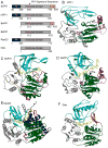
Figure 1
Figure 1. Structures of superfamily 1B helicases
(A) Domain maps of SF1B helicases. N-terminal domains are shown in blue, helicase domains in gray, C-terminal domains in orange, and the Pif1 signature sequence in pink. (B) Structure of hPIF1-HD (PDB: 5FHH) [33]. Domain 1A is shown in green, 1B in red, 2A in gray, 2B in cyan, and the Pif1 signature sequence in pink. ADP-AlF4 is shown in purple. (C) Structure of BsPif1 (PDB: 5FTE) [32]. Colors are the same as in B with the C-terminal domain in orange, AMPPNP in purple, and DNA is in yellow. (D) Structure of BaPif1 (PDB: 5FHD) [33]. Colors are as in B with DNA shown in yellow. (E) Structure of D. radiodurans RecD2 (PDB: 3GPL) [34]. Colors are the same as in B with the N-terminal domain in blue, AMPPNP in purple, and DNA in yellow. (F) Structure of Bacteriophage T4 Dda (PDB: 3UPU) [35]. Colors are as in B with DNA in yellow.
Figure 2
Figure 2. Structure of the Pif1 signature sequence
Hydrophobic amino acids in the BaPif1 (PDB: 5FHD) [33] signature sequence (pink) interact with amino acids in domains 1A (green) and 1B (red) stabilizing this region of the protein. Ile118 which is essential for helicase activity [33] and corresponds to L319 in hPIF1 is shown in blue. DNA is shown in yellow.
Figure 3
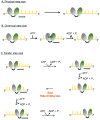
Figure 3. Step sizes of for helicases
The two RecA-like domains of a helicase are shown (gray and green) and the yellow lattice represents DNA. (A) A helicase with a physical step size of one nucleotide is depicted. The enzyme moves by one nucleotide in each step. (B) A helicase with chemical and physical step sizes of one nucleotide is shown. Binding of ATP results in movement of the rear domain forward by one nucleotide closing the cleft between the two RecA-like domains. Release of ADP and Pi results in movement of the leading domain forward by one nucleotide as the cleft opens. (C) The scheme illustrates movement by a helicase with physical and chemical step sizes of one nucleotide and a kinetic step size of 2 nucleotides. The enzyme moves in single nucleotide steps, each powered by the hydrolysis of a single ATP molecule. After two forward steps, a slow kinetic step occurs which limits the overall rate, resulting in an observed step size of 2 nucleotides. After the slow kinetic step, ATP hydrolysis and forward movement resume.
Figure 4
Figure 4. Substrates utilized for G4DNA unfolding studies
(A) Some experiments for measuring G4DNA unfolding rely on trapping the unfolded duplex as dsDNA to prevent refolding of the G4DNA structure. Experiments typically start with the enzyme prebound to the G4DNA substrate and unfolding is initiated by addition of ATP, Mg2+, and a DNA trap (green) complementary to the G4DNA. G4DNA substrate and dsDNA product can be separated by electrophoresis. * indicates either a fluorescent or radiolabel. Since refolding is a unimolecular process and trapping is a bimolecular process, trapping can be inefficient. (B) Alternatively, G4DNA unfolding can be monitored by measuring the unwinding of a reporter duplex following the G4DNA. Appearance of ssDNA provides a “report” on the unfolding of the G4DNA. A gap of 2 nucleotides (red) between the G4DNA and reporter duplex is necessary for folding of both the G4DNA and dsDNA to occur [83]. (C) Based on smFRET studies, some helicases have been reported to remain anchored at a ssDNA/dsDNA junction while repetitively reeling in the ssDNA and unfolded G4DNA. Green and red dots represent Cy3 and Cy5.

References

    1. Crouch JD, Brosh RM., Jr Mechanistic and biological considerations of oxidatively damaged DNA for helicase-dependent pathways of nucleic acid metabolism. Free Radic Biol Med. 2017;107:245–257. - PMC - PubMed
    1. Byrd AK, Raney KD. Superfamily 2 helicases. Front Biosci. 2012;17:2070–2088. - PMC - PubMed
    1. Raney KD, Byrd AK, Aarattuthodiyil S. Structure and Mechanisms of SF1 DNA Helicases. Adv Exp Med Biol. 2013;767:17–46. - PMC - PubMed
    1. Bochman ML, Judge CP, Zakian VA. The Pif1 family in prokaryotes: what are our helicases doing in your bacteria? Mol Biol Cell. 2011;22:1955–1959. - PMC - PubMed
    1. Bessler JB, Torredagger JZ, Zakian VA. The Pif1p subfamily of helicases: region-specific DNA helicases? Trends Cell Biol. 2001;11:60–65. - PubMed