Comparative Study
. 2017 Nov 3;292(44):18169-18177.
doi: 10.1074/jbc.M117.811752. Epub 2017 Sep 12.

The metal chaperone Atox1 regulates the activity of the human copper transporter ATP7B by modulating domain dynamics

Affiliations
Comparative Study

The metal chaperone Atox1 regulates the activity of the human copper transporter ATP7B by modulating domain dynamics

Corey H Yu et al. J Biol Chem. .

Abstract

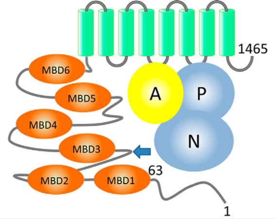
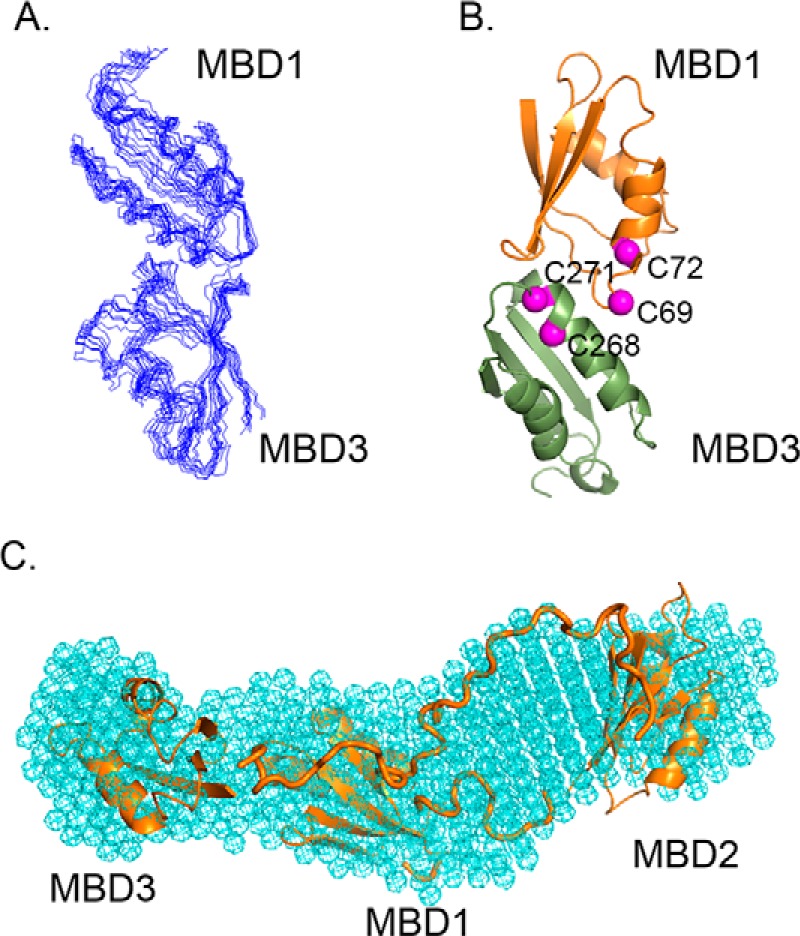
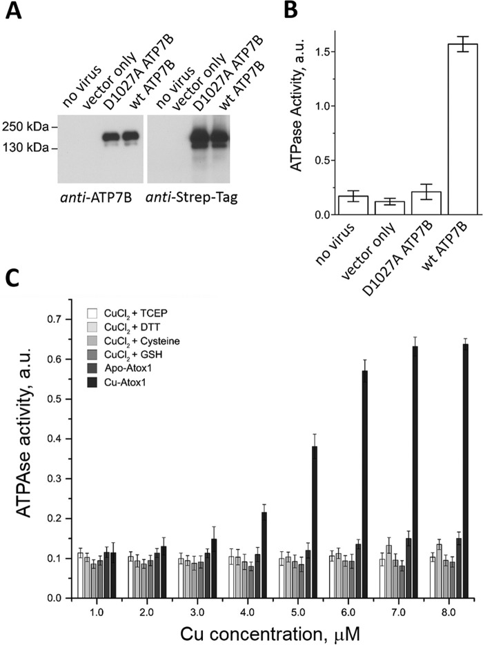
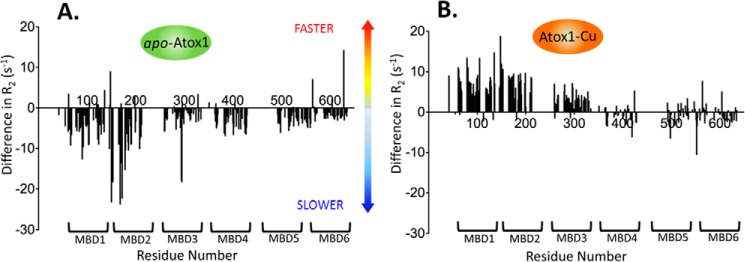
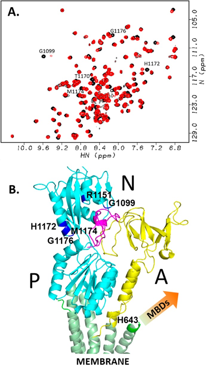
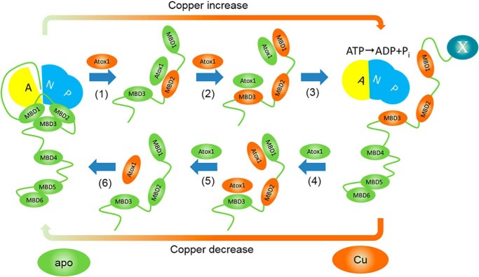
The human transporter ATP7B delivers copper to the biosynthetic pathways and maintains copper homeostasis in the liver. Mutations in ATP7B cause the potentially fatal hepatoneurological disorder Wilson disease. The activity and intracellular localization of ATP7B are regulated by copper, but the molecular mechanism of this regulation is largely unknown. We show that the copper chaperone Atox1, which delivers copper to ATP7B, and the group of the first three metal-binding domains (MBD1-3) are central to the activity regulation of ATP7B. Atox1-Cu binding to ATP7B changes domain dynamics and interactions within the MBD1-3 group and activates ATP hydrolysis. To understand the mechanism linking Atox1-MBD interactions and enzyme activity, we have determined the MBD1-3 conformational space using small angle X-ray scattering and identified changes in MBD dynamics caused by apo-Atox1 and Atox1-Cu by solution NMR. The results show that copper transfer from Atox1 decreases domain interactions within the MBD1-3 group and increases the mobility of the individual domains. The N-terminal segment of MBD1-3 was found to interact with the nucleotide-binding domain of ATP7B, thus physically coupling the domains involved in copper binding and those involved in ATP hydrolysis. Taken together, the data suggest a regulatory mechanism in which Atox1-mediated copper transfer activates ATP7B by releasing inhibitory constraints through increased freedom of MBD1-3 motions.

Keywords: ATPase; copper transport; membrane protein; membrane transport; nuclear magnetic resonance (NMR); protein NMR; protein dynamic.


Conflict of interest statement

The authors declare that they have no conflicts of interest with the contents of this article

Figures

Figure 1.
Figure 1.
Domain organization of ATP7B. ATP7B includes the N-terminal peptide (ATP7B1–63, residues labeled), six metal-binding domains (MBD1–MBD6, orange), and eight transmembrane helices (green). The N and P domains (blue) together hydrolyze ATP, with participation of the A domain (yellow). The arrow indicates the site of the spontaneous ATP7B proteolysis in the cell.
Figure 2.
Figure 2.
Conformational space of the MBD1–3 group revealed by molecular docking and SAXS. A, ensemble of the 10 top-scoring MBD1-MBD3 models produced by HADDOCK. B, ribbon diagram of MBD1–MBD3 complex with the α-carbons of the cysteine residues in the invariant CXXC motifs shown in magenta. C, overall shape of the MBD1–3 group determined by SAXS (cyan mesh) with MBDs 1–3 model including connecting loops (orange) fitted into the SAXS shape using SUPCOMB (54).
Figure 3.
Figure 3.
ATPase activity of ATP7B is stimulated by Atox1–Cu, but not free copper. A, expression of ATP7B in baculovirus-infected Sf9 cells detected by Western blot with antibodies against the ATP7B C terminus (left panel) and against the Strep tag (right panel). B, ATPase activity of microsomes prepared from noninfected Sf9 cells (1 μg of total protein/data point), cells infected with the virus encoding no exogenous protein (mock), D1027A-ATP7B, or wild-type ATP7B (n = 3). a.u., absorption at 600 nm. D1027A is a catalytically inactive variant of ATP7B used as a control. C, effect of free copper with various reducing agents (2 mm TCEP, 2 mm DTT, 10 mm cysteine, or 2 mm GSH) and of Atox1–Cu on the ATPase activity of Sf9 microsomes expressing wild-type ATP7B. Atox1 was added at 1:1 molar ratio with copper (Cu–Atox1). Equal amount of apo–Atox1 was used as a control for each experimental point (apo–Atox1).
Figure 4.
Figure 4.
Copper transfer from Atox1 increases mobility of MBDs 1–3. Difference in the transverse relaxation rate values (R2) between free MBD1–6 and MBD1–6 with either apo–Atox1 (A) or Atox1–Cu (B) is plotted as a function of the amino acid residue number. Negative difference corresponds to lower domain mobility in the presence of apo–Atox1 or Atox1–Cu, positive difference (e.g. for MBD1–3, B) to higher mobility.
Figure 5.
Figure 5.
ATP7B33–63 peptide interacts with the N domain. A, 1H,15N-HSQC spectra of 0.5 mm N domain with (red) and without (black) 2.8 mm ATP7B33–63. B, the model structure of ATP7B33–63 peptide bound to the N domain of ATP7B (Protein Data Bank code 2ARF) calculated from the chemical shift perturbation data (A) and aligned to the homology model of ATP7B (12) based on the X-ray structure of copper ATPase CopA from Legionella pneumophila (58), by minimizing RMSD for the N domain. The N and P domains are in cyan, the A domain is in yellow, transmembrane helices are in green, and the ATP7B33–63 peptide is in magenta. Amino acid residues in the N domain with a combined chemical shift change Δδ > 0.03 (cf. A) are shown in blue and labeled. Metal-binding domains, which are absent from the homology model, are not shown (arrow).
Figure 6.
Figure 6.
Proposed mechanism of ATP7B regulation by Atox1–Cu. At low copper, MBD1–3 associate with each other, and the N-terminal peptide is bound at the A-N domain interface, preventing ATP hydrolysis. (1) Copper transfer from Atox1 to MBD2 breaks up interactions between MBDs 1–3, and then apo–Atox1 dissociates. (2) Copper transfer from Atox1–Cu to MBD1 and MBD3 stabilizes the open conformation of MBD1–3. (3) Transition of MBD1–3 to the open conformation dislodges the N-terminal peptide (ATP7B1–63) from its binding site at the interface of the N and A domains, activating the enzyme and, possibly, inducing trafficking by making ATP7B1–63 accessible for interaction with a hypothetical partner protein X (right panel). (4) At low Atox1–Cu/apo–Atox1 ratios, apo–Atox1 removes copper from the MBDs, including, finally, MBD2 (5). (6) MBD1–3 reassociate, and ATP7B1–63 peptide binds at the A–N interface inhibiting ATP7B. For clarity, only regulatory copper transfer events are shown.

References

    1. Festa R. A., and Thiele D. J. (2011) Copper: an essential metal in biology. Curr. Biol. 21, R877–R883 - PMC - PubMed
    1. Lutsenko S., Barnes N. L., Bartee M. Y., and Dmitriev O. Y. (2007) Function and regulation of human copper-transporting ATPases. Physiol. Rev. 87, 1011–1046 - PubMed
    1. Dmitriev O. Y. (2011) Mechanism of tumor resistance to cisplatin mediated by the copper transporter ATP7B. Biochem. Cell Biol. 89, 138–147 - PubMed
    1. Kuo M. T., Chen H. H., Song I. S., Savaraj N., and Ishikawa T. (2007) The roles of copper transporters in cisplatin resistance. Cancer Metastasis Rev. 26, 71–83 - PubMed
    1. Li X. P., Yin J. Y., Wang Y., He H., Li X., Gong W. J., Chen J., Qian C. Y., Zheng Y., Li F., Yin T., Gong Z. C., Zhou B. T., Zhang Y., Xiao L., et al. (2014) The ATP7B genetic polymorphisms predict clinical outcome to platinum-based chemotherapy in lung cancer patients. Tumour Biol. 35, 8259–8265 - PubMed

Publication types

MeSH terms

Associated data