. 2017 Sep 12;7(1):11300.
doi: 10.1038/s41598-017-10749-5.

Adaptation of influenza A (H7N9) virus in primary human airway epithelial cells

Affiliations

Adaptation of influenza A (H7N9) virus in primary human airway epithelial cells

Daniel Tsung-Ning Huang et al. Sci Rep. .

Abstract

Influenza A (H7N9) is an emerging zoonotic pathogen with pandemic potential. To understand its adaptation capability, we examined the genetic changes and cellular responses following serial infections of A (H7N9) in primary human airway epithelial cells (hAECs). After 35 serial passages, six amino acid mutations were found, i.e. HA (R54G, T160A, Q226L, H3 numbering), NA (K289R, or K292R for N2 numbering), NP (V363V/I) and PB2 (L/R332R). The mutations in HA enabled A(H7N9) virus to bind with higher affinity (from 39.2% to 53.4%) to sialic acid α2,6-galactose (SAα2,6-Gal) linked receptors. A greater production of proinflammatory cytokines in hAECs was elicited at later passages together with earlier peaking at 24 hours post infection of IL-6, MIP-1α, and MCP-1 levels. Viral replication capacity in hAECs maintained at similar levels throughout the 35 passages. In conclusion, during the serial infections of hAECs by influenza A(H7N9) virus, enhanced binding of virion to cell receptors with subsequent stronger innate cell response were noted, but no enhancement of viral replication could be observed. This indicates the existence of possible evolutional hurdle for influenza A(H7N9) virus to transmit efficiently from human to human.


Conflict of interest statement

The authors declare that they have no competing interests.

Figures

Figure 1
Figure 1
Illustration of how serial passages of A(H7N9) influenza virus were performed on hAECs.
Figure 2
Figure 2
The replication of influenza A(H7N9) virus during serial passage in hAECs. During the 35 passages of A(H7N9) virus in hAECs, viral replication varied but did not change substantially.
Figure 3
Figure 3
Sialic acid-linked receptors binding properties at individual passage in hAECs. Virus supernatants of passage 1, 15, and 35 were used to infect hAECs. Infected cells were fixed and stained with NP (representing H7N9 virus), Sambucus nigra lectin (SNA, substrate of α2,6-sialic acid), Maackia Amurensis II (MALII, substrate of α2,3-sialic acid) and DAPI at 5 hpi. Each was observed for 8 fields under ZEISS, LSM 510 META Confocal Microscope. Four patterns were observed, including virus located in cells expressing (Fig. 3A) SAα2,3-Gal receptors only, (Fig. 3B) SAα2,6-Gal receptors only, (Fig. 3C) both SAα2,3- and SAα2,6-Gal receptors, and (Fig. 3D) neither SAα2,3- nor SAα2,6-Gal receptors. The white arrows indicate the specific localization panels, and the star signature represents virus infection in cells expressing both SAα2,3- and SAα2,6-Gal receptors.
Figure 4
Figure 4
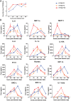
Cytokine responses to H7N9-P0, H7N9-P2 and H7N9-P35 infection in hAECs. Virus supernatants were collected at 6, 24, 48, 72 and 96 hpi and analyzed by TCID50 assay for virus titer determination and procataplex immunoassay for cytokines and chemokines. (Figure 4A) Virus replication kinetics of H7N9-P0, H7N9-P2 and H7N9-P35 were similar. (Figure 4B) Cytokine and chemokine expression panels at 0(H7N9-P0), 2nd (H7N9-P2) and 35th (H7N9-P35) passages in hAECs. Blue line: H7N9-P0, Orange line: H7N9-P2, Red line: H7N9-P35. Statistics method: Two-way ANOVA. *P < 0.01, **P < 0.001 at 24 hpi.

References

    1. Gao R, et al. Human infection with a novel avian-origin influenza A (H7N9) virus. The New England journal of medicine. 2013;368:1888–1897. doi: 10.1056/NEJMoa1304459. - DOI - PubMed
    1. Bao CJ, et al. Live-animal markets and influenza A (H7N9) virus infection. The New England journal of medicine. 2013;368:2337–2339. doi: 10.1056/NEJMc1306100. - DOI - PubMed
    1. Lam TT, et al. Dissemination, divergence and establishment of H7N9 influenza viruses in China. Nature. 2015;522:102–105. doi: 10.1038/nature14348. - DOI - PubMed
    1. Chen GW, Gong YN, Shih SR. Influenza A virus plasticity-A temporal analysis of species-associated genomic signatures. Journal of the Formosan Medical Association = Taiwan yi zhi. 2015;114:456–463. doi: 10.1016/j.jfma.2015.01.015. - DOI - PubMed
    1. Liu D, et al. Origin and diversity of novel avian influenza A H7N9 viruses causing human infection: phylogenetic, structural, and coalescent analyses. Lancet (London, England) 2013;381:1926–1932. doi: 10.1016/S0140-6736(13)60938-1. - DOI - PubMed

Publication types