. 2017 Sep 12;7(1):11247.
doi: 10.1038/s41598-017-10982-y.

Physicochemical stability and transfection efficiency of cationic amphiphilic copolymer/pDNA polyplexes for spinal cord injury repair

Affiliations

Physicochemical stability and transfection efficiency of cationic amphiphilic copolymer/pDNA polyplexes for spinal cord injury repair

So-Jung Gwak et al. Sci Rep. .

Abstract

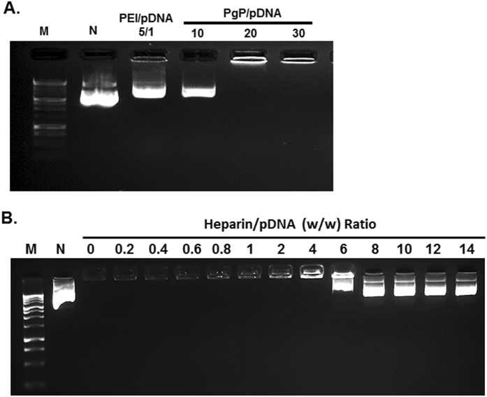
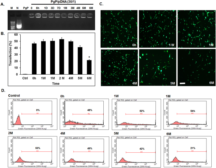
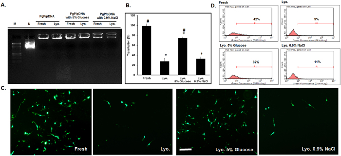
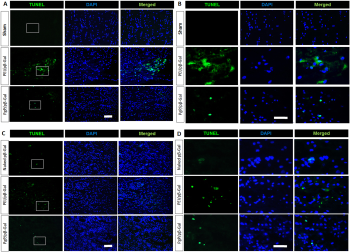
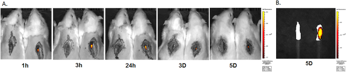
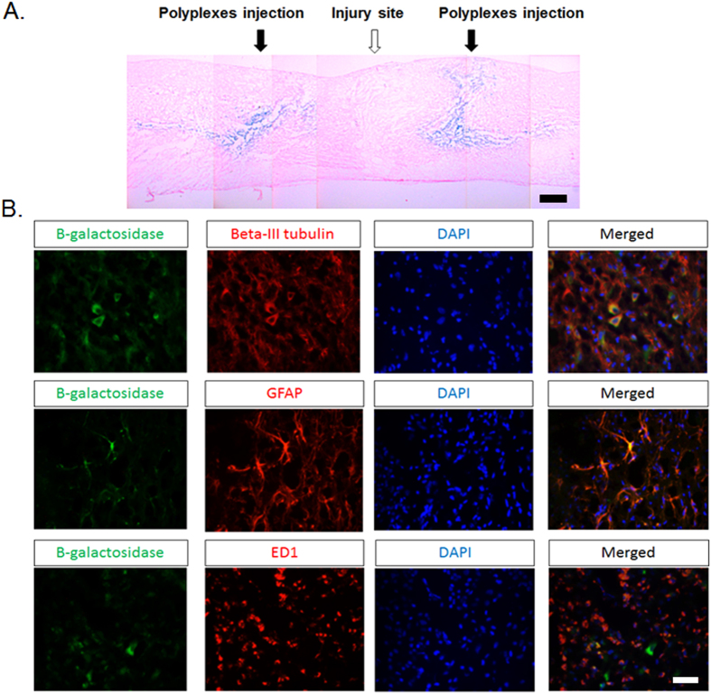
Multiple age-related and injury-induced characteristics of the adult central nervous system (CNS) pose barriers to axonal regeneration and functional recovery following injury. In situ gene therapy is a promising approach to address the limited availability of growth-promoting biomolecules at CNS injury sites. The ultimate goal of our work is to develop, a cationic amphiphilic copolymer for simultaneous delivery of drug and therapeutic nucleic acids to promote axonal regeneration and plasticity after spinal cord injury. Previously, we reported the synthesis and characterization of a cationic amphiphilic copolymer, poly (lactide-co-glycolide)-graft-polyethylenimine (PgP) and its ability to efficiently transfect cells with pDNA in the presence of serum. We also demonstrated the efficacy of PgP as a therapeutic siRhoA carrier in a rat compression spinal cord injury model. In this work, we show that PgP/pDNA polyplexes provide improved stability in the presence of competing polyanions and nuclease protection in serum relative to conventional branched polyethylenimine control. PgP/pDNA polyplexes maintain bioactivity for transfection after lyophilization/reconstitution and during storage at 4 °C for up to 5 months, important features for commercial and clinical application. We also demonstrate that PgP/pDNA polyplexes loaded with a hydrophobic fluorescent dye are retained in local neural tissue for up to 5 days and that PgP can efficiently deliver pβ-Gal in a rat compression SCI model.


Conflict of interest statement

The authors declare that they have no competing interests.

Figures

Figure 1
Figure 1
Heparin competition assay of PgP/pDNA polyplexes (2 µg pDNA). (A) PgP/pDNA polyplexes at varying N/P ratios and bPEI/pDNA polyplex at N/P ratio 5/1 were prepared and incubated in the presence of heparin (heparin/polyplexes ratio of 3/1 (w/w)) at 37 °C for 30 min. (B) PgP/pDNA polyplexes (2 µg pDNA) at N/P ratio of 30/1 incubated with solutions containing heparin at varying concentration (0–14 heparin/pDNA (w/w) ratios) at 37 °C for 30 min. M: Molecular marker, N: naked DNA.
Figure 2
Figure 2
Stability of PgP/pDNA polyplexes (N/P ratio 30/1, 2 µg pDNA) after incubation in medium containing 50% serum at 37 °C. Molecular weight marker (M, Lane 1), naked DNA (N, lane 2), fetal bovine serum only (FBS, lane 3), and PgP/pDNA polyplexes at various time points during incubation in 50% serum for 0, 0.5, 1, 3, 6, 24, and 72 hrs (lane 4–10).
Figure 3
Figure 3
Long-term stability of PgP/pDNA polyplexes (N/P ratio of 30/1, 2 µg pDNA) in water at 4 °C. (A) Gel retardation assay of PgP/pDNA polyplexes stored at 4 °C for different time periods. Molecular weight marker (M, Lane 1), naked pDNA (N, lane 2), PgP only (lane 3), PgP/pDNA polyplexes at various time points during storage at 4 °C for 0, 6hrs, 1, 3, and 7 days, and 1, 3, 4, 5, and 6 months (lane 4–13). (B) Transfection efficacy of PgP/pGFP polyplexes stored at 4 °C in B35 cells in media containing 10% serum. *p < 0.05 compared with freshly prepared polyplexes. (C) Representative images and (D) Flow cytometry histogram of GFP + B35 cells at 2 days post-transfection with PgP/pGFP polyplexes stored at 4 °C. Scale bar indicates 200 µm. (Fresh: freshly prepared polyplexes, W: week, and M: Month).
Figure 4
Figure 4
Stability and transfection efficiency of PgP/pGFP polyplexes after lyophilization. (A) Stability of PgP/pGFP polyplexes (N/P ratio 30/1) freshly prepared and after lyophilization alone or with the inclusion of cryoprotectants. Molecular weight maker (Lane 1), naked plasmid GFP (lane 2), fresh polyplexes (lane 3), reconstituted polyplexes after lyophilization (lane 4), fresh polyplexes with 5% glucose (lane 5), reconstituted polyplexes after lyophilization with 5% glucose (lane 6), fresh polyplexes with 0.9% NaCl (lane 7), reconstituted polyplexes after lyophilization with 0.9% NaCl (lane 8). (B) Transfection efficacy of freshly prepared and lyophilized PgP/pGFP polyplexes in B35 cells. *p < 0.05 compared with freshly prepared polyplexes. # p < 0.05 compared with lyophilized polyplexes without cryoprotectant. (C) Representative images and (D) flow cytometry histogram of GFP+ B35 cells at 2 days after transfection with PgP/pGFP polyplexes. Fresh: Freshly prepared polyplexes, Lyo.: Lyophilized PgP/pGFP, Lyo. 5% Glucose: Lyophilized PgP/pGFP with 5% glucose, Lyo. 0.9% NaCl: Lyophilized PgP/pGFP with 0.9% NaCl. Scale bar indicates 200 µm.
Figure 5
Figure 5
Cytotoxicity of PgP/pβ-Gal polyplexes at N/P ratio of 30/1 in normal rat spinal cord. At 2 and 7 days post-injection, rats were sacrificed and spinal cords explanted, embedded, sectioned, and toxicity was evaluated by TUNEL staining. Apoptotic cells were stained in green with blue DAPI nuclear counterstaining. (A and B) show representative images of TUNEL+ cells in spinal cord at 2 days post-injection (Top: sham control, Middle: bPEI/pβ-Gal polyplexes at N/P ratio of 5/1, Bottom: PgP/pβ-Gal polyplexes at N/P ratio of 30/1). (C and D) show representative images of TUNEL+ cells in spinal cord at 7 days post-injection (Top: naked pDNA, Middle: bPEI/pβ-Gal polyplexes at N/P ratio of 5/1, Bottom: PgP/pβ-Gal polyplexes at N/P ratio of 30/1), (A and C) Original magnification 200X (scale bar indicates 100 µm), (B and D) Enlarged images of highlighted interest region, original magnification 400X (scale bar indicates 50 µm).
Figure 6
Figure 6
Retention of DiR-PgP/pDNA polyplexes (N/P ratio of 30/1) after local injection in SCI lesion site. (A) Visualization of DiR-PgP/pβ-gal polyplexes at 1, 3, 24 hours, and 3, and 5 days following injection by live animal fluorescence imaging system (IVIS). Left: Uninjected SCI animal (Control), Right: DiR-PgP/pβ-gal polyplexes injected animal (B) Ex vivo fluorescent imaging of DiR-PgP/pDNA polyplexes at 5 days post-injection by IVIS.
Figure 7
Figure 7
(A) Representative image of β-Gal expression (blue staining) in SCI model 7 days after injection of PgP/pβ-gal polyplexes at N/P ratio 30/1. Scale bar indicates 400 µm. (B) Double immunohistochemical staining for β-Gal+ cells (green) and beta-III tubulin (red, Left), GFAP (red, Middle) and ED-1 (red, Right) in SCI region. Merged images show co-localization of β-Gal+ cells and beta-III tubulin+ neurons, GFAP+ astrocytes, and ED-1+ microglia cells/infiltrated macrophages. Scale bar indicates 50 µm.

References

    1. Tator CH. Update on the pathophysiology and pathology of acute spinal cord injury. Brain Pathol. 1995;5:407–13. doi: 10.1111/j.1750-3639.1995.tb00619.x. - DOI - PubMed
    1. Modell HI. Charting a course for advances in physiology education. Am J Physiol. 1989;256:S1–2. - PubMed
    1. McDonald JW, Sadowsky C. Spinal-cord injury. Lancet. 2002;359:417–25. doi: 10.1016/S0140-6736(02)07603-1. - DOI - PubMed
    1. Gwak, S. J., Yun, Y., Yoon, D. H., Kim, K. N., & Ha, Y. Therapeutic Use of 3 beta-[N-(N′,N′-Dimethylaminoethane) Carbamoyl] Cholesterol-Modified PLGA Nanospheres as Gene Delivery Vehicles for Spinal Cord Injury. Plos One. 11 (2016). - PMC - PubMed
    1. Yao L, et al. Improved axonal regeneration of transected spinal cord mediated by multichannel collagen conduits functionalized with neurotrophin-3 gene. Gene Therapy. 2013;20:149–57. doi: 10.1038/gt.2013.42. - DOI - PubMed

Publication types

MeSH terms