. 2017 Sep 12;7(1):11420.
doi: 10.1038/s41598-017-11645-8.

Adolescent THC Exposure Causes Enduring Prefrontal Cortical Disruption of GABAergic Inhibition and Dysregulation of Sub-Cortical Dopamine Function

Affiliations

Adolescent THC Exposure Causes Enduring Prefrontal Cortical Disruption of GABAergic Inhibition and Dysregulation of Sub-Cortical Dopamine Function

Justine Renard et al. Sci Rep. .

Abstract

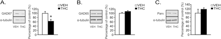
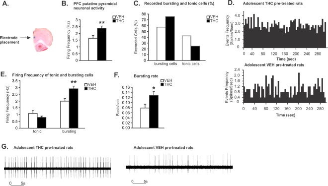
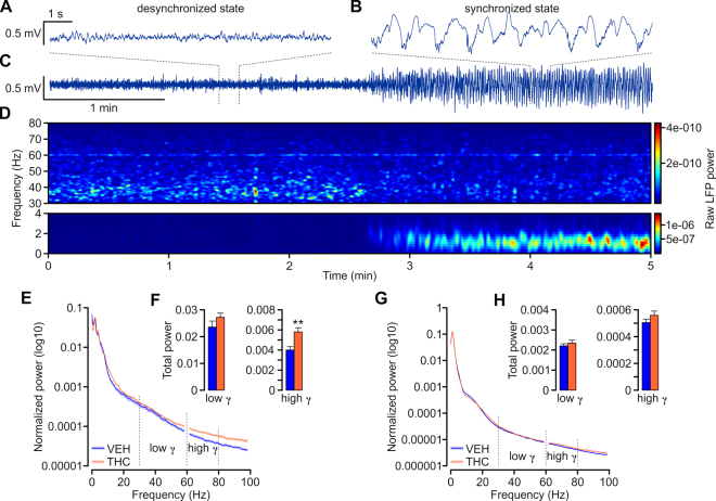
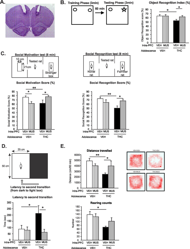
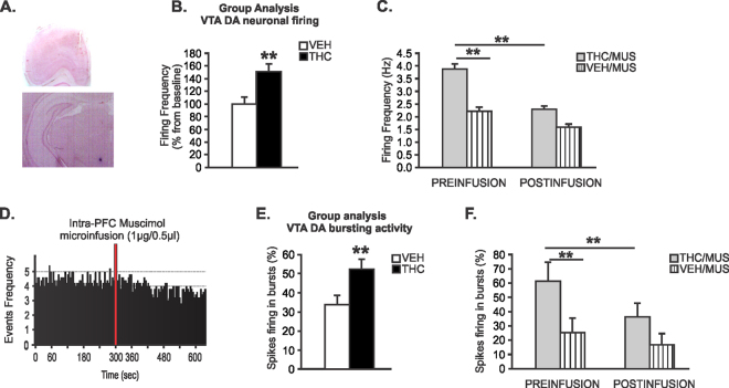
Chronic adolescent marijuana use has been linked to the later development of psychiatric diseases such as schizophrenia. GABAergic hypofunction in the prefrontal cortex (PFC) is a cardinal pathological feature of schizophrenia and may be a mechanism by which the PFC loses its ability to regulate sub-cortical dopamine (DA) resulting in schizophrenia-like neuropsychopathology. In the present study, we exposed adolescent rats to Δ-9-tetra-hydrocannabinol (THC), the psychoactive component in marijuana. At adulthood, we characterized the functionality of PFC GABAergic neurotransmission and its regulation of sub-cortical DA function using molecular, behavioral and in-vivo electrophysiological analyses. Our findings revealed a persistent attenuation of PFC GABAergic function combined with a hyperactive neuronal state in PFC neurons and associated disruptions in cortical gamma oscillatory activity. These PFC abnormalities were accompanied by hyperactive DAergic neuronal activity in the ventral tegmental area (VTA) and behavioral and cognitive abnormalities similar to those observed in psychiatric disorders. Remarkably, these neuronal and behavioral effects were reversed by pharmacological activation of GABAA receptors in the PFC. Together, these results identify a mechanistic link between dysregulated frontal cortical GABAergic inhibition and sub-cortical DAergic dysregulation, characteristic of well-established neuropsychiatric endophenotypes.


Conflict of interest statement

The authors declare that they have no competing interests.

Figures

Figure 1
Figure 1
Long-term effects of chronic THC exposure during adolescence on mPFC GABAergic markers. (A) Representative western blot for GAD67 expression in the mPFC (left). A significant decrease in GAD67 expression is observed between adolescent VEH and adolescent THC pretreated rats. (B) Representative western blot for GAD65 expression in the mPFC (left). No significant changes in GAD65 were found between groups. (C) Representative western blot for parvalbumin (PV) expression in the mPFC (left). No significant changes in PV were found between groups. n = 8 rats, t-tests; *Indicated p < 0.05. Error bars represent the standard error of the means (SEMs). Western blots for GAD 67, GAD65 and parvalbumin levels are shown in Supplementary Figure 1.
Figure 2
Figure 2
Long-term effects of chronic THC exposure during adolescence on spontaneous mPFC putative pyramidal neuron activity. (A) Microphotograph of a representative mPFC neuronal recording placement. (B) Adolescent THC pretreated rats displayed increasing spontaneous PFC putative pyramidal neuronal firing frequency. (C) Greater proportion of bursting neurons was observed in adolescent THC exposed rats when compared to VEH controls (75.41% vs. 57.58%). (D) Representative rastergrams showing spontaneous activity of putative PFC pyramidal neurons in THC (top) vs. VEH pretreated rats (bottom). (E) Firing frequencies of bursting cells, not tonic cells, were significantly higher in adolescent THC exposed rats when compared to VEH controls. (F) In the bursting cells population, the bursting rate of putative pyramidal neurons of adolescent THC-exposed rats was significantly higher than in VEH controls. (G) Representative examples showing bursting activity of putative PFC pyramidal neurons in THC (left) vs. VEH pretreated rats (right). Two-tailed t-tests; **indicated p < 0.01; *indicated p < 0.05. Error bars represent the standard error of the means (SEMs).
Figure 3
Figure 3
Adolescent THC-treatment lead to increased high gamma (61–80 Hz) power in the mPFC of adult rat. (AC) Example recording traces from PFC of urethane anesthetized rat showing different cortical states. Desynchronized state (A) was characterized by small-amplitude fast oscillations while synchronized state (B) by large-amplitude slow oscillations. (C) Five-minute recording showing spontaneous alternation from the desynchronized to synchronized state. (D) Spectrogram calculated for a five-minute recording presented in C showing the temporal changes in the power at different frequencies. Note that upon transition from desynchronized to synchronized state gamma power decreased while the slow delta oscillation gradually emerged. The power values are color-coded as indicated on the right-hand side insets. A peak at around 60 Hz reflect power line frequency and the LFP power values for frequencies between 59–61 Hz were excluded from further analysis. (E,G) Average normalized power spectra corresponding to prefrontal LFP of VEH- (blue) and THC-treated (orange) rats in desynchronized (E) and synchronized (G) states. Note the increased power of high-gamma band (61–80 Hz) in THC-treated rats. (F,H) Bar graphs summarizing the average total power of the low- and high-gamma calculated for desynchronized (F) and synchronized (H) states of VEH (blue) and THC-treated (orange) rats. The high-gamma of THC-treated rats was significantly more powerful than VEH. Two-tailed t-tests; **indicated p < 0.01. Error bars represent the standard error of the means (SEMs).
Figure 4
Figure 4
Effects of Intra-mPFC MUS on adolescent THC-induced behavioral abnormalities. (A) Microphotograph of a representative Intra-mPFC injector placement. (B) THC exposure during adolescence induced short-term memory deficits in the object recognition task. Intra-mPFC MUS treatment reversed adolescent THC-induced short-term memory deficits relative to intra-mPFC VEH controls. (C) THC exposure during adolescence induced lower social motivation (left) and social cognition (right) index in the social interaction task. Intra-mPFC MUS treatment reversed adolescent THC-induced social motivation (left) and social cognition (right) deficits relative to intra-mPFC VEH controls. (D) THC exposure during adolescence increased anxiety levels in the light dark box test. Intra-mPFC MUS reduced adolescent THC-induced anxiety relative to intra-mPFC VEH controls. (E) THC exposure during adolescence decreased locomotion (top) and rearing counts (bottom) in the open field test. Intra-mPFC MUS reduced adolescent THC-induced hypolocomotion relative to intra-mPFC VEH controls (top) but had no effects on rearing counts. Anova 2 factors **indicated p < 0.01; *indicated p < 0.05. Error bars represent the standard error of the means (SEMs).
Figure 5
Figure 5
Effects of Intra-mPFC MUS on adolescent THC-induced sub-cortical hyperdopaminergia. (A) microphotograph of a representative mPFC microsyringe and VTA neuronal recording placements. (B) Increased VTA putative DA neuronal firing frequency in adolescent THC-pretreated rats relative to adolescent VEH-pretreated rats. (C) Before intra-mPFC MUS microinfusion, sub-population of VTA putative DA neurons firing frequency in adolescent THC pretreated rats were significantly increased compared to adolescent VEH pretreated rats. Intra-mPFC MUS reduced firing frequency of VTA putative DA in adolescent THC-pretreated rats relative to adolescent VEH-pretreated rats. (D) Representative rastergram showing the spontaneous activity of 1 putative DA neuron treated with intra-mPFC muscimol in adolescent THC pretreated rat. (E) Increased VTA putative DA neurons bursting levels in adolescent THC vs. VEH pretreated rats. (F) Before intra-mPFC MUS microinfusion, sub-population of VTA putative DA neurons spikes firing in bursts in adolescent THC pretreated rats were significantly increased compared to adolescent VEH pretreated rats. Intra-mPFC MUS reduced spikes firing in bursts of VTA putative DA in adolescent THC-pretreated rats relative to adolescent VEH-pretreated rats. Two-way repeated measures ANOVA or Two-tailed t-tests; **indicated p < 0.01. Error bars represent the standard error of the means (SEMs).

References

    1. Andréasson S, Allebeck P, Engström A, Rydberg U. Cannabis and schizophrenia. A longitudinal study of Swedish conscripts. Lancet. 1987;2:1483–1486. doi: 10.1016/S0140-6736(87)92620-1. - DOI - PubMed
    1. Arseneault L, et al. Cannabis use in adolescence and risk for adult psychosis: longitudinal prospective study. BMJ. 2002;325:1212–1213. doi: 10.1136/bmj.325.7374.1212. - DOI - PMC - PubMed
    1. Arseneault L, Cannon M, Witton J, Murray RM. Causal association between cannabis and psychosis: Examination of the evidence. British Journal of Psychiatry. 2004;184:110–117. doi: 10.1192/bjp.184.2.110. - DOI - PubMed
    1. Stefanis NC, et al. Early adolescent cannabis exposure and positive and negative dimensions of psychosis. Addiction. 2004;99:1333–1341. doi: 10.1111/j.1360-0443.2004.00806.x. - DOI - PubMed
    1. Renard J, Krebs MO, Le Pen G, Jay TM. Long-term consequences of adolescent cannabinoid exposure in adult psychopathology. Front Neurosci. 2014;8:1–14. doi: 10.3389/fnins.2014.00361. - DOI - PMC - PubMed

Publication types

MeSH terms

Grants and funding