. 2017:2017:8379539.
doi: 10.1155/2017/8379539. Epub 2017 Aug 16.

Resveratrol-Enriched Rice Attenuates UVB-ROS-Induced Skin Aging via Downregulation of Inflammatory Cascades

Affiliations

Resveratrol-Enriched Rice Attenuates UVB-ROS-Induced Skin Aging via Downregulation of Inflammatory Cascades

Lalita Subedi et al. Oxid Med Cell Longev. 2017.

Erratum in

Abstract

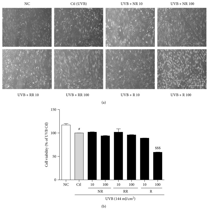
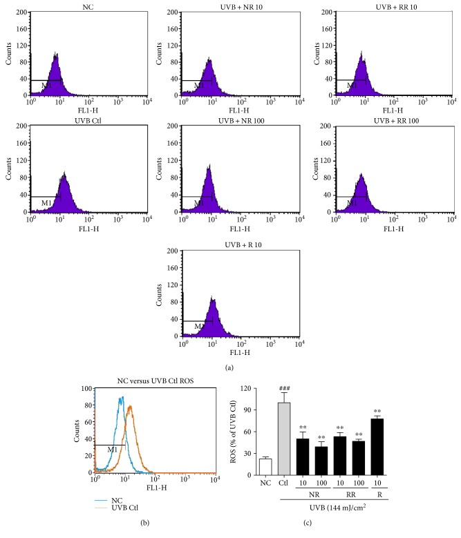
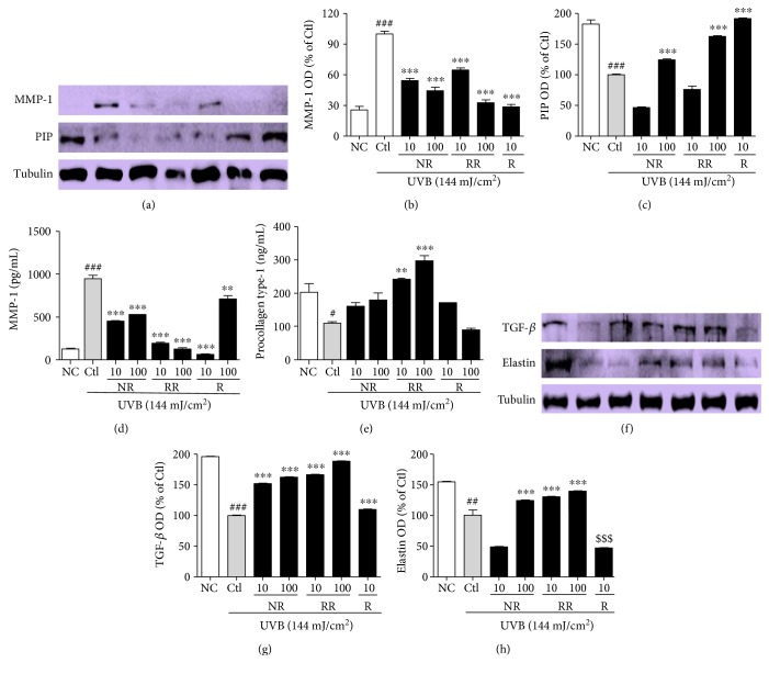
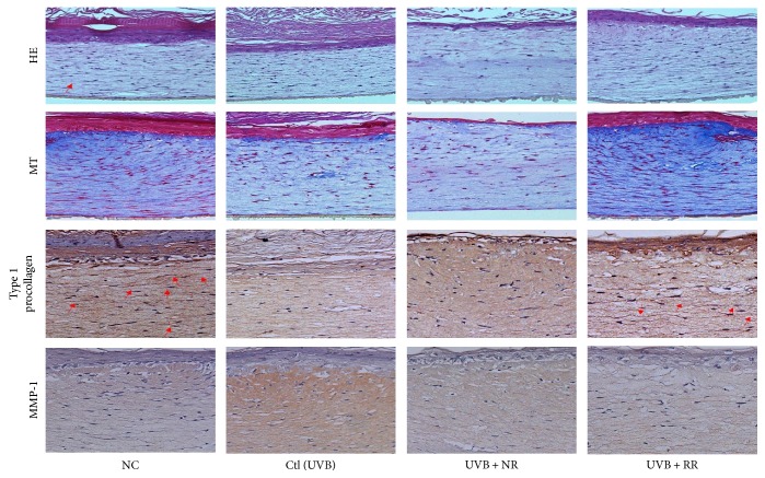
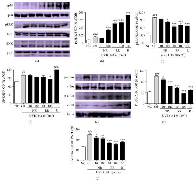
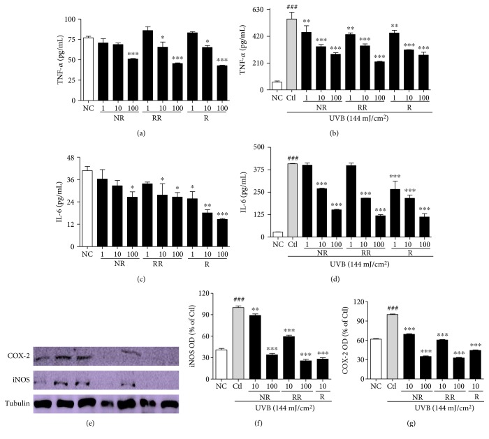
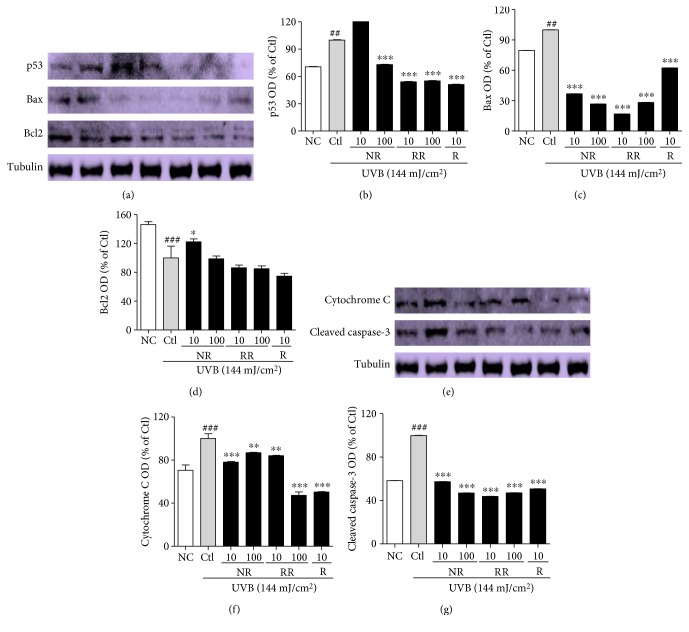
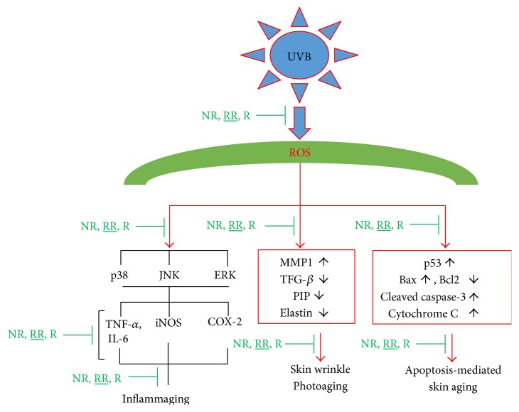
The skin is the outermost protective barrier between the internal and external environments in humans. Chronic exposure to ultraviolet (UV) radiation is a major cause of skin aging. UVB radiation penetrates the skin and induces ROS production that activates three major skin aging cascades: matrix metalloproteinase- (MMP-) 1-mediated aging; MAPK-AP-1/NF-κB-TNF-α/IL-6, iNOS, and COX-2-mediated inflammation-induced aging; and p53-Bax-cleaved caspase-3-cytochrome C-mediated apoptosis-induced aging. These mechanisms are collectively responsible for the wrinkling and photoaging characteristic of UVB-induced skin aging. There is an urgent requirement for a treatment that not only controls these pathways to prevent skin aging but also avoids the adverse effects often encountered when applying bioactive compounds in concentrated doses. In this study, we investigated the efficacy of genetically modified normal edible rice (NR) that produces the antiaging compound resveratrol (R) as a treatment for skin aging. This resveratrol-enriched rice (RR) overcomes the drawbacks of R and enhances its antiaging potential by controlling the abovementioned three major pathways of skin aging. RR does not exhibit the toxicity of R alone and promisingly downregulates the pathways underlying UVB-ROS-induced skin aging. These findings advocate the use of RR as a nutraceutical for antiaging purposes.


Figures

Figure 1
Figure 1
Normal human dermal fibroblast (NHDF). (a) Cell morphology and (b) cell viability after 72 h of treatment with or without 144 mJ/cm2 ultraviolet B (UVB) and 10 or 100 μg/mL of the following samples: normal rice (NR), resveratrol-enriched rice (RR), and resveratrol (R). All data are presented as the mean ± SEM of three independent experiments. #p < 0.05 versus the normal control. $$$p < 0.001 indicates the significant toxicity versus a UVB-irradiated control. NC is normal control, Ctl is UVB control, NR is normal rice, RR is resveratrol-enriched rice, and R is resveratrol. NR and RR were treated in μg/mL, and R was treated in μM.
Figure 2
Figure 2
The levels of ROS in NHDFs treated as indicated for 24 h were measured using flow cytometry with DCFH-DA dye. The number of cells is plotted against the dichlorofluorescein fluorescence detected by the FL-1 channel (a). The relative ROS production of the cells is shown in each histogram (b). Values are mean ± SEM. The labels # and ∗ indicate significant differences (p < 0.05) when compared with the normal control and UV (+) control, respectively. ###p < 0.001 versus the normal control, ∗∗p < 0.01 versus the UVB-irradiated control. NC is normal control, Ctl is UVB control, NR is normal rice, RR is resveratrol-enriched rice, and R is resveratrol. NR and RR were treated in μg/mL, and R was treated in μM.
Figure 3
Figure 3
UVB-MMP1-mediated protein expression and the secretion of MMP-1 and PIP were measured using Western blot analysis and an ELISA kit, respectively. NHDF cells were incubated for 72 h with or without UVB exposure and treated with or without NR, RR, and R. (a) MMP-1 and PIP expression, accompanied by the corresponding (b, c) densitometric analysis results (d, e) and the quantities MMP-1 and PIP secreted into the treated medium supernatant. (f) TGF-β and elastin expression, accompanied by the corresponding (g, h) densitometric analysis results, taking the UVB-treated control as 100%. All data are presented as the mean ± SEM of three independent experiments. #p < 0.05, ##p < 0.01, ###p < 0.001 versus the NC and ∗∗p < 0.01, ∗∗∗p < 0.001 versus the UVB control. NC is normal control, Ctl is UVB control, NR is normal rice, RR is resveratrol-enriched rice, and R is resveratrol. $$$p < 0.001 versus UVB control represents the inhibition of elastin expression by resveratrol treatment. NR and RR were treated in μg/mL, and R was treated in μM.
Figure 4
Figure 4
Photomicrographs of Masson's trichrome-stained sections and PIP-1 and MMP-1 production in reconstructed skin after treatment with UVB and samples (NR, RR, and R) at a concentration of 1% (w/v). NC is normal control, Ctl is UVB control, NR is normal rice, RR is resveratrol-enriched rice, and R is resveratrol. NR and RR were treated in μg/mL, and R was treated in μM.
Figure 5
Figure 5
Protein expression levels of MAPKs and AP-1. (a) MAPK signal intensities from multiple experiments. (b, c, d) Bar graphs represent quantitative densitometric results of upper bands. (e) AP-1 protein intensities from multiple experiments. (f, g) Bar graphs represent quantitative densitometric results of upper bands. All data are presented as the mean ± SEM of three independent experiments. ##p < 0.01, ###p < 0.001 versus the NC and p < 0.05, ∗∗p < 0.01, ∗∗∗p < 0.001 versus the UVB-irradiated control. NC is normal control, Ctl is UVB control, NR is normal rice, RR is resveratrol-enriched rice, and R is resveratrol. $$$p < 0.001 versus UVB control represents the resveratrol-induced pJNK expression. NR and RR were treated in μg/mL, and R was treated in μM.
Figure 6
Figure 6
Proinflammatory cytokines (TNF-α and IL-6) secretion and inflammatory markers (iNOS and COX-2) expression. NHDF cells were treated with or without UVB (144 mJ/cm2) and with NR, RR, and R for 72 h. Proinflammatory cytokines were measured in cell supernatant using an ELISA kit, and protein expression of iNOS and COX-2 was measured using Western blot analysis. (a, b) TNF-α secretion in NHDF cells without UVB-conditioned medium and with UVB-conditioned medium. (c, d) IL-6 secretion in NHDF cells without UVB-conditioned medium and with UVB-conditioned medium. (e) iNOS and COX-2 protein expression from multiple experiments. (f, g) Bar graphs represent quantitative densitometric results of upper bands. All data are presented as the mean ± SEM of three independent experiments. ###p < 0.001 versus the NC and p < 0.05, ∗∗p < 0.01, ∗∗∗p < 0.001 versus the UVB-irradiated control. NC is normal control, Ctl is UVB control, NR is normal rice, RR is resveratrol-enriched rice, and R is resveratrol. NR and RR were treated in μg/mL, and R was treated in μM.
Figure 7
Figure 7
Apoptosis-induced skin aging and related protein expression in NHDF cells. NHDF cells were treated with or without UVB (144 mJ/cm2) and with NR, RR, and R for 72 h. Protein expression was measured using Western blot analysis. (a) p53, Bax, and Bcl2 protein expression from multiple experiments. (b, c, d) Bar graphs represent quantitative densitometric results of upper bands. (e) Cytosolic cytochrome C and cleaved caspase-3 protein expression from multiple experiments. (f, g) Bar graphs represent quantitative densitometric results of upper bands. Tubulin was used as a loading control. All data are presented as the mean ± SEM of three independent experiments. ##p < 0.01, ###p < 0.001 versus the NC and p < 0.05, ∗∗p < 0.01, ∗∗∗p < 0.001 versus the UVB-irradiated control. NC is normal control, Ctl is UVB control, NR is normal rice, RR is resveratrol-enriched rice, and R is resveratrol. NR and RR were treated in μg/mL, and R was treated in μM.
Figure 8
Figure 8
Scheme of the UVB-ROS-mediated skin aging and the protective role of resveratrol rice against its toxicity to prevent skin aging.

References

    1. Pendegrass C. J., Goodship A. E., Price J. S., Blunn G. W. Nature’s answer to breaching the skin barrier: an innovative development for amputees. Journal of Anatomy. 2006;209(1):59–67. doi: 10.1111/j.1469-7580.2006.00595.x. - DOI - PMC - PubMed
    1. Wlaschek M., Tantcheva-Poor I., Naderi L., et al. Solar UV irradiation and dermal photoaging. Journal of Photochemistry and Photobiology B: Biology. 2001;63(1–3):41–51. doi: 10.1016/S1011-1344(01)00201-9. - DOI - PubMed
    1. Rittie L., Fisher G. J. Natural and sun-induced aging of human skin. Cold Spring Harbor Perspectives in Medicine. 2015;5(1, article a015370) doi: 10.1101/cshperspect.a015370. - DOI - PMC - PubMed
    1. Sherratt M. J. Tissue elasticity and the ageing elastic fibre. Age (Dordrecht, Netherlands) 2009;31(4):305–325. doi: 10.1007/s11357-009-9103-6. - DOI - PMC - PubMed
    1. Hwang K. A., Yi B. R., Choi K. C. Molecular mechanisms and in vivo mouse models of skin aging associated with dermal matrix alterations. Laboratory Animal Research. 2011;27(1):1–8. doi: 10.5625/lar.2011.27.1.1. - DOI - PMC - PubMed