

Review
doi: 10.1007/82_2017_62.
Tim-3, Lag-3, and TIGIT
Affiliations
- PMID: 28900677
- PMCID: PMC5902028
- DOI: 10.1007/82_2017_62
Item in Clipboard
Review
Tim-3, Lag-3, and TIGIT
Curr Top Microbiol Immunol.
2017.
Abstract
Co-inhibitory receptors play a key role in regulating T cell responses and maintaining immune homeostasis. Their inhibitory function prevents autoimmune responses but also restricts the ability of T cells to mount effective immune responses against tumors or persistent pathogens. T cells express a module of co-inhibitory receptors, which display great diversity in expression, structure, and function. Here, we focus on the co-inhibitory receptors Tim-3, Lag-3, and TIGIT and how they regulate T cell function, maintenance of self-tolerance, their role in regulating ongoing T cell responses at peripheral tissues, and their synergistic effects in regulating autoimmunity and antitumor responses.
Figures
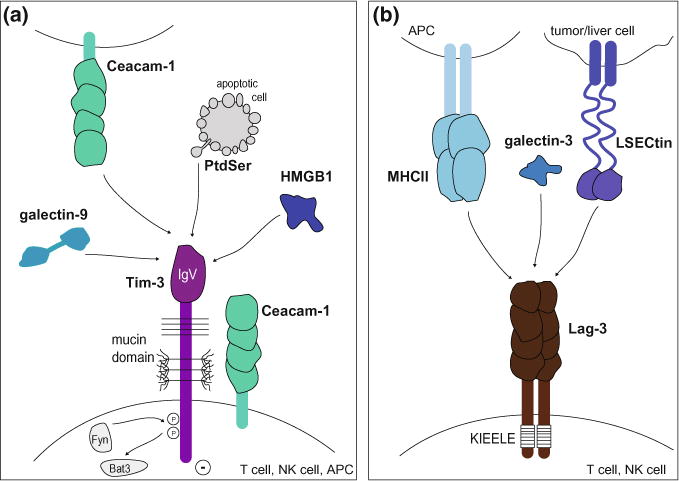
Tim-3 and Lag-3 pathways. a The Tim-3 pathway. Tim-3 is composed of an extracellular IgV domain, a mucin stalk with N- and O-linked glycosylation sites, and an intracellular tail with conserved tyrosine residues. It is expressed on T cells, NK cells, and APCs and binds to cell surface receptors (Ceacam-1 and phosphatidyl serine (PtdSer)) and soluble ligands (galectin-9 and HMGB1). Ligand binding triggers phosphorylation of two conserved tyrosine residues and release of Bat3 from the cytoplasmic tail of Tim-3, allowing Tim-3 to exert its inhibitory function. b The Lag-3 pathway. Lag-3 is composed of four extracellular Ig-like domains, a transmembrane domain, and a cytoplasmic tail containing a unique KIEELE motif. It is expressed on T cells and NK cells and binds to MHC class II on APCs, galectin-3, and LSECtin on tumor cells or liver cells
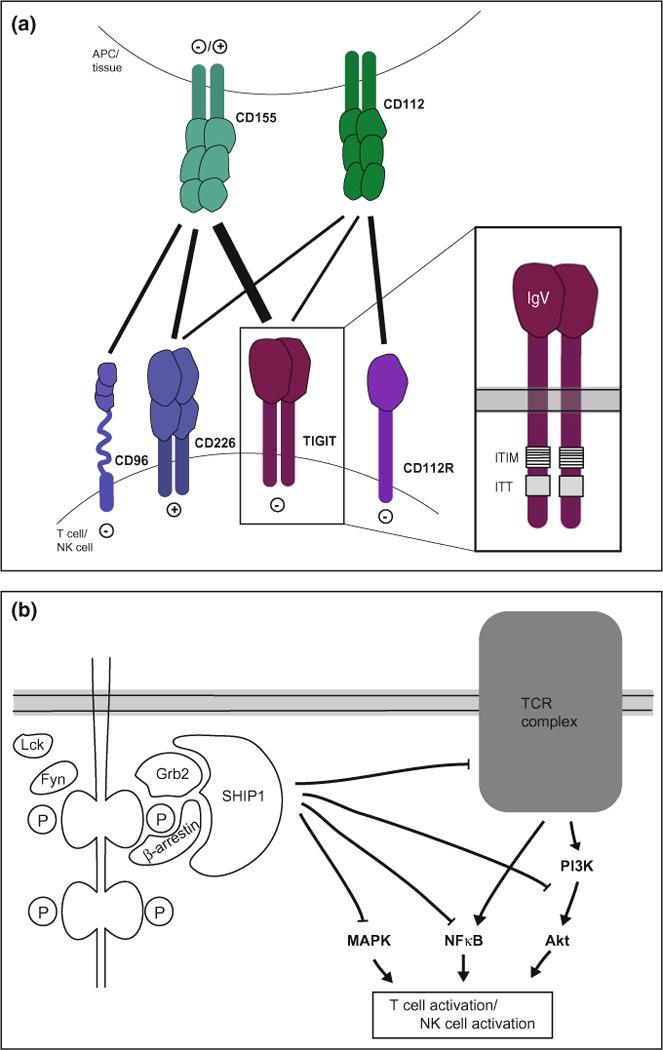
TIGIT pathway. a TIGIT forms part of a complex network where TIGIT, the co-stimulatory receptor CD226, and the co-inhibitory receptors CD96 and CD112R are expressed on T and NK cells and their ligands CD155 and CD112 are expressed on APCs and in tissue. TIGIT is composed of an extracellular IgV domain and a cytoplasmic tail containing an ITIM and ITT-like motif. b Upon ligand binding, the ITIM and ITT-like motifs in the TIGIT tail are phosphorylated and recruit SHIP1 via the adaptor proteins Grb2 or β-arrestin. SHIP1 inhibits signaling through the MAPK, NFκB, and Akt pathways, thus inhibiting activation

Co-inhibitory receptors in chronic infections and cancer. Antigen persistence drives T cells into a state of exhaustion/dysfunction characterized by hierarchical loss of cytokine production as well as impairment of cytotoxicity. As T cells enter the state of T cell exhaustion they progressively express PD-1 and upregulate Lag-3, Tim-3, and TIGIT
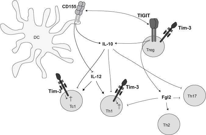
The Tim-3 and TIGIT pathways specifically inhibit pro-inflammatory responses in autoimmunity. Tim-3 is selectively expressed on Th1 and Tc1 cells, which drive tissue inflammation and autoimmunity. Tim-3 regulates their response by inducing apoptotic cell death or dysfunction by binding to its ligands. TIGIT expressed on Tregs induces IL-10 as well as Fgl2, which selectively inhibit pathogenic Th1 and Th17 responses. TIGIT expressing effector and regulatory T cells engage CD155 on APC thereby dampening IL-12 and enhancing IL-10 secretion and thus inhibiting inflammatory responses
References
-
- Baitsch L, Baumgaertner P, Devevre E, Raghav SK, Legat A, Barba L, Wieckowski S, Bouzourene H, Deplancke B, Romero P, Rufer N, Speiser DE. Exhaustion of tumor-specific CD8(+) T cells in metastases from melanoma patients. J Clin Invest. 2011;121(6):2350–2360. doi: 10.1172/JCI46102. - DOI - PMC - PubMed
Publication types
MeSH terms
Substances
Grants and funding
LinkOut - more resources
Full Text Sources
Other Literature Sources
Research Materials
