

Osteitis fibrosa cystica-a forgotten radiological feature of primary hyperparathyroidism
- PMID: 28900835
- PMCID: PMC5671544
- DOI: 10.1007/s12020-017-1414-2
Osteitis fibrosa cystica-a forgotten radiological feature of primary hyperparathyroidism
Abstract
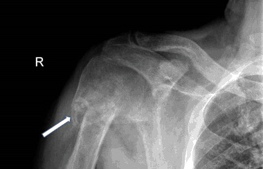
Although bone disease and stone disease are the universally accepted classical manifestations of primary hyperparathyroidism, clinical parathyroid bone disease is rarely seen today in the United States (<5% of patients) and Western Europe. Nevertheless, in a given patient, classical skeletal involvement can be the first sign of primary hyperparathyroidism, but not recognized because it is not usually included, anymore, in the differential diagnosis of this manifestation of skeletal disease. We describe four cases of primary hyperparathyroidism in which the first clinical manifestation of the disease was a pathological fracture that masqueraded as a malignancy. The presence of large osteolytic lesions gave rise to the initial diagnosis of a primary or metastatic cancer. In none of the reported cases was primary hyperparathyroidism with osteitis fibrosa considered as the diagnosis. It would seem to us that this course is best explained by the fact that in many countries such manifestations of primary hyperparathyroidism have become a rarity. In fact, the incidence of osteitis fibrosa among patients with primary hyperparathyroidism in the US is estimated as so rare, that in majority of medical centers routine x-ray examinations of the bones in these patients is not recommended. The X-ray or computed tomography scan findings of osteitis fibrosa cystica include lytic or multilobular cystic changes. Multiple bony lesions representing brown tumors may be misdiagnosed on computed tomography scan as metastatic carcinoma, bone cysts, osteosarcoma, and especially giant-cell tumor. Distinguishing between primary hyperparathyroidism and malignancy is made readily by the concomitant measurement of parathyroid hormone which in primary hyperparathyroidism, again, will be markedly elevated. In the hypercalcemias of malignancy, such elevations of parathyroid hormone are virtually never seen.
Conclusion: When radiographic evidence of a lytic lesion and hypercalcemia are present, primary hyperparathyroidism should always be considered in the differential diagnosis.
Keywords: Osteitis fibrosa cystica; Primary hyperparathyroidism; differential diagnosis.
Conflict of interest statement
Conflict of interest
The authors declare that they have no competing interests.
Informed consent
Informed consent to the use of their medical data was obtained from all individual patients described in the study.
Figures


References
-
- Silverberg SJ, Bilezikian JP. Evaluation and management of primary hyperparathyroidism. J. Clin. Endocrinol. Metab. 1996;81:2036–2040. - PubMed
-
- Bilezikian JP, Silverberg SJ, Shane E, et al. Characterization and evaluation of asymptomatic primary hyperparathyroidism. J. Bone Miner. Res. 1991;6(Suppl 2):S85. - PubMed
-
- J.P. Bilezikian, Primary hyperparathyroidism. Endotext (2017). http://www.endotext.org/chapter/primary-hyperparathyroidism/, 15 Jan 2017
Publication types
MeSH terms
Substances
Grants and funding
LinkOut - more resources
Full Text Sources
Other Literature Sources
