. 2017 Aug;26(8):1405-1417.
doi: 10.1177/0963689717720278.

PPAR-δ Agonist With Mesenchymal Stem Cells Induces Type II Collagen-Producing Chondrocytes in Human Arthritic Synovial Fluid

Affiliations

PPAR-δ Agonist With Mesenchymal Stem Cells Induces Type II Collagen-Producing Chondrocytes in Human Arthritic Synovial Fluid

Bruce E Heck et al. Cell Transplant. 2017 Aug.

Abstract

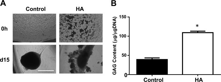
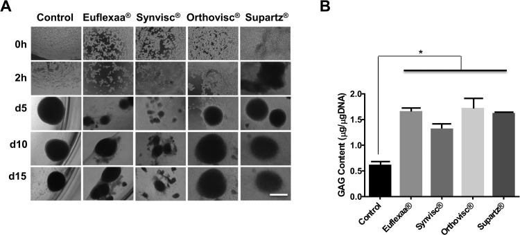
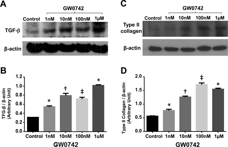
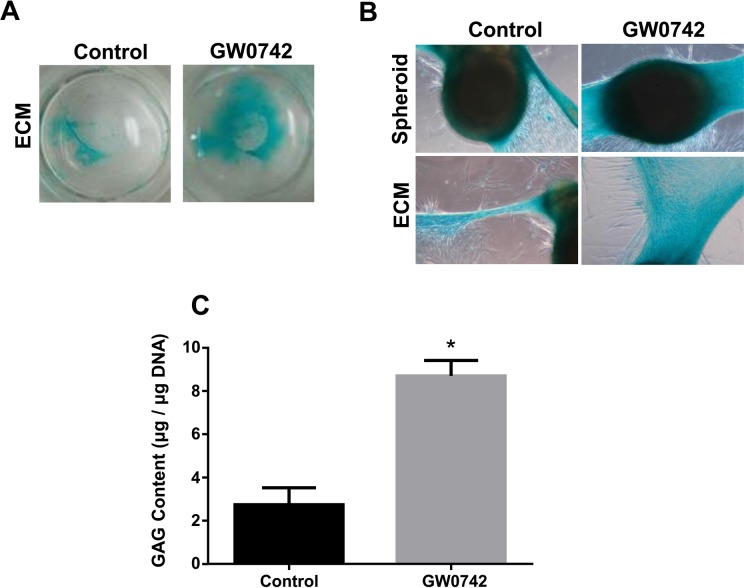
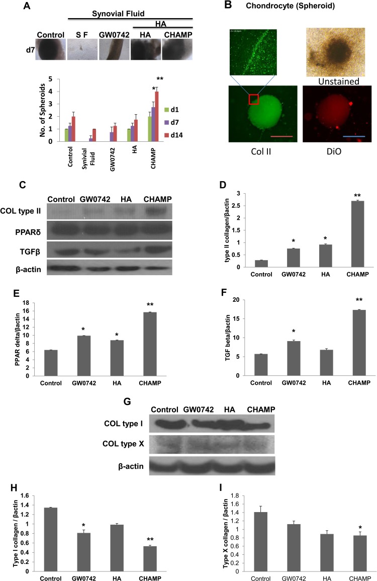
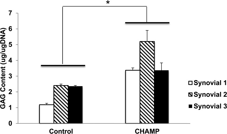
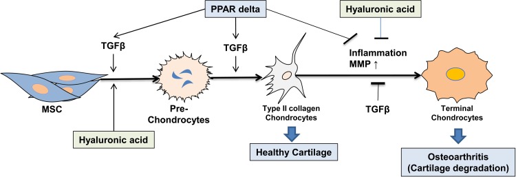
Osteoarthritis (OA) is an inflammatory joint disease characterized by degeneration of articular cartilage within synovial joints. An estimated 27 million Americans suffer from OA, and the population is expected to reach 67 million in the United States by 2030. Thus, it is urgent to find an effective treatment for OA. Traditional OA treatments have no disease-modifying effect, while regenerative OA therapies such as autologous chondrocyte implantation show some promise. Nonetheless, current regenerative therapies do not overcome synovial inflammation that suppresses the differentiation of mesenchymal stem cells (MSCs) to chondrocytes and the expression of type II collagen, the major constituent of functional cartilage. We discovered a synergistic combination that overcame synovial inflammation to form type II collagen-producing chondrocytes. The combination consists of peroxisome proliferator-activated receptor (PPAR) δ agonist, human bone marrow (hBM)-derived MSCs, and hyaluronic acid (HA) gel. Interestingly, those individual components showed their own strong enhancing effects on chondrogenesis. GW0742, a PPAR-δ agonist, greatly enhanced MSC chondrogenesis and the expression of type II collagen and glycosaminoglycan (GAG) in hBM-MSC-derived chondrocytes. GW0742 also increased the expression of transforming growth factor β that enhances chondrogenesis and suppresses cartilage fibrillation, ossification, and inflammation. HA gel also increased MSC chondrogenesis and GAG production. However, neither GW0742 nor HA gel could enhance the formation of type II collagen-producing chondrocytes from hBM-MSCs within human OA synovial fluid. Our data demonstrated that the combination of hBM-MSCs, PPAR-δ agonist, and HA gel significantly enhanced the formation of type II collagen-producing chondrocytes within OA synovial fluid from 3 different donors. In other words, the novel combination of PPAR-δ agonist, hBM-MSCs, and HA gel can overcome synovial inflammation to form type II collagen cartilage within human OA synovial fluid. This novel articularly injectable formula could improve OA treatment in the future clinical application.

Keywords: PPAR-δ agonist; hyaluronic acid; mesenchymal stem cells; osteoarthritis; synovial inflammation; type II collagen.


Conflict of interest statement

Declaration of Conflicting Interests: The author(s) declared no potential conflicts of interest with respect to the research, authorship, and/or publication of this article.

Figures

Figure 1.
Figure 1.
Culturing in hyaluronic acid (HA) gel plus peroxisome proliferator–activated receptor (PPAR) δ agonist enhances chondrogenesis and the formation of type II collagen and transforming growth factor (TGF) β. (A) Human bone marrow–derived mesenchymal stem cells (hBM-MSCs) were cultured either on a plastic wall or in 70 μL of HA gel during chondrogenesis. Cell pictures (100×) were taken after 0- and 15-d chondrogenesis. (B) The amount of glycosaminoglycan (GAG) produced by chondrocytes was quantified by modified dimethyl-methylene (DMM) method and normalized by DNA content. *P < 0.01 versus control. The image is the representative of 3 independent experiments. Data presented is the result of 3 independent experiments. Scale bar: 80 μm.
Figure 2.
Figure 2.
(A) Human bone marrow–derived mesenchymal stem cells (hBM-MSCs) were cultured in 70 μL of clinical hyaluronic acid (HA) gel during chondrogenesis in 96-well plates. Cell pictures (100×) were taken at 0 and 2 h and on 5, 10, and 15 d on the gels. (B) hBM-MSCs were cultured in 70 μL of different HA gels during chondrogenesis in 96-well plates. On day 15, the content of glycosaminoglycan (GAG) of chondrocytes was measured with modified dimethyl-methylene (DMM) blue assay at 525 nm. The amount of GAG was extrapolated from a standard curve using shark chondroitin sulfate. n = 3. *P < 0.05 versus control. Image is the representation of 3 independent experiments. Data presented are the result of 3 independent experiments. Scale bar: 80 μm.
Figure 3.
Figure 3.
Human bone marrow–derived mesenchymal stem cells (hBM-MSCs) were cultured in 70 μL of Euflexxa during chondrogenesis in 96-well plates. On day 7, the expression of type II collagen and transforming growth factor (TGF) β in chondrocytes was detected by immunoblotting using antibodies to type II collagen (A) and TGF-β (B; anti-β-actin antibody as control). The relative levels of type II collagen (C) and TGF-β (D) to β-actin were quantified by densitometry (ImageJ [from the National Institutes of Health in Bethesda, MD, USA https://imagej.nih.gov/ij/]) and presented in bar graphs (n = 3). *P < 0.05 versus control. †P < 0.005 versus control. ‡P < 0.001 versus control. Image is the representative of 3 independent experiments. Data presented are the result of 3 independent experiments.
Figure 4.
Figure 4.
GW0742 enhances glycosaminoglycan (GAG) production. Human bone marrow–derived mesenchymal stem cells (hBM-MSCs) in chondrogenic medium were treated with vehicle (dimethyl sulfoxide [DMSO]) or GW0742 (0.1 μM) every 2 d in a 24-well plate. On day 14, GAGs were stained with 1% Alcian blue. The images of Alcian blue staining of the whole well or individual spheroids were taken by a light microscope. (C) The amount of GAG produced by chondrocytes was quantified by modified dimethyl-methylene (DMM) method and normalized by DNA content. *P < 0.005 versus control. Image is the representative of 3 independent experiments. Data presented are the result of 3 independent experiments.
Figure 5.
Figure 5.
Human bone marrow–derived mesenchymal stem cells (hBM-MSCs) were incubated in chondrogenic medium without (control) and with GW0742, hyaluronic acid (HA), or Chondrogenic Hyaluronic Acid–Mesenchymal Stem Cells–PPAR-δ agonist (CHAMP) in 50% of human osteoarthritis (OA) synovial fluid for 14 d. The numbers of chondrocyte spheroid (>40 mm) were counted for bar graphs (A). The spheroids were stained with fluorescent agent (DiO) and collagen II were stained with fluorescein isothiocyanate (FITC) to confirm chondrocytes. The magnified image was taken using confocal microscope (B). On day 14, chondrocytes were processed for immunoblotting using antibodies to type II collagen, peroxisome proliferator–activated receptor (PPAR) δ, transforming growth factor (TGF) β (C–F), type I collagen, type X collagen, and β-actin (G–I). Protein band densities were measured to obtain bar graphs (C–I). Mean ± standard error, n = 3. *P < 0.05 versus control. **P < 0.01 versus control. Image is the representative of 3 independent experiments. Data presented are the result of 3 independent experiments. Scale bar: 80 μm.
Figure 6.
Figure 6.
The amount of glycosaminoglycan (GAG) produced by chondrocytes was quantified by modified dimethyl-methylene (DMM) method and normalized by DNA content with 3 different synovial fluids from patients. *P < 0.01 versus control.
Figure 7.
Figure 7.
Working model: transforming growth factor (TGF) β, peroxisome proliferator–activated receptor (PPAR) δ and hyaluronic acid (HA) greatly enhance mesenchymal stem cell (MSC) chondrogenesis to type II collagen-producing chondrocytes that constitute healthy cartilage. MSCs, PPAR-δ, TGF-β, and HA suppress inflammation and matrix metalloproteases (MMPs) in inflamed osteoarthritis (OA) joints, thus blocking terminal chondrocyte differentiation, which should reduce cartilage degradation and calcification that occur inside OA joint.

Similar articles

Cited by

References

    1. Knutsen G, Drogset JO, Engebretsen L, Grontvedt T, Isaksen V, Ludvigsen TC, Roberts S, Solheim E, Strand T, Johansen O. A randomized trial comparing autologous chondrocyte implantation with microfracture. Findings at five years. J Bone Joint Surg Am. 2007;89(10):2105–2112. - PubMed
    1. McNickle AG, L’Heureux DR, Yanke AB, Cole BJ. Outcomes of autologous chondrocyte implantation in a diverse patient population. Am J Sports Med. 2009;37(7):1344–1350. - PubMed
    1. Pelttari K, Steck E, Richter W. The use of mesenchymal stem cells for chondrogenesis. Injury. 2008;39(suppl 1):S58–S65. - PubMed
    1. Farkouh ME, Kirshner H, Harrington RA, Ruland S, Verheugt FW, Schnitzer TJ, Burmester GR, Mysler E, Hochberg MC, Doherty M, et al. ; TARGET Study Group. Comparison of lumiracoxib with naproxen and ibuprofen in the Therapeutic Arthritis Research and Gastrointestinal Event Trial (TARGET), cardiovascular outcomes: randomised controlled trial. Lancet. 2004;364(9435):675–684. - PubMed
    1. Lazzaroni M, Bianchi Porro G. Gastrointestinal side-effects of traditional non-steroidal anti-inflammatory drugs and new formulations. Aliment Pharmacol Ther. 2004;20(Suppl 2):48–58. - PubMed

LinkOut - more resources