. 2017 Aug;26(8):1365-1379.
doi: 10.1177/0963689717720282.

Engineered Microvasculature in PDMS Networks Using Endothelial Cells Derived from Human Induced Pluripotent Stem Cells

Affiliations

Engineered Microvasculature in PDMS Networks Using Endothelial Cells Derived from Human Induced Pluripotent Stem Cells

Amogh Sivarapatna et al. Cell Transplant. 2017 Aug.

Abstract

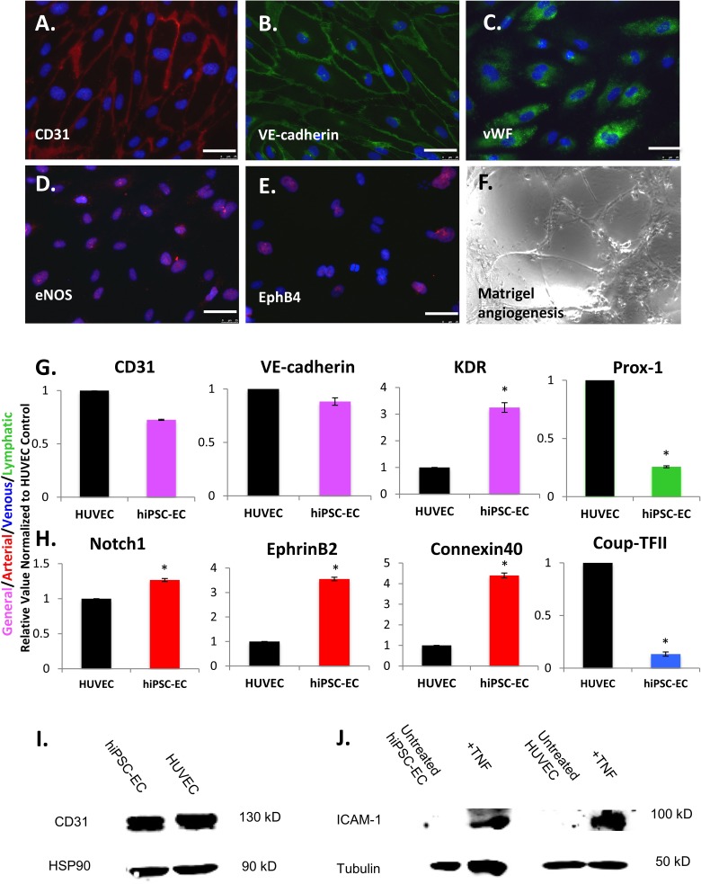
In this study, we used a polydimethylsiloxane (PDMS)-based platform for the generation of intact, perfusion-competent microvascular networks in vitro. COMSOL Multiphysics, a finite-element analysis and simulation software package, was used to obtain simulated velocity, pressure, and shear stress profiles. Transgene-free human induced pluripotent stem cells (hiPSCs) were differentiated into partially arterialized endothelial cells (hiPSC-ECs) in 5 d under completely chemically defined conditions, using the small molecule glycogen synthase kinase 3β inhibitor CHIR99021 and were thoroughly characterized for functionality and arterial-like marker expression. These cells, along with primary human umbilical vein endothelial cells (HUVECs), were seeded in the PDMS system to generate microvascular networks that were subjected to shear stress. Engineered microvessels had patent lumens and expressed VE-cadherin along their periphery. Shear stress caused by flowing medium increased the secretion of nitric oxide and caused endothelial cells s to align and to redistribute actin filaments parallel to the direction of the laminar flow. Shear stress also caused significant increases in gene expression for arterial markers Notch1 and EphrinB2 as well as antithrombotic markers Kruppel-like factor 2 (KLF-2)/4. These changes in response to shear stress in the microvascular platform were observed in hiPSC-EC microvessels but not in microvessels that were derived from HUVECs, which indicated that hiPSC-ECs may be more plastic in modulating their phenotype under flow than are HUVECs. Taken together, we demonstrate the feasibly of generating intact, engineered microvessels in vitro, which replicate some of the key biological features of native microvessels.

Keywords: arterial; endothelial cells; human induced pluripotent stem cells (hiPSCs); microfluidics; microvessels; shear stress.


Conflict of interest statement

Declaration of Conflicting Interests: The author(s) declared the following potential conflicts of interest with respect to the research, authorship, and/or publication of this article: LEN is a founder and shareholder in Humacyte, Inc., which is a regenerative medicine company. LEN’s spouse has equity in Humacyte, and LEN serves on Humacyte’s Board of Directors. LEN is an inventor on patents that are licensed to Humacyte and that produce royalties for LEN.

Figures

Fig. 1.
Fig. 1.
In silico modeling of microvascular network using COMSOL shows (A) velocity magnitude (m/s) distribution profile in the device is symmetrical after encountering bifurcation points near the inlet and outlet. (B) Pressure (Pa) gradient exists in the device, decreasing uniformly from high to low (from inlet to outlet, respectively) after gravity-driven loading of medium. (C) Simulated wall shear stress (dyne/cm2) profile, at a flow rate of 10 μL/min, at inlet of device (left) and middle of device (right). (D) Syringe pump setup showing syringe-containing medium (top) connecting to inlet of polydimethylsiloxane (PDMS) device (middle) with the outlet leading to a 15 cc conical tube reservoir (bottom) to collect spent medium. Right shows blown up image of fully connected PDMS device.
Fig. 2.
Fig. 2.
Quantitative real-time reverse transcription polymerase chain reaction (qRT-PCR) showing the time course for the upregulation of arterial endothelial cell (EC)-specific genes (A) Connexin 40, (B) protein jagged-2 (Jag2), and (C) tyrosine kinase with immunoglobulin and epidermal growth factor homology domains (Tie2) over 5 d (D1–D5). (D) qRT-PCR showing the time course for upregulation of Notch1, and (E) Western blot demonstrating the protein-level expression of Notch1-activated (NICD, 100 kD band) from day 0 (D0) of differentiation all the way through day (D5) compared to human umbilical vein endothelial cell (HUVEC) and human aortic endothelial cells HAEC controls (left to right, heat shock protein 90 [HSP90] used as loading control, bottom). (F) qRT-PCR showing the time course for upregulation of the arterial marker EphrinB2 from day 0 to day 5 (D1-D5). Asterisks in A-F indicate P < 0.02 compared to day 1 of differentiation. (G) Western blot demonstrating the protein-level expression of EphrinB2 (37 kD band) from day 0 (D5) of differentiation all the way through day 5 (D5) compared to HUVEC and HAEC controls. Bottom graph shows quantitative densitometry of EphrinB2 expression relative to HAECs over 5 d, with asterisks indicating P < 0.05 compared to HAECs, demonstrating a significant increase on days 4 and 5. For qRT-PCR, values from 3 independent experiments from the triplicate polymerase chain reaction (PCR) reactions for genes of interest were normalized against average glyceraldehyde 3-phosphate dehydrogenase (GAPDH) Ct values from the same complementary DNA (cDNA) sample. Fold change of gene of interest (GOI) transcript levels between samples equals 2-ΔΔCt, where ΔCt=Ct(GOI) − Ct(GAPDH), and ΔΔCt=ΔCt(ATII) − ΔCt(ATII). For all panels, data represent at least 3 observations for each experiment and are expressed as mean values ± standard error of mean [SEM].
Fig. 3.
Fig. 3.
(A) Schematic for directed differentiation protocol of human induced pluripotent stem cell-derived endothelial cells (hiPSC-ECs) in vitro in 5 d via a 2D monolayer approach using the small molecule glycogen synthase kinase 3β inhibitor CHIR99021. Quantitative real-time reverse transcription polymerase chain reaction (qRT-PCR) showing the time course for the upregulation of the endothelial cell (EC)-specific genes (B) CD3, (C) vascular endothelial (VE)-cadherin, and (D) kinase insert domain receptor (KDR) over 5 d (D1-D5). Asterisks indicate P < 0.05 compared to D1. Phase contrast images of (E) early mesodermal cells at day 2 (F) endothelial or endothelial-progenitor cells at day 5 and (G) isolated P0 hiPSC-ECs (via CD31+ magnetic bead selection) at confluence, showing typical EC cobblestone morphology. Immunofluorescence staining of typical differentiating hiPSC colony at day 5 showing (H) nuclei via 4’,6-diamidino-2-phenylindole (DAPI, blue) (I) emerging VE-cadherin positive cells (red) and (J) merge of DAPI and VE-cadherin, scale bar = 200 μm. (K) Western blot showing no expression of VE-cadherin (132 kD band) in hiPSCs (left) and strong expression on day 5 of differentiation (D5, right) with HSP90 (bottom) used as loading controls. (L) Western blot showing no expression of CD31 (130 kD band) in hiPSCs (left), and strong expression on day 5 of differentiation (D5, right) with HSP90 (bottom) used as loading controls. For qRT-PCR, values from 3 independent experiments from the triplicate PCR reactions for genes of interest were normalized against average GAPDH Ct values from the same complementary DNA (cDNA) sample. Fold change of GOI transcript levels between samples equals 2-ΔΔCt, where ΔCt=Ct(GOI) − Ct(GAPDH), and ΔΔCt=ΔCt(ATII) − ΔCt(ATII). (Error bar indicates ± standard error of mean [SEM] and n = 3 independent experiments).
Fig. 4.
Fig. 4.
Immunofluorescence analysis of selected endothelial cell (EC) markers in human induced pluripotent stem cell-derived endothelial cells (hiPSC-ECs) after differentiation, isolation, and expansion for 2 to 3 passages on fibronectin-coated plates shows expression of (A) CD31, (B) vascular endothelial (VE)-cadherin, (C) von Willebrand factor (vWF), (D) endothelial nitric oxide synthase (eNOS), and (E) Ephrin type-B receptor 4 (EphB4). A functional assay demonstrates that hiPSC-ECs (F) form networks when cultured on Matrigel-coated plates for 24 h. Scale bars = 25 μm. Quantitative real-time reverse transcription polymerase chain reaction (qRT-PCR) analysis of endothelial cells (EC) gene expression in hiPSC-ECs, relative to human umbilical vein endothelial cells (HUVECs), for (G) general EC markers CD31, vascular endothelial (VE)-cadherin and kinase insert domain receptor (KDR; pink), lymphatic marker prospero homeobox protein 1 (Prox-1; green), and (H) arterial markers Notch1, EphrinB2, and Connexin 40 (red) and venous marker chicken ovalbumin upstream promoter transcription factor 2 (Coup-TFII; blue), with asterisks indicating P < 0.05 compared to HUVECs. Western blot showing (I) strong CD31 expression (130 kD band) in isolated hiPSC-ECs compared to HUVECs with HSP90 loading control and (J) expression of intracellular adhesion molecule 1 (ICAM-1; 100 kD band) in hiPSC-ECs after treatment with tumor necrosis factor (left) and in HUVECs (right) with tubulin-loading controls, demonstrating comparable levels of induction. (Error bar indicates ± standard error of mean [SEM] and n = 3 independent experiments. Asterisks indicate P < 0.05 compared to HUVECs).
Fig. 5.
Fig. 5.
Physical model of (A) polydimethylsiloxane (PDMS)-microfluidic device attached to glass slide with blunt needles (green) plugged at the inlet and outlet used for seeding of cells and medium replenishment (left), phase-contrast image showing seeding of human umbilical vein endothelial cells (HUVECs; center) and attached and proliferating HUVECs after 4 d of culture in device (right). Immunofluorescence analysis of microvessels generated from HUVECs for (B) von Willebrand factor (vWF; green, top left and bottom left, scale bars = 100 μm and 50 μm, respectively) and vascular endothelial (VE)-cadherin (red, top right and bottom right, scale bars = 100 μm and 50 μm, respectively). (C) 3D confocal micrographs of microvessels generated from hiPSC-ECs showing4, 6-diamidino-2-phenylindole (DAPI) for lumens (blue, left) and VE-cadherin showing intact microvascular junctions (scale bars = 100 μm).
Fig. 6.
Fig. 6.
Phase contrast imaging of (A) static human induced pluripotent stem cell-derived endothelial cell (hiPSC-EC) microvessel morphology compared to (B) morphology of hiPSC-ECs after 24 h of flow. Immunofluorescence analysis of (C) F-actin staining (red) of static hiPSC-ECs compared to (D) F-actin after 24 h of flow. Immunofluorescence staining of (E) EphrinB2 expression (red) in static hiPSC-EC microvessels compared to (F) EphrinB2 expression after 24 h of flow. Live cell immunofluorescence analysis of (G) nitric oxide (NO) production in static hiPSC-EC microvessels compared to (H) NO production after 24 h of flow. Flow condition was 10 μl/mL for 24 h. Scale bars = 100 μm.
Fig. 7.
Fig. 7.
Quantitative real-time reverse transcription polymerase chain reaction (qRT-PCR) showing gene expression changes under shear compared to static controls for Kruppel-like factor 2 (KLF2), KLF4, Ephrin type-B receptor 4 (EphB4), Notch1 and EphrinB2 in (A) human induced pluripotent stem cell-derived endothelial cell (hiPSC-EC) microvessels and (B) human umbilical vein endothelial cell (HUVEC) microvessels. Values from 3 independent experiments from the triplicate polymerase chain reaction (PCR) reactions for genes of interest were normalized against average glyceraldehyde 3-phosphate dehydrogenase (GAPDH) Ct values from the same complementary DNA (cDNA) sample. Fold change of gene of interest (GOI) transcript levels between samples equals 2-ΔΔCt, where ΔCt = Ct(GOI) − Ct(GAPDH), and ΔΔCt=ΔCt(ATII) − ΔCt(ATII). Data represented as relative value (shear over static). Asterisks indicate P < 0.05. For all panels, data represent at least 3 observations for each experiment and are expressed as mean values ± standard error of mean.

Similar articles

Cited by

References

    1. Nolan DJ, Ginsberg M, Israely E, Palikuqi B, Poulos MG, James D, Ding BS, Schachterle W, Liu Y, Rosenwaks Z, et al. Molecular signatures of tissue-specific microvascular endothelial cell heterogeneity in organ maintenance and regeneration. Dev Cell. 2013;26(2):204–219. - PMC - PubMed
    1. Mendez JJ, Ghaedi M, Sivarapatna A, Dimitrievska S, Shao Z, Osuji CO, Steinbacher DM, Leffell DJ, Niklason LE. Mesenchymal stromal cells form vascular tubes when placed in fibrin sealant and accelerate wound healing in vivo. Biomaterials. 2015;40:61–71. - PMC - PubMed
    1. Wells RG. The role of matrix stiffness in regulating cell behavior. Hepatology. 2008;47(4):1394–1400. - PubMed
    1. Lacorre DA, Baekkevold ES, Garrido I, Brandtzaeg P, Haraldsen G, Amalric F, Girard JP. Plasticity of endothelial cells: rapid dedifferentiation of freshly isolated high endothelial venule endothelial cells outside the lymphoid tissue microenvironment. Blood. 2004;103(11):4164–4172. - PubMed
    1. Gimbrone MA, Cotran RS, Folkman J. Human vascular endothelial cells in culture: growth and DNA synthesis. J Cell Biol. 1974;60(3):673–684. - PMC - PubMed

Publication types

Substances

LinkOut - more resources