. 2017 Nov;16(5):6757-6763.
doi: 10.3892/mmr.2017.7426. Epub 2017 Sep 5.

miR‑21 attenuates contrast‑induced renal cell apoptosis by targeting PDCD4

Affiliations

miR‑21 attenuates contrast‑induced renal cell apoptosis by targeting PDCD4

Kun Wang et al. Mol Med Rep. 2017 Nov.

Abstract

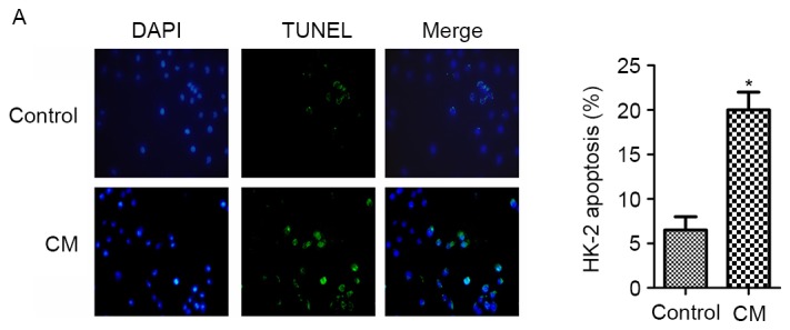
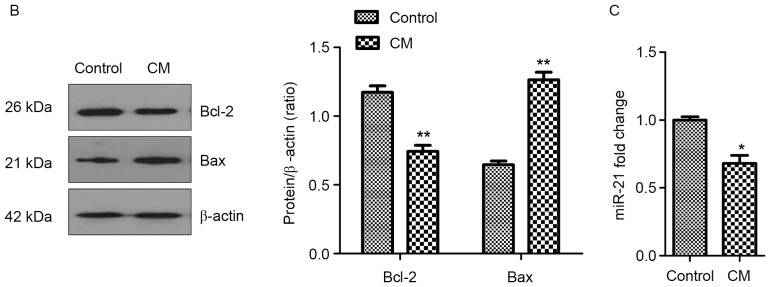
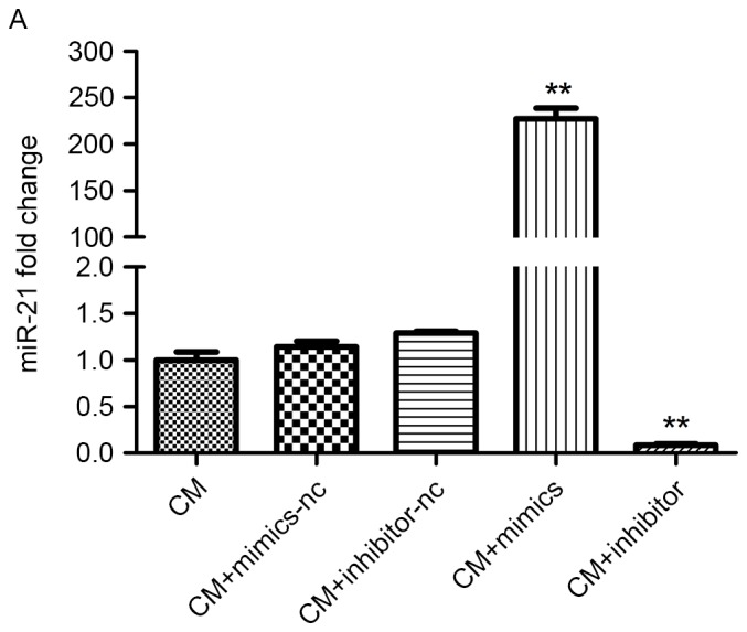
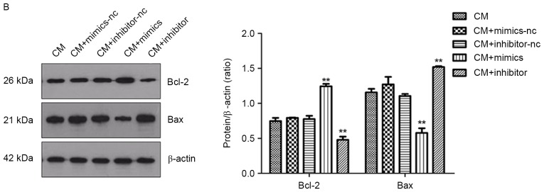
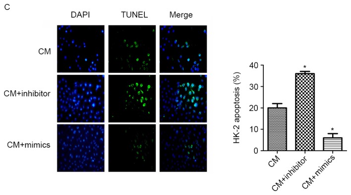
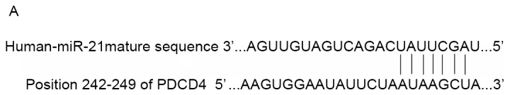
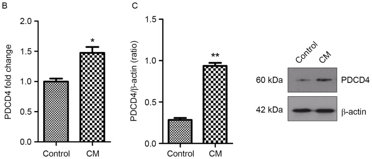
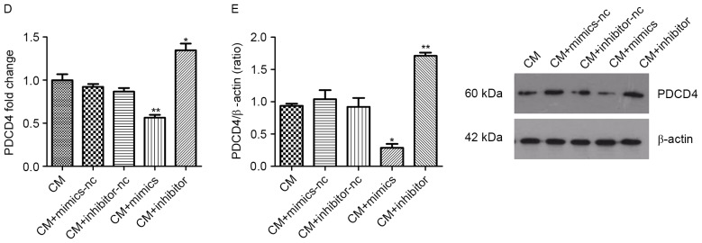
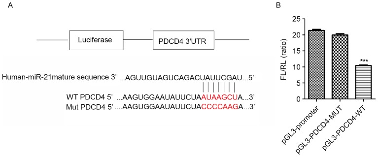
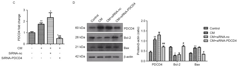
Contrast medium (CM) is widely used in cardiac catheterization; however, it may induce acute kidney injury or renal failure, although the underlying mechanism remains to be elucidated. MicroRNA‑21 (miR‑21) is involved in renal disease and has been indicated to regulate cellular apoptosis and fibrosis, although its role in CM‑induced renal cell injury is unknown. The present study examined the expression and potential targets of miR‑21 in human renal proximal tubular epithelial (HK‑2) cells following CM treatment. CM induced renal cell apoptosis and decreased miR‑21 expression. The expression level of the apoptosis regulator protein, B‑cell lymphoma 2 (Bcl‑2) was upregulated, whereas that of the apoptosis regulator, Bcl‑2‑associated X protein (Bax) was downregulated upon transfection of miR‑21 mimics; miR‑21 overexpression additionally directly inhibited the expression of programmed cell death protein 4 (PDCD4), as determined by a dual luciferase reporter assay, and PDCD4 silencing reduced the rate of HK‑2 cell apoptosis. The results of the present study indicated that miR‑21 protected renal cells against CM‑induced apoptosis by regulating PDCD4 expression.


Figures

Figure 1.
Figure 1.
CM induces HK-2 cell apoptosis and inhibits miR-21 expression. (A) Apoptosis (green cells) was measured via the TUNEL assay. Magnification, ×400. (B) Bcl-2 and Bax protein expression, as detected by western blotting. (C) MiR-21 expression, determined by the reverse transcription-quantitative polymerase chain reaction. Cells were treated with 150 mgI/ml Ultravist in the CM groups. *P<0.05, **P<0.01 vs. control group (n=3). CM, contrast medium; miR, microRNA; TUNEL, terminal deoxynucleotidyl transferase dUTP nick-end labeling; Bcl-2, B-cell lymphoma 2; Bax, Bcl-2-associated X protein.
Figure 1.
Figure 1.
CM induces HK-2 cell apoptosis and inhibits miR-21 expression. (A) Apoptosis (green cells) was measured via the TUNEL assay. Magnification, ×400. (B) Bcl-2 and Bax protein expression, as detected by western blotting. (C) MiR-21 expression, determined by the reverse transcription-quantitative polymerase chain reaction. Cells were treated with 150 mgI/ml Ultravist in the CM groups. *P<0.05, **P<0.01 vs. control group (n=3). CM, contrast medium; miR, microRNA; TUNEL, terminal deoxynucleotidyl transferase dUTP nick-end labeling; Bcl-2, B-cell lymphoma 2; Bax, Bcl-2-associated X protein.
Figure 2.
Figure 2.
Effect of miR-21 on HK-2 cell apoptosis under CM treatment. (A) MiR-21 expression in cells transfected with miR-21 mimic, inhibitor, or negative control miR was detected using the reverse transcription-quantitative polymerase chain reaction. (B) Bcl-2 and Bax protein expression in cells transfected with miR-21 mimic, inhibitor or negative control miR was measured by western blotting. (C) Detection of apoptosis (green cells) with the TUNEL assay. Magnification, ×400. Cells were treated with 150 mgI/ml Ultravist in the CM groups. *P<0.05, **P<0.01 vs. CM group (n=3). CM, contrast medium; miR, microRNA; HK-2, human renal proximal tubular epithelial; TUNEL, terminal deoxynucleotidyl transferase dUTP nick-end labeling; Bcl-2, B-cell lymphoma 2; Bax, Bcl-2-associated X protein.
Figure 2.
Figure 2.
Effect of miR-21 on HK-2 cell apoptosis under CM treatment. (A) MiR-21 expression in cells transfected with miR-21 mimic, inhibitor, or negative control miR was detected using the reverse transcription-quantitative polymerase chain reaction. (B) Bcl-2 and Bax protein expression in cells transfected with miR-21 mimic, inhibitor or negative control miR was measured by western blotting. (C) Detection of apoptosis (green cells) with the TUNEL assay. Magnification, ×400. Cells were treated with 150 mgI/ml Ultravist in the CM groups. *P<0.05, **P<0.01 vs. CM group (n=3). CM, contrast medium; miR, microRNA; HK-2, human renal proximal tubular epithelial; TUNEL, terminal deoxynucleotidyl transferase dUTP nick-end labeling; Bcl-2, B-cell lymphoma 2; Bax, Bcl-2-associated X protein.
Figure 3.
Figure 3.
miR-21 inhibits PDCD4 expression. (A) Target gene analysis results, exhibiting the miR-21 binding sites in the 3′ UTR of the human PDCD4 transcript. PDCD4 mRNA and protein levels in HK-2 cells treated with 150 mgI/ml Ultravist were determined by (B) reverse transcription-quantitative polymerase chain reaction and (C) western blotting, respectively. PDCD4 mRNA and protein expression in cells treated with 150 mgI/ml Ultravist following transfection with miR-21 mimic, inhibitor, or negative control miR was also measured by (D) reverse transcription-quantitative polymerase chain reaction and (E) western blotting, respectively. *P<0.05, **P<0.01 vs. CM group (n=3). miR, microRNA; PDCD4, programmed cell death protein 4; UTR, untranslated region; HK-2, human renal proximal tubular epithelial; CM, contrast medium.
Figure 3.
Figure 3.
miR-21 inhibits PDCD4 expression. (A) Target gene analysis results, exhibiting the miR-21 binding sites in the 3′ UTR of the human PDCD4 transcript. PDCD4 mRNA and protein levels in HK-2 cells treated with 150 mgI/ml Ultravist were determined by (B) reverse transcription-quantitative polymerase chain reaction and (C) western blotting, respectively. PDCD4 mRNA and protein expression in cells treated with 150 mgI/ml Ultravist following transfection with miR-21 mimic, inhibitor, or negative control miR was also measured by (D) reverse transcription-quantitative polymerase chain reaction and (E) western blotting, respectively. *P<0.05, **P<0.01 vs. CM group (n=3). miR, microRNA; PDCD4, programmed cell death protein 4; UTR, untranslated region; HK-2, human renal proximal tubular epithelial; CM, contrast medium.
Figure 4.
Figure 4.
miR-21 inhibits renal cell apoptosis by directly targeting PDCD4 expression. (A) Plasmid with and without the miR-21 binding site in the 3′ UTR sequence of PDCD4. The WT and MUT sequences were AUAAGCU and CCCCAAG, respectively (bold red letters). (B) Targeting of PDCD4 by miR-21, as determined with the dual-luciferase reporter assay. ***P<0.001 vs. MUT construct. (C) PDCD4 expression in HK-2 cells treated with siRNA against PDCD4. (D) PDCD4, Bcl-2, and Bax expression in HK-2 cells treated with siRNA against PDCD4, as determined by western blotting. *P<0.05, **P<0.01 vs. control group; #P<0.05, ##P<0.01 vs. CM group (n=3). miR, microRNA; PDCD4, programmed cell death protein 4; UTR, untranslated region; WT, wild-type; MUT, mutant; HK-2, HK-2, human renal proximal tubular epithelial; CM, contrast medium; siRNA, small interfering RNA; Bcl-2, B-cell lymphoma 2; Bax, Bcl-2-associated X protein; nc, negative control.
Figure 4.
Figure 4.
miR-21 inhibits renal cell apoptosis by directly targeting PDCD4 expression. (A) Plasmid with and without the miR-21 binding site in the 3′ UTR sequence of PDCD4. The WT and MUT sequences were AUAAGCU and CCCCAAG, respectively (bold red letters). (B) Targeting of PDCD4 by miR-21, as determined with the dual-luciferase reporter assay. ***P<0.001 vs. MUT construct. (C) PDCD4 expression in HK-2 cells treated with siRNA against PDCD4. (D) PDCD4, Bcl-2, and Bax expression in HK-2 cells treated with siRNA against PDCD4, as determined by western blotting. *P<0.05, **P<0.01 vs. control group; #P<0.05, ##P<0.01 vs. CM group (n=3). miR, microRNA; PDCD4, programmed cell death protein 4; UTR, untranslated region; WT, wild-type; MUT, mutant; HK-2, HK-2, human renal proximal tubular epithelial; CM, contrast medium; siRNA, small interfering RNA; Bcl-2, B-cell lymphoma 2; Bax, Bcl-2-associated X protein; nc, negative control.

Similar articles

Cited by

References

    1. Nash K, Hafeez A, Hou S. Hospital-acquired renal insufficiency. Am J Kidney Dis. 2002;39:930–936. doi: 10.1053/ajkd.2002.32766. - DOI - PubMed
    1. Tsai TT, Patel UD, Chang TI, Kennedy KF, Masoudi FA, Matheny ME, Kosiborod M, Amin AP, Messenger JC, Rumsfeld JS, Spertus JA. Contemporary incidence, predictors, and outcomes of acute kidney injury in patients undergoing percutaneous coronary interventions: Insights from the NCDR Cath-PCI registry. JACC Cardiovasc Interv. 2014;7:1–9. doi: 10.1016/j.jcin.2013.06.016. - DOI - PMC - PubMed
    1. Hernandez GT, Nasri H. World Kidney Day 2014: Increasing awareness of chronic kidney disease and aging. J Renal Inj Prev. 2014;3:3–4. - PMC - PubMed
    1. Nicola R, Shaqdan KW, Aran K, Mansouri M, Singh A, Abujudeh HH. Contrast-induced nephropathy: Identifying the risks, choosing the right agent and reviewing effective prevention and management methods. Curr Probl Diagn Radiol. 2015;44:501–504. doi: 10.1067/j.cpradiol.2015.04.002. - DOI - PubMed
    1. Perrin T, Descombes E, Cook S. Contrast-induced nephropathy in invasive cardiology. Swiss Med Wkly. 2012;142:w13608. - PubMed

MeSH terms