. 2018 Jul;106(5):1788-1798.
doi: 10.1002/jbm.b.33994. Epub 2017 Sep 13.

Prevascularization of 3D printed bone scaffolds by bioactive hydrogels and cell co-culture

Affiliations

Prevascularization of 3D printed bone scaffolds by bioactive hydrogels and cell co-culture

Mitchell A Kuss et al. J Biomed Mater Res B Appl Biomater. 2018 Jul.

Abstract

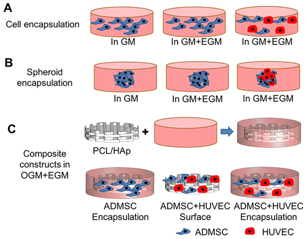
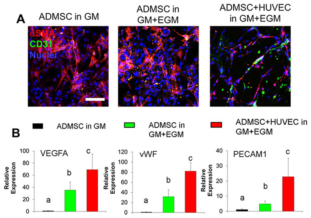
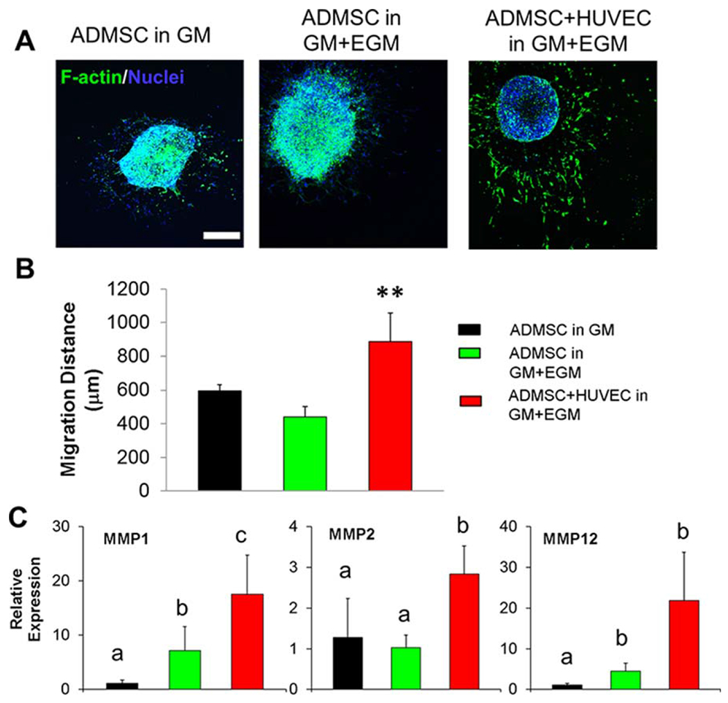
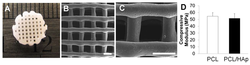
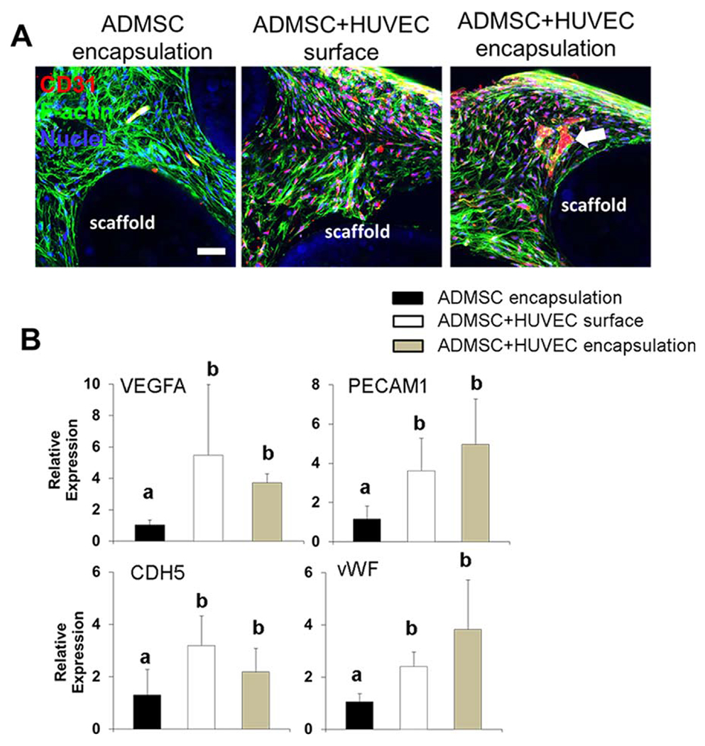
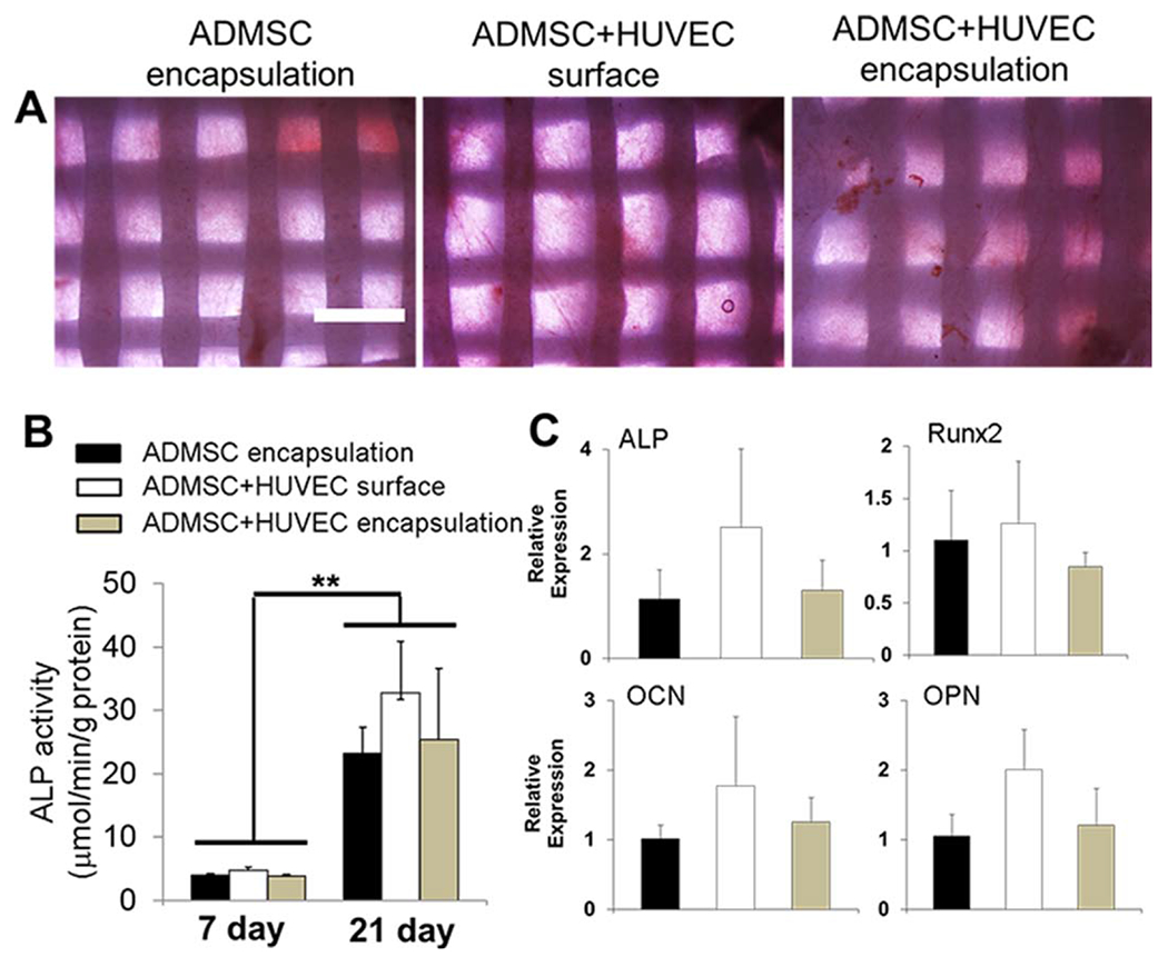
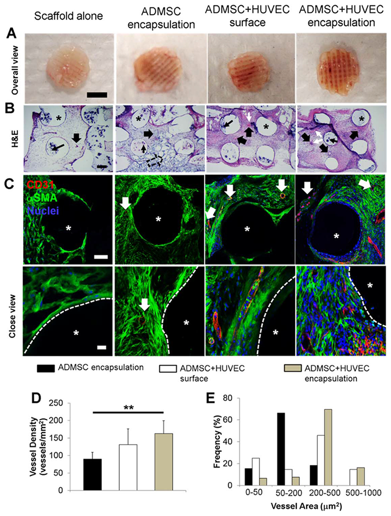
Vascularization is a fundamental prerequisite for large bone construct development and remains one of the main challenges of bone tissue engineering. Our current study presents the combination of 3D printing technique with a hydrogel-based prevascularization strategy to generate prevascularized bone constructs. Human adipose derived mesenchymal stem cells (ADMSC) and human umbilical vein endothelial cells (HUVEC) were encapsulated within our bioactive hydrogels, and the effects of culture conditions on in vitro vascularization were determined. We further generated composite constructs by forming 3D printed polycaprolactone/hydroxyapatite scaffolds coated with cell-laden hydrogels and determined how the co-culture affected vascularization and osteogenesis. It was demonstrated that 3D co-cultured ADMSC-HUVEC generated capillary-like networks within the porous 3D printed scaffold. The co-culture systems promoted in vitro vascularization, but had no significant effects on osteogenesis. The prevascularized constructs were subcutaneously implanted into nude mice to evaluate the in vivo vascularization capacity and the functionality of engineered vessels. The hydrogel systems facilitated microvessel and lumen formation and promoted anastomosis of vascular networks of human origin with host murine vasculature. These findings demonstrate the potential of prevascularized 3D printed scaffolds with anatomical shape for the healing of larger bone defects. © 2017 Wiley Periodicals, Inc. J Biomed Mater Res Part B: Appl Biomater, 106B: 1788-1798, 2018.

Keywords: 3D printing; adipose derived stem cells; bone tissue engineering; human umbilical vein endothelial cells; vascularization.


Figures

FIGURE 1.
FIGURE 1.
Schematic of experimental design. (A) Homogeneous cell encapsulation within Me-HA/Me-Gel hydrogels: ADMSC or ADMSC-HUVEC and hydrogel precursors were loaded into silicone molds and subsequently exposed to 365 nm UV light for 60 s. The cell-hydrogel constructs were maintained in GM, hybrid media (GM and EGM) for 14 days; (B) Cellular spheroid encapsulation within hydrogels: ADMSC or ADMSC-HUVEC cell spheroids were fabricated first and then encapsulated within the hydrogels. The spheroid laden constructs were conditioned in GM or hybrid media for 7 days; (C) Composite constructs: PCL/HAp scaffolds were first 3D printed, then the printed scaffolds were incorporated with cell laden hydrogels. ADMSC or ADMSC-HUVEC were encapsulated. ADMSC-HUVEC were also surface seeded onto the printed PCL/Hap scaffolds. All of the constructs were conditioned in hybrid media with OGM and EGM for 21 days before further characterization or subcutaneous implantation.
FIGURE 2.
FIGURE 2.
3D Co-culture of ADMSC and HUVEC within the hydrogel promoted capillary network formation and vascularization gene expression. (A) Representative immunohistochemical (IHC) staining for αSMA (red), CD31 (green) and nuclei (blue) within cell laden hydrogels after 14-day culture; (B) qPCR analysis of VEGFA, vWF and PECAM1 in cell laden hydrogels after 14-day culture. Relative gene expression is presented as normalized to 18S and expressed relative to ADMSC in GM (n = 3; bars that do not share letters are significantly different from each other; p < 0.05). ADMSC or ADMSC-HUVEC were encapsulated and conditioned in GM or GM and EGM.
FIGURE 3.
FIGURE 3.
3D co-culture of ADMSC and HUVEC spheroids within hydrogel promoted cell migration. (A) Representative images of phalloidin staining for F-actin (green) and nuclei (blue) within spheroid laden hydrogels after 7-day culture (scale bar: 500 μm); (B) cell migration distance from encapsulated spheroids (**p < 0.01); (C) qPCR analysis of MMP1, MMP2 and MMP12 in spheroid laden hydrogels after 7-day culture. Relative gene expression is presented as normalized to 18S and expressed relative to ADMSC in GM (n = 3; bars that do not share letters are significantly different from each other; p < 0.05). ADMSC or ADMSC-HUVEC spheroids were encapsulated and conditioned in GM or GM and EGM.
FIGURE 4.
FIGURE 4.
3D printed PCL/HAp scaffold. (A) Representative image of 3D printed PCL/HAp scaffold with 10% of HAp nanocrystals; (B, C) SEM images of printed PCL/HAp scaffold (scale bar: 500 μm). The PCL/HAp scaffold had regular pores of around 500 μm and strands with a thickness of 350–450 μm; (D) Compressive modulus of 3D printed PCL and PCL/HAp scaffolds.
FIGURE 5.
FIGURE 5.
In vitro vascularization within 3D printed scaffolds. (A) Representative IHC staining for F-actin (green), CD31 (red) and nuclei (blue) within cell laden hydrogels around 3D printed PCL/HAp scaffolds after 21-day culture (scale bar: 50 μm; white arrow indicates the formation of microvessel like structure); (B) qPCR analysis. Relative gene expression is presented as normalized to 18 S and expressed relative to scaffolds with ADMSC alone (n = 3; bars that do not share letters are significantly different from each other; p < 0.05). ADMSC or ADMSC-HUVEC were encapsulated and conditioned in hybrid media with OGM and EGM.
FIGURE 6.
FIGURE 6.
Co-culture of ADMSC and HUVEC within 3D printed scaffolds did not affect osteogenic differentiation in vitro. (A) Representative images for ALP staining after 21-day culture (scale bar: 1 mm); (B) ALP activity (**p < 0.01); (C) qPCR analysis. Relative gene expression is presented as normalized to 18S and expressed relative to scaffolds with ADMSC alone. (n = 3). ADMSC or ADMSC-HUVEC were encapsulated and conditioned in hybrid media with OGM and EGM.
FIGURE 7.
FIGURE 7.
In vivo vascularization within 3D printed scaffolds. (A) Overall view of explanted scaffolds after 4-week subcutaneous implantation (scale bar: 4 mm); (B) Representative images of H&E staining (scale bar: 200 μm). The wide, solid arrows indicate the formed microvessels with lumen structure and red blood cells inside. These microvessels can be of either human or mouse origin. The black, narrow arrows indicate the HAp particles. White, narrow arrows indicate the murine erythrocytes within blood vessels. The remnants of hydrogels are denoted by hollow arrow, and the asterisks indicate the PCL/HAp scaffold strand areas; (C) IHC staining for human CD31 (red), αSMA (green), and nuclei (blue) within 3D printed scaffolds. The white, solid arrows indicate the stained cells/microvessles positive for human CD31. Very few human CD31 positive cells were observed in scaffold alone group, indicating that the formed capillaries were of mouse origin. The asterisks indicate the PCL/HAp scaffold strand areas; (D) Microvessel density of the scaffolds. The density was measured in vessels/mm2. (**p < 0.01); (E) Microvessel area distribution in the scaffolds. The microvessel density and microvessel area distribution were measure by quantifying the formed CD31 positive vascular-network.

Similar articles

Cited by

References

    1. Griffin KS, Davis KM, McKinley TO, Anglen JO, Chu TMG, Boerckel JD, Kacena MA. Evolution of bone grafting: Bone grafts and tissue engineering strategies for vascularized bone regeneration. Clin Rev Bone Miner Metab 2015;13:232–244.
    1. Samorezov JE, Alsberg E. Spatial regulation of controlled bioactive factor delivery for bone tissue engineering. Adv Drug Deliv Rev 2015;84:45–67. - PMC - PubMed
    1. Giannoudis PV, Dinopoulos H, Tsiridis E. Bone substitutes: an update. Injury 2005;36:S20–27. - PubMed
    1. Verrier S, Alini M, Alsberg E, Buchman SR, Kelly D, Laschke MW, Menger MD, Murphy WL, Stegemann JP, Schütz M, Miclau T, Stoddart MJ, Evans C. Tissue engineering and regenerative approaches to improving the healing of large bone defects. European Cells and Materials 2016;32:87–110. - PubMed
    1. Bose S, Roy M, Bandyopadhyay A. Recent advances in bone tissue engineering scaffolds Trends Biotechnol 2012;30:546–554. - PMC - PubMed

Publication types

MeSH terms