. 2018 Sep;23(5):1079-1093.
doi: 10.1111/adb.12559. Epub 2017 Sep 13.

α6 subunit-containing nicotinic receptors mediate low-dose ethanol effects on ventral tegmental area neurons and ethanol reward

Affiliations

α6 subunit-containing nicotinic receptors mediate low-dose ethanol effects on ventral tegmental area neurons and ethanol reward

Scott C Steffensen et al. Addict Biol. 2018 Sep.

Abstract

Dopamine (DA) neuron excitability is regulated by inhibitory GABAergic synaptic transmission and modulated by nicotinic acetylcholine receptors (nAChRs). The aim of this study was to evaluate the role of α6 subunit-containing nAChRs (α6*-nAChRs) in acute ethanol effects on ventral tegmental area (VTA) GABA and DA neurons. α6*-nAChRs were visualized on GABA terminals on VTA GABA neurons, and α6*-nAChR transcripts were expressed in most DA neurons, but only a minority of VTA GABA neurons from GAD67 GFP mice. Low concentrations of ethanol (1-10 mM) enhanced GABAA receptor (GABAA R)-mediated spontaneous and evoked inhibition with blockade by selective α6*-nAChR antagonist α-conotoxins (α-Ctxs) and lowered sensitivity in α6 knock-out (KO) mice. Ethanol suppression of VTA GABA neuron firing rate in wild-type mice in vivo was significantly reduced in α6 KO mice. Ethanol (5-100 mM) had no effect on optically evoked GABAA R-mediated inhibition of DA neurons, and ethanol enhancement of VTA DA neuron firing rate at high concentrations was not affected by α-Ctxs. Ethanol conditioned place preference was reduced in α6 KO mice compared with wild-type controls. Taken together, these studies indicate that relatively low concentrations of ethanol act through α6*-nAChRs on GABA terminals to enhance GABA release onto VTA GABA neurons, in turn to reduce GABA neuron firing, which may lead to VTA DA neuron disinhibition, suggesting a possible mechanism of action of alcohol and nicotine co-abuse.

Keywords: VTA; ethanol; nicotine.


Figures

Figure 1
Figure 1
GAD-67 and α-conotoxin (Ctx) MII-labeled α6*-nAChRs are co-expressed on presynaptic terminals attached to GAD-67-producing cells in the mouse VTA. (A) The four confocal images show two representative dissociated VTA GABA neurons from a GAD-67 GFP mouse evincing diffuse GFP labeling (green), as well as dense GFP labeling at putative boutons. Biotin α-Ctx MII labeling (red; Cy5) in the same neurons shows α6*nAChRs co-expressed on presynaptic terminals attached to GAD-67-producing cells. VGAT labeling (blue; Alexa Fluor® 405) on the same neurons shows punctate boutons on VTA GABA neurons. The merge shows α6*-nAChRs on GABA boutons attached to VTA GABA neurons. Scale bar represents 10 μm. (B) Single-cell, quantitative real-time PCR (qRT-PCR) was used to examine nAChR subunit expression in VTA GABA neurons (GFP+, TH). Relative fluorescence acquired using a FAM-TAMRA hydrolysis probe is a measure of cDNA quantity for each nAChR target relative to PCR cycle number. 18S is used as a control housekeeping gene. Inset: This agarose gel illustrates the expected cDNA amplicon size for α6 (71 base pairs) taken from the PCR reaction, compared to the 50 base pair ladder (left lane). This representative VTA GABA neuron from a GAD-67 GFP mouse expressed α6, α4, and β2 nAChR subunit mRNA. (C) This representative VTA DA neuron from a GAD-67 GFP mouse expressed α6 and β2, but not α4 subunit mRNA. It is important to point out that as single cell PCR has some level of false negatives, actual expression levels are likely higher than reported. Therefore, ~45% expression in GABA cells and ~55–65% in DA cells suggests that α4, and β2 are likely present in the vast majority of DA cells and most of the GABA cells, even though it may not be detected in every neuron. (D) Summary of nAChR mRNA expression in VTA GABA and DA neurons evaluated for single-cell qRT-PCR. α6*-nAChR subunit mRNA was expressed in ~10% of GABA neurons and 45% of DA neurons, while α4 and β2 mRNA were expressed in ~50% of VTA neurons. These data support the expression of α6*-nAChRs in a subpopulation of midbrain GABA neurons.
Figure 2
Figure 2
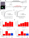
Ethanol enhances mIPSC frequency and amplitude in VTA GABA neurons. (A) Images show an acutely-dissociated VTA neuron from a GAD-67 GFP mouse using phase-contrast (Aa) and GPF-filtered (Ab) microscopic modes of illumination. (B) A typical trace shows spontaneous mIPSCs (in the presence of 0.5 μM TTX). They were sensitive to a selective GABA(A)R blocker bicuculline indicating that they were mediated by GABA. (C) Bath exposure of 5 mM ethanol increased the frequency of GABA mIPSCs. Further analysis revealed that 5 mM ethanol increases the frequency (D) and the amplitude (E) of spontaneous mIPSCs. (F,G) Ethanol enhanced GABA mIPSC frequency to VTA GABA neurons at all concentrations tested and amplitude at 5, 30 and 60 mM ethanol. Pre-treatment with the α-Ctx P1A (10 nM) prevented the ethanol (30 mM)-induced increase in mIPSC frequency (H) and amplitude (I) in WT mice. Similarly, 30 mM ethanol did not affect mIPSC frequency and amplitude in α6 KO mice. Vertical bars represent means ± SEM. Asterisks *,** indicate significance levels P<0.05 and 0.01, respectively. Values in parentheses represent n values.
Figure 3
Figure 3
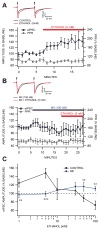
Effects of ethanol and α-Ctxs on evoked IPSCs (eIPSCs) in VTA GABA Neurons. Evoked IPSCs were recorded in VTA GABA neurons in the horizontal slice of GAD-67 GFP mice in the presence of APV/CNQX (or kynurenic acid) to block GLUR-mediated synaptic transmission, atropine to block muscarinic cholinergic effects and CGP55845 to block GABAB receptors. (A) Only one neuron/slice was tested for each of the concentrations of ethanol. (A) Inset shows representative superimposed recordings of IPSCs evoked in VTA GABA neurons at a paired-pulse interval of 50 msec before and after superfusion of 5 mM ethanol. Ethanol enhanced eIPSC amplitudes. The graph plots the time course of eIPSC amplitudes (integrated over 1 min intervals) as well as the paired-pulse ratio (PPR) for all cells studied with ethanol at 5 mM. Note the enhancement of eIPSC amplitude at this low concentration. There was no obvious effect of ethanol on PPR. (B) Insets show representative superimposed recordings of eIPSCs during superfusion ofthe α-Ctx MII and after ethanol + MII. In the presence of MII, ethanol had little effect on eIPSC amplitudes in this representative VTA GABA neuron. (C) Summary of the effects of ethanol (1–100 mM) on eIPSCs with block at lower and higher concentrations by MII. Vertical bars represent means ± SEM. Asterisks **,*** indicate significance levels P<0.01 and 0.001, respectively.
Figure 4
Figure 4
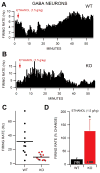
A role for α6*-nAChRs in ethanol effects on VTA GABA neuron firing rate in vivo and in the slice preparation ex vivo. For in vivo studies, VTA GABA neurons were identified by stereotaxic coordinates and by spontaneous electrophysiological criteria under isoflurane anesthesia (Steffensen et al., 1998). These included in mice: Relatively fast firing rate (>10Hz); ON-OFF phasic non-bursting activity under isoflurane anesthesia; spike duration less than 200 μsec; and activation by generalized sensory stimulation (Ludlow et al., 2009; Steffensen et al., 2011). In some experiments, multiple spike discharges were evoked in putative GABA neurons by electrical stimulation of the internal capsule, as previously reported (Steffensen et al., 1998). (A) This ratemeter record shows that intraperitoneal administration of 1.5 g/kg ethanol inhibited the firing rate of this representative VTA GABA neuron in an isoflurane-anesthetized WT mouse. The baseline firing rate of this neuron was approximately 5 Hz. (B) This ratemeter record shows that intraperitoneal administration of 1.5 g/kg ethanol increased the firing rate of this representative VTA GABA neuron recorded in a α6 KO mouse. The baseline firing rate for this neuron was 4 Hz. This example was chosen to match the firing rate of the example in (A). (C) However, on average, VTA GABA neurons were characterized by significantly faster firing rates than those recorded in α6 KO mice. Horizontal bar denotes mean. (D) Knock-out mice were resistant to the typical inhibition of firing rate in WTs by this dose of ethanol. In fact, they were slightly excited on average. Vertical bars represent means ± SEM. Asterisk * indicates significance level P<0.05. Values in parentheses represent n values.
Figure 5
Figure 5
Lack of effects of ethanol on GABAAR-mediated inhibition to DA neurons in the slice preparation of VGAT ChR2 transgenic mice. VTA GABA and DA neuron spikes were recorded in cell-attached, voltage clamp mode. Dopamine neurons were characterized by the expression of TH transcripts and response to the GABA spike command waveform (in cells where whole cell recordings were made). In VGAT::ChR2 mice, blue light stimulation was used to evaluate ethanol effects on optically-evoked IPSCs (oIPSCs). (A) Inset shows a representative recording of a VTA GABA neuron that is activated by a 200 msec light pulse (denoted by blue bar), evincing multiple spikes during the stimulation. The peri-stimulus interval spike histogram shows this spike’s cumulated activity over 12 stimulation epochs, demonstrating the marked activation and subsequent inhibition of spontaneous GABA neuron spikes after the optical stimulation. (B) Inset shows a representative recording of a VTA DA neuron whose spontaneous activity is inhibited by a 200 msec light pulse, evincing inhibition of DA neuron spontaneous spiking. The peri-stimulus interval spike histogram shows this spike’s cumulated activity over 12 stimulation epochs, demonstrating the marked inhibition of spontaneous DA spikes. (C) In whole cell mode, optical stimulation (5.0 msec) evoked fast, large inward sodium currents in GABA neurons and relatively slow, small Cl currents in DA neurons. (D) oIPSCs were markedly reduced by bicuculline and moderately reduced by eticlopride. (E) In the presence of eticlopride, ethanol (10 – 100 mM) had no effect on oIPSC amplitudes or area under the curve in DA neurons. Vertical bars represent means ± SEM. Asterisks **,*** indicate significance levels P<0.01 and P<0.001, respectively. Values in parentheses represent n values.
Figure 6
Figure 6
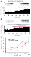
Lack of a role for α6*-nAChRs in ethanol enhancement of VTA DA neuron firing rate in the slice preparation. Using cell-attached mode, spontaneous spike activity was measured in horizontal slices of GAD-67 GFP mice in the presence of eticlopride. Only one neuron/slice was tested for each of the concentrations of ethanol unless indicated. Insets a,b are representative 5 sec recordings of separate VTA DA neuron spikes recorded in GAD-67 GFP mice at the times indicated on each of the ratemeter graphs. (A) Superfusion of 100 mM ethanol enhanced the firing rate of this representative VTA DA neuron recorded in a GAD-67 GFP mouse. (B) In a neuron from a separate slice, superfusion of 100 nM α-Ctx MII had no effect on 100 mM ethanol enhancement of the firing rate of this representative DA neuron. (C) Summary of the effects of MII on ethanol effects across ethanol concentrations 5–100 mM. MII had no significant effects on ethanol enhancement of VTA DA neuron firing rate.
Figure 7
Figure 7
α6*-nAChRs mediate ethanol reward. (A) Results show WT mice have a preference for the ethanol paired chamber at 0.1, 0.5 and 2.0 g/kg ethanol, but was significantly reduced in the α6 KO mice at the 2.0 g/kg level. (B) Locomotor activity in WT and KO mice was inhibited similarly by ethanol during the 20 min conditioning sessions compared to saline at the 2.0 g/kg dose level. Asterisks *,**,*** indicate significance levels P<0.05, P<0.01 and P<0.001, respectively compared to saline and ## indicate significance level P<0.01 between WT and KO mice at the 2.0 g/kg level. Values in parentheses represent n values.

Similar articles

Cited by

References

    1. Allison DW, Ohran AJ, Stobbs SH, Mameli M, Valenzuela CF, Sudweeks SN, Ray AP, Henriksen SJ, Steffensen SC. Connexin-36 gap junctions mediate electrical coupling between ventral tegmental area GABA neurons. Synapse. 2006;60:20–31. - PubMed
    1. Allison DW, Wilcox RS, Ellefsen KL, Askew CE, Hansen DM, Wilcox JD, Sandoval SS, Eggett DL, Yanagawa Y, Steffensen SC. Mefloquine effects on ventral tegmental area dopamine and GABA neuron inhibition: a physiologic role for connexin-36 GAP junctions. Synapse. 2011a;65:804–813. - PMC - PubMed
    1. Allison DW, Wilcox RS, Ellefsen KL, Askew CE, Hansen DM, Wilcox JD, Sandoval SS, Eggett DL, Yanagawa Y, Steffensen SC. Mefloquine effects on ventral tegmental area dopamine and GABA neuron inhibition: A physiologic role for connexin-36 gap junctions. Synapse 2011b - PMC - PubMed
    1. Azam L, Winzer-Serhan UH, Chen Y, Leslie FM. Expression of neuronal nicotinic acetylcholine receptor subunit mRNAs within midbrain dopamine neurons. J Comp Neurol. 2002;444:260–274. - PubMed
    1. Berrios J, Stamatakis AM, Kantak PA, McElligott ZA, Judson MC, Aita M, Rougie M, Stuber GD, Philpot BD. Loss of UBE3A from TH-expressing neurons suppresses GABA co-release and enhances VTA-NAc optical self-stimulation. Nature communications. 2016;7:10702. - PMC - PubMed

Publication types