. 2017 Oct;98(10):2582-2588.
doi: 10.1099/jgv.0.000925.

Simian varicella virus inhibits the interferon gamma signalling pathway

Affiliations

Simian varicella virus inhibits the interferon gamma signalling pathway

Werner J D Ouwendijk et al. J Gen Virol. 2017 Oct.

Abstract

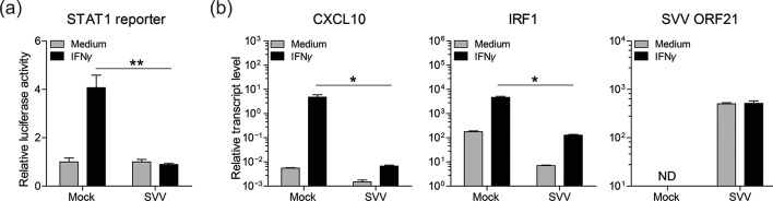
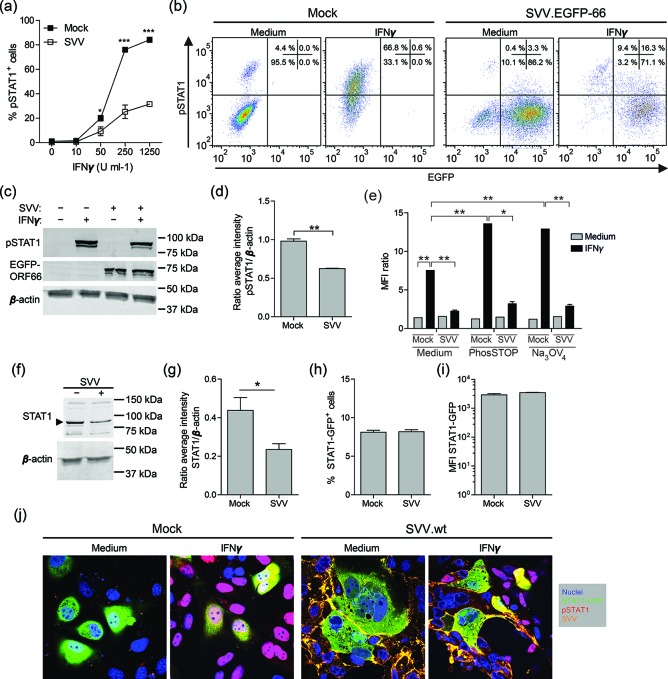
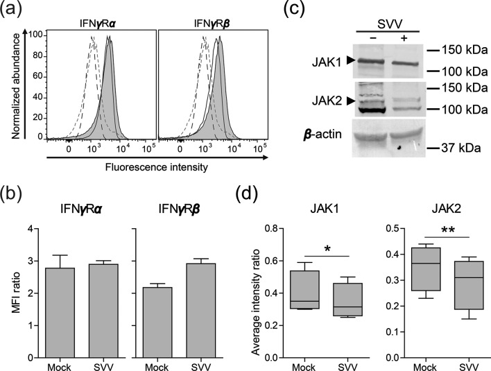
The alphaherpesvirus simian varicella virus (SVV) causes varicella and zoster in nonhuman primates. Herpesviruses evolved elaborate mechanisms to escape host immunity, but the immune evasion strategies employed by SVV remain ill-defined. We analysed whether SVV impairs the cellular response to key antiviral cytokine interferon-γ (IFNγ). SVV infection inhibited the expression of IFNγ-induced genes like C-X-C motif chemokine 10 and interferon regulatory factor 1. Phosphorylation and nuclear translocation of the signal transducer and activator of transcription 1 (STAT1) was blocked in SVV-infected cells, which did not involve cellular and viral phosphatases. SVV infection did not downregulate IFNγ receptor α and β chain expression on the cell surface. Instead, STAT1, Janus tyrosine kinases 1 (JAK1) and JAK2 protein levels were significantly decreased in SVV-infected cells. Collectively, these results demonstrate that SVV targets three proteins in the IFNγ signal transduction pathway to escape the antiviral effects of IFNγ.

Keywords: JAK1; JAK2; STAT1; interferon-γ; simian varicella virus.


Conflict of interest statement

The authors declare that there are no conflicts of interest.

Figures

Fig. 1.
Fig. 1.
SVV inhibits the expression of IFNγ-stimulated genes. (a) BSC-1 cells were transiently transfected to express firefly luciferase under a STAT1-responsive promotor [19, 20] and renilla luciferase under a constitutively active SV40 promotor. The next day, cells were mock- or SVV-infected for 72 h and subsequently cultured with or without recombinant human IFNγ (1000 U ml−1) for an additional 6 h, after which luciferase activity was measured. The relative STAT1 promotor activity of IFNγ-stimulated cells compared to medium control cells, after normalization to renilla luciferase activity, is shown. Bars represent average ± standard error of the mean (SEM) of three replicates measured in duplicate. **P<0.01 by unpaired Student’s t-test. (b) BSC-1 cells were mock- or SVV-infected for 72 h and subsequently cultured with or without recombinant human IFNγ (1000 U ml−1) for an additional 24 h. The transcript levels of C-X-C motif chemokine 10 (CXCL10), interferon regulatory factor 1 (IRF1) and SVV open reading frame 21 (ORF21) were determined in cells by real-time PCR. Relative transcript levels were normalized to the cellular oncostatin-M control transcript and expressed as log10 values. Bars represent average ± sem of two experiments, each performed in duplicate. nd, not detected. *P<0.05 by the Mann–Whitney U test.
Fig. 2.
Fig. 2.
SVV inhibits IFNγ-induced phosphorylation of STAT1. (a) BSC-1 cells were mock- or SVV-infected and 72 h later (72 h p.i.), stimulated with the indicated dose of IFNγ for 20 min and finally analysed for intracellular phosphorylated STAT1 (pSTAT1) expression by flow cytometry. The frequency of pSTAT1-positive mock- or SVV-infected BSC-1 cells is shown. Data represent average values ± sem from two independent experiments. *P<0.05, ***P<0.001 by two-way ANOVA test with Bonferroni correction. (b) Flow cytometric analysis of intracellular pSTAT1 and EGFP expression by BSC-1 cells infected with SVV expressing EGFP-ORF66 fusion protein (SVV.EGFP-66) (72 h p.i.), and control mock-infected BSC-1 cells, upon incubation with and without 1000 U ml−1 IFNγ for 20 min. Quadrants were set based on isotype control stained cells and the frequencies of cells within each quadrant are indicated. (c) Mock- and SVV.EGFP-66−infected BSC-1 cells were incubated with and without 1000 U ml−1 IFNγ for 20 min and total cell lysates were analysed by Western blotting for pSTAT1 (two bands, STAT1α=91 kDa and STAT1β=84 kDa), EGFP and β-actin protein expression. (d) The signal intensity of pSTAT1 and β-actin was quantified and the average ratio pSTAT1/β-actin from three experiments is shown. (e) Mock- and SVV.EGFP-66−infected BSC-1 cells (72 h p.i.) were stimulated with medium or 1000 U ml−1 IFNγ for 20 min in the presence or absence of phosphatase inhibitors phosSTOP (Roche) and sodium orthovanadate (Na3OV4). Intracellular phosphorylated STAT1 (pSTAT1) expression was determined by flow cytometry and presented as the median fluorescence intensity (MFI) ratio of cells stained for pSTAT1 and cells stained using isotype control antibodies. (f) Mock- and SVV.EGFP-66−infected BSC-1 cells were lysed at 72 h p.i. and probed with antibodies specific for STAT1 and β-actin by Western blotting. The arrowheads indicate bands of the appropriate size corresponding to STAT1 (two bands, STAT1α=91 kDa and STAT1β=84 kDa). (g) The signal intensity of STAT1 protein was quantified and normalized to the corresponding β-actin protein abundance (STAT1/β-actin ratio). (h–i) Flow cytometric analysis of GFP fluorescence intensity of BSC-1 cells transiently transfected to express STAT1-GFP and mock- and SVV-infected (48 h p.i.). The frequency of STAT1-GFP-expressing cells (h) and the MFI of STAT1-GFP (i) are shown. (j) Transiently transfected BSC-1 cells expressing STAT1-GFP (green) were mock- or wild-type SVV (SVV.wt)-infected (48 h p.i.), incubated with medium or 1000 U ml−1 IFNγ for 20 min and stained for pSTAT1 (red) and SVV (orange). Nuclei were counterstained with DAPI (blue). Representative confocal microscopy images from two independent experiments are shown. Magnification: 400×. (d, e, g, h) Data represent average values ± sem from three (d, e, h) and four (g) independent experiments. *P<0.05, **P<0.01, ***P<0.001 by unpaired Student’s t-test.
Fig. 3.
Fig. 3.
SVV infection downregulates JAK1 and JAK 2 protein expression. (a) Histograms showing the expression of the IFNγ receptor alpha (IFNγRα) and beta (IFNγRβ) subunits in mock- and SVV.EGFP-66-infected BSC-1 cells (72 h p.i.) as determined by flow cytometry. Filled (virus-infected) and open (mock-infected) solid lines indicate cells stained with the indicated antibodies. Dashed grey (virus-infected) and black (mock-infected) lines indicate cells stained with isotype control antibodies. (b) The MFI ratio of cells stained for IFNγRα or IFNγRβ, and cells stained using isotype control antibodies, is presented. Data represent average values ± sem from two independent experiments. (c) Mock- and SVV.EGFP-66−infected BSC-1 cells were lysed at 72 h p.i. and probed with antibodies specific for JAK1, JAK2 and β-actin by Western blotting. The arrowheads indicate bands of the appropriate size corresponding to JAK1 (130 kDa) and JAK2 protein (128 kDa). (d) Signal intensities of JAK1 and JAK2 proteins were quantified and normalized to corresponding β-actin protein abundance (e.g. the JAK1/β-actin ratio). Box plots indicate the average intensity ratios from four independent experiments. *P<0.05 and **P<0.01 by paired Student’s t-test.

Similar articles

Cited by

References

    1. Clarkson MJ, Thorpe E, Mccarthy K. A virus disease of captive vervet monkeys (Cercopithecus aethiops) caused by a new herpesvirus. Arch Gesamte Virusforsch. 1967;22:219–234. doi: 10.1007/BF01240517. - DOI - PubMed
    1. Gray WL. Simian varicella in Old World monkeys. Comp Med. 2008;58:22–30. - PMC - PubMed
    1. Messaoudi I, Barron A, Wellish M, Engelmann F, Legasse A, et al. Simian varicella virus infection of rhesus macaques recapitulates essential features of varicella zoster virus infection in humans. PLoS Pathog. 2009;5:e1000657. doi: 10.1371/journal.ppat.1000657. - DOI - PMC - PubMed
    1. Mahalingam R, Smith D, Wellish M, Wolf W, Dueland AN, et al. Simian varicella virus DNA in dorsal root ganglia. Proc Natl Acad Sci USA. 1991;88:2750–2752. doi: 10.1073/pnas.88.7.2750. - DOI - PMC - PubMed
    1. Kolappaswamy K, Mahalingam R, Traina-Dorge V, Shipley ST, Gilden DH, et al. Disseminated Simian varicella virus infection in an irradiated rhesus macaque (Macaca mulatta) J Virol. 2007;81:411–415. doi: 10.1128/JVI.01825-06. - DOI - PMC - PubMed

LinkOut - more resources