Review
. 2017 Sep 13;17(9):2095.
doi: 10.3390/s17092095.

Whispering-Gallery Mode Resonators for Detecting Cancer

Affiliations
Review

Whispering-Gallery Mode Resonators for Detecting Cancer

Weeratouch Pongruengkiat et al. Sensors (Basel). .

Abstract

Optical resonators are sensors well known for their high sensitivity and fast response time. These sensors have a wide range of applications, including in the biomedical fields, and cancer detection is one such promising application. Sensor diagnosis currently has many limitations, such as being expensive, highly invasive, and time-consuming. New developments are welcomed to overcome these limitations. Optical resonators have high sensitivity, which enable medical testing to detect disease in the early stage. Herein, we describe the principle of whispering-gallery mode and ring optical resonators. We also add to the knowledge of cancer biomarker diagnosis, where we discuss the application of optical resonators for specific biomarkers. Lastly, we discuss advancements in optical resonators for detecting cancer in terms of their ability to detect small amounts of cancer biomarkers.

Keywords: biosensor; cancer; evanescent wave; instrumentation; label-free; optical resonator; optical waveguide; sensor platform; whispering-gallery mode.


Conflict of interest statement

The authors declare no conflicts of interest.

Figures

Figure 1
Figure 1
Graphs plotted between 5-year survival rates versus stages of cancers. It is clear that at the initial stage of cancer development, patients have significantly larger chances of being cured. (a) Ovarian stromal tumor [5], (b) Cervical cancer [6].
Figure 2
Figure 2
Figure shows how the number of articles on fix in figure label optical resonators evolved between 1999–2016. The graph shows increasing trends in this area of study. Remark: The data were gathered from the Web of Science database (www.web of knowledge.com) with the keyword “Optical Resonator”, and then the category filter “Optics” was applied [19].
Figure 3
Figure 3
Various types of optical coupling. (a) Tapered; (b) prism; (c) angled fiber; (d) planar waveguide side; (e) free-space; (f) polished half-block coupler.
Figure 4
Figure 4
Comparison of mechanical wave and electromagnetic wave WGM. (a) Diagram shows how WGM occurs: sound travels from one side of the gallery to another in the structure of St. Paul’s Cathedral where WGM was first observed; (b) WGM of light: light is totally internally reflected within the small curved cavity [54,75]. The phenomenon can be observed as light trapped inside the spherical structure [76].
Figure 5
Figure 5
Diagram shows the optical resonator system and analysis. (a) Optical resonator system: the laser guided by a coupling waveguide excites the resonator. The detector is equipped to measure the intensity dip of the resonant wavelength at the end of the coupling waveguide (Detector 1) or scattered light (Detector 2). (b) Graph shows the absence of light at resonant wavelength. When the analyte binds on the surface, it causes a wavelength shift that can be observed by both modes.
Figure 6
Figure 6
Example of microsphere configuration. Shown is an example of a microsphere with tapered coupling. The microsphere utilizes the WGM principle to resonate light inside its cavity. The analyte binding on the microsphere surface causes the change in the refractive index. The resonant wavelength is also shifted as a result.
Figure 7
Figure 7
Diagram shows an example common microring optical resonator setting. A tunable laser source provides the optical input through the coupling waveguide. The coupling illuminates the resonator structure. The coupling wavelength can be observed using a photodetector and an analytic instrument such as a computer. The wavelength shifts when the resonator is bound with the analyte, altering the refractive index.
Figure 8
Figure 8
Microtoroid resonator fabrication process. (1) SiO2 is deposited on a silicon wafer; (2) Hydrofluoric acid etching is applied to create the disk structure on top of the wafer; (3) XeF2 etching is used to create a post structure; (4) CO2 laser illuminates the structure to smoothen the toroid structure.
Figure 9
Figure 9
Analysis of HER2 biomarker. (a) OFRR schematic. The cross-section diagram visualizes the layer of the OFRR inner core surface; (b) spectral shift at (a) buffer flow, (b) sample flow, (c) sample binding and (d) buffer washes. Reprinted with permission from [138].
Figure 10
Figure 10
Diagram of HRAS and FGR3 detection study. Surface functionalization on the microring resonator is visualized. Reprinted with permission from [131].
Figure 11
Figure 11
Schematic shows the surface functionalization of OFRR for detecting CA15-3; the system shows that the sample is drawn by the syringe pump while the OFRR performs the analysis. Reprinted with permission from [30].
Figure 12
Figure 12
The multiple microsphere resonator system: the objective lens is utilized as the coupling waveguide, while two differently sized microspheres are paired with different receptors. CCD detects the fluorescence behavior of the system. Reprinted with permission from [136].
Figure 13
Figure 13
Calibration curves of microsphere performance. (a) CA-125 detection (38 μm) shows the linear relation between the applied concentration and the resonance wavelength; (b) TNF-α (53 μm) also shows the same relation. Reprinted with permission from [136].
Figure 14
Figure 14
The configuration of ovarian cancer biomarker detection. (a) The system is based on a previous experiment but uses prism coupling for expanding the excitation area; (b) Fluorescence image revealing the illumination of some spheres specific for different markers. Reprinted with permission from [27].
Figure 15
Figure 15
Diagram shows the functionalized surface of a microring resonator. DNA oligomer is immobilized on the surface. Telomerase extracted from a cancer cell is introduced into the system along with dNTPs. The result shows that the system can detect telomerase activity. Reprinted with permission from [38].
Figure 16
Figure 16
3D model shows the mechanism of BSA binding with gold nanoshell particle at the equator of a dielectric microsphere with WGM. The WGM-h enhancement and adsorption of the biomolecule caused the resonant wavelength shift. Reprinted with permission from [141].

Similar articles

Cited by

References

    1. Cancer Research UK Worldwide Cancer Statistics. [(accessed on 28 June 2017)]; Available online: http://www.cancerresearchuk.org/health-professional/cancer-statistics/wo....
    1. American Cancer Society Survival Rates for Hodgkin Lymphoma By Stage. [(accessed on 28 June 2017)]; Available online: https://www.cancer.org/cancer/hodgkin-lymphoma/detection-diagnosis-stagi....
    1. American Cancer Society How Are Lung Carcinoid Tumors Staged? [(accessed on 28 June 2017)]; Available online: https://www.cancer.org/cancer/lung-carcinoid-tumor/detection-diagnosis-s....
    1. Cancer Research UK Stages of Cancer. [(accessed on 28 June 2017)]; Available online: http://www.cancerresearchuk.org/about-cancer/what-is-cancer/stages-of-ca....
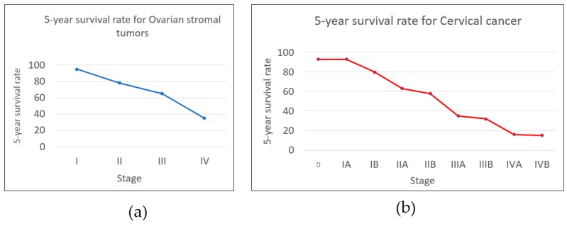
    1. American Cancer Society Survival Rates for Ovarian Cancer, by Stage. [(accessed on 28 June 2017)]; Available online: https://www.cancer.org/cancer/ovarian-cancer/detection-diagnosis-staging....