. 2017 Sep 28;549(7673):528-532.
doi: 10.1038/nature23910. Epub 2017 Sep 13.

Maternal gut bacteria promote neurodevelopmental abnormalities in mouse offspring

Affiliations

Maternal gut bacteria promote neurodevelopmental abnormalities in mouse offspring

Sangdoo Kim et al. Nature. .

Abstract

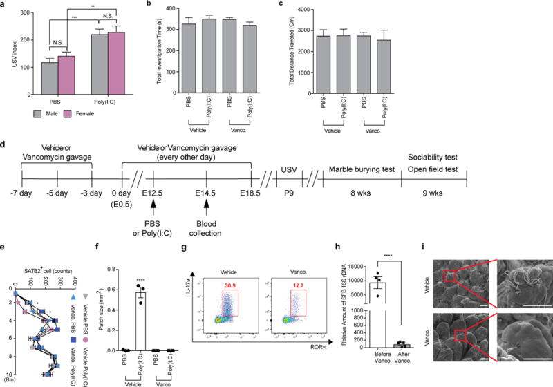
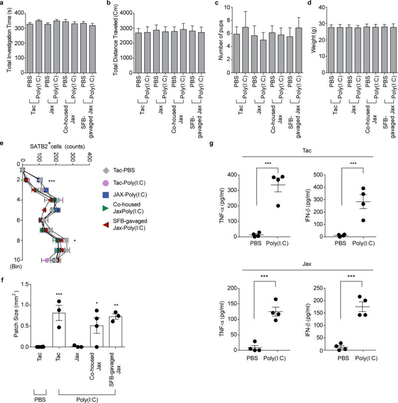
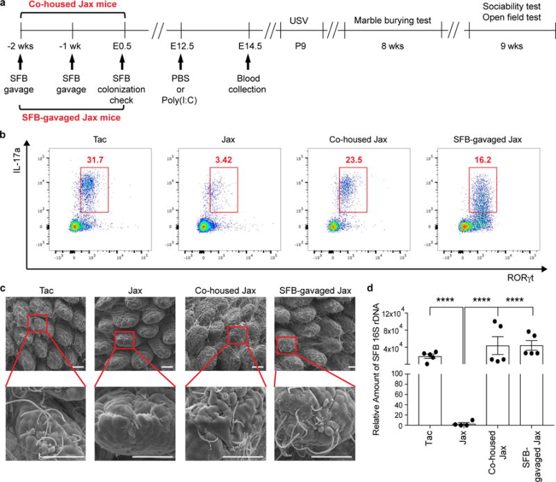
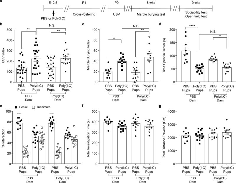
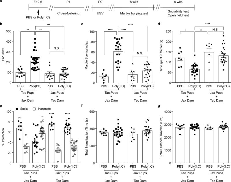
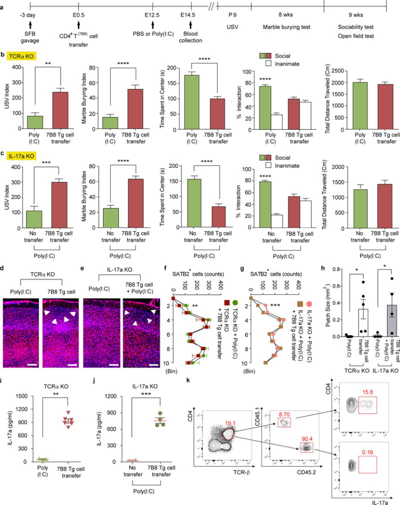
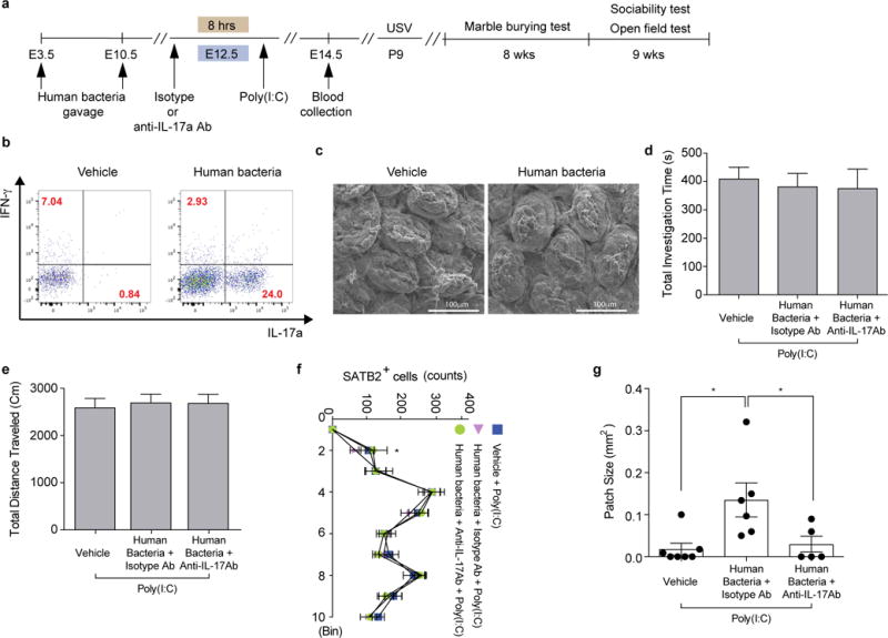
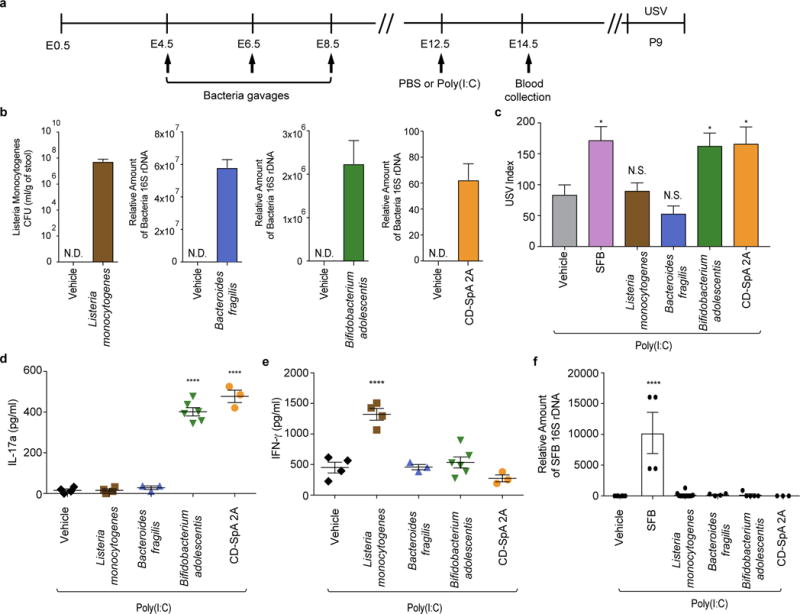
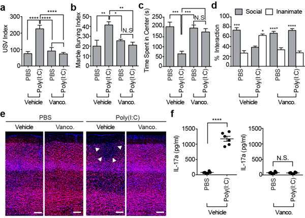
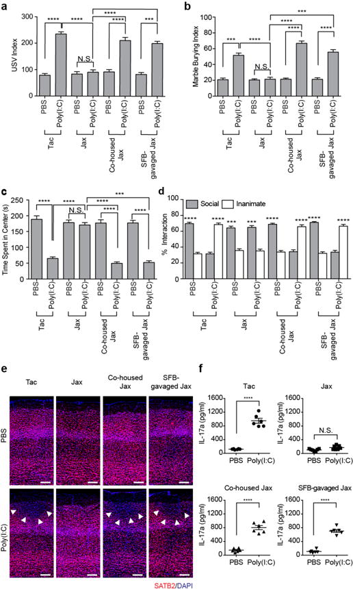
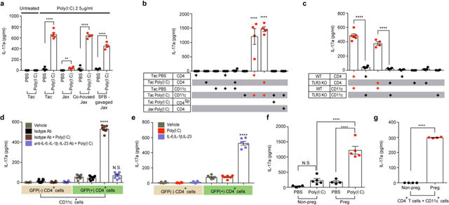
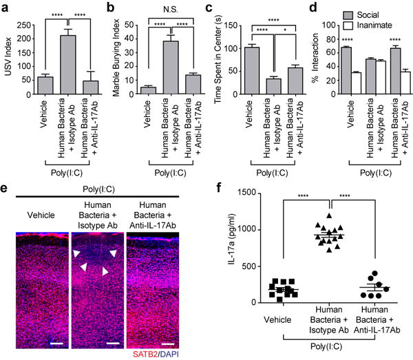
Maternal immune activation (MIA) contributes to behavioural abnormalities associated with neurodevelopmental disorders in both primate and rodent offspring. In humans, epidemiological studies suggest that exposure of fetuses to maternal inflammation increases the likelihood of developing autism spectrum disorder. In pregnant mice, interleukin-17a (IL-17a) produced by T helper 17 (TH17) cells (CD4+ T helper effector cells involved in multiple inflammatory conditions) induces behavioural and cortical abnormalities in the offspring exposed to MIA. However, it is unclear whether other maternal factors are required to promote MIA-associated phenotypes. Moreover, the underlying mechanisms by which MIA leads to T cell activation with increased IL-17a in the maternal circulation are not well understood. Here we show that MIA phenotypes in offspring require maternal intestinal bacteria that promote TH17 cell differentiation. Pregnant mice that had been colonized with mouse commensal segmented filamentous bacteria or human commensal bacteria that induce intestinal TH17 cells were more likely to produce offspring with MIA-associated abnormalities. We also show that small intestine dendritic cells from pregnant, but not from non-pregnant, females secrete IL-1β, IL-23 and IL-6 and stimulate T cells to produce IL-17a upon exposure to MIA. Overall, our data suggest that defined gut commensal bacteria with a propensity to induce TH17 cells may increase the risk of neurodevelopmental disorders in the offspring of pregnant mothers undergoing immune system activation owing to infections or autoinflammatory syndromes.


Conflict of interest statement

The authors declare no competing financial interests. Readers are welcome to comment on the online version of the paper.

Figures

Extended Data Figure 1
Extended Data Figure 1. Maternal vancomycin-treatment prevented induction of behavioral abnormalities in MIA offspring
a, USV index (n=27/29 (PBS;male/female); n=28/21 (Poly(I:C);male/female); 6 independent experiments). b-c, Total investigation time (b) and total distance traveled (c) during the sociability test (n=13/15 (vehicle;PBS/poly(I:C)); n=12/16 (vancomycin;PBS/poly(I:C)); 3-4 independent experiments). d, Schematic of the experimental design. e-f, Quantification of SATB2+ cells (e) in the cortex divided into ten equal bins representing different depths of the cortex or of the cortical patch size (f) in the primary somatosensory cortex (S1) (n=3/4 (PBS;vehicle/vancomycin); n=3/4 (poly(I:C);vehicle/vancomycin); 2 independent experiments). g, Flow cytometry of CD4+ T cells (gated on TCR-β+CD4+) stained intracellularly for IL-17a and RORγt. Mononuclear cells were collected at E14.5 from the ilea of poly(I:C)-treated mice with/without vancomycin treatment; Representative FACS plot from 3 independent experiments. h, qPCR analysis measuring relative SFB levels in B6 mice before/after vancomycin treatments (n=4-5/group). i, Representative SEM images of epithelial surfaces in the ilea of the vehicle-/vancomycin-treated mice from 2 independent experiments. Scale bars, 30 μm. *p<0.05, **p<0.01, ***p<0.001, ****p<0.0001 as calculated by two-way (a,e) and one-way (b,c,f) ANOVA with Tukey post-hoc tests. N.D., not determined; N.S., not significant. Graphs indicate mean +/− s.e.m.
Extended Data Figure 2
Extended Data Figure 2. MIA in SFB-absent Jax mothers does not induce changes in the total activity of the adult offspring, properties of the litter and maternal cytokine production
a-b, Total investigation time (a) and total distance traveled (b) during the sociability test. c, Litter size upon weaning (n=59/125 (Tac;PBS/poly(I:C)); n=51/50 (Jax;PBS/poly(I:C)); n=55/81 (Co-housed Jax;PBS/poly(I:C)); n=55/89 (SFB-gavaged Jax;PBS/poly(I:C)). d, Weight of male offspring from the groups described in (c) (n=32/50 (Tac;PBS/poly(I:C)); n=29/27 (Jax;PBS/poly(I:C)); n=29/29 (Co-housed Jax;PBS/poly(I:C)); n=33/30 (SFB-gavaged Jax;PBS/poly(I:C)). Data in a, b, and d are from 7-8 independent experiments. e-f, Quantification of SATB2+ cells (e) in the cortex divided into ten equal bins representing different depth and of patch size (f) in the S1 (n=4 (Tac;PBS); n=3/3/4/3 (Tac/Jax/Co-housed Jax/SFB-gavaged Jax;poly(I:C)). g, Maternal plasma concentrations of TNF-α and IFN-β at 3 hrs after PBS/poly(I:C) injection into Tac/Jax dams at E12.5; n=4/group. *p<0.05, **p<0.01, ***p<0.001 as calculated by two-way (e) and one-way ANOVA (a-d,g,f) with Tukey post-hoc tests and Student’s t-test (g). N.D., not determined. Graphs indicate mean +/− s.e.m.
Extended Data Figure 3
Extended Data Figure 3. SFB colonization leads to increased levels of gut Th17 cells in Jax pregnant mice
a, Schematic of the experimental design. b. Flow cytometry of CD4+ T cells (gated on TCR-β+CD4+) stained intracellularly for IL-17a and RORγt. Mononuclear cells were collected at E14.5 from the ilea of poly(I:C)-treated Tac/Jax/co-housed Jax/SFB-gavaged Jax mothers. c, Representative SEM images of epithelial surfaces in the ilea of Tac/Jax/co-housed Jax/SFB-gavaged Jax mothers. Scale bars, 30 μm. Data representative of 3 (b) and 2 (c) independent experiments. d, qPCR analysis for SFB levels in the fecal samples of the groups described in (a) (n=4-5/group). ****p<0.0001 as calculated by one-way (d) ANOVA with Tukey post-hoc test. Graphs indicate mean +/− s.e.m.
Extended Data Figure 4
Extended Data Figure 4. Poly(I:C)-induced inflammation during pregnancy, not after giving birth, is critical in inducing MIA-associated behavioral abnormalities in offspring
a, Schematic of the experimental design for cross-fostering experiments. b, USV index (n=21/20 (PBS dams;PBS/poly(I:C) pups); n=22/15 (poly(I:C) dams;PBS/poly(I:C) pups); 2-4 independent experiments). c-g Marble-burying index (c), time spent in the center of an open field (d), and % interaction (e), total investigation time (f), and total distance traveled (g) during the sociability test (n=9/14 (PBS dams;PBS/poly(I:C) pups); n=12/10 (poly(I:C) dams;PBS/poly(I:C) pups); 2 independent experiments). **p<0.01, ****p <0.0001 as calculated by one-way (b-d,f-g) and two-way (e) ANOVA with Tukey post-hoc tests. N.S., not significant. Graphs indicate mean +/− s.e.m.
Extended Data Figure 5
Extended Data Figure 5. Composition of maternal gut microbiota during pregnancy, not after giving birth, is critical in inducing MIA-associated behavioral abnormalities in offspring
a, Schematic of the experimental design for cross-fostering experiments. b, USV index (n=9/36 (Tac pups with Jax dams;PBS/poly(I:C)); n=10/24 (Jax pups with Tac dams;PBS/poly(I:C)); 2-4 independent experiments). c-g, Marble-burying index (c), time spent in the center of an open field (d), and % interaction (e), total investigation time (f), and total distance traveled (g) during the sociability test (n=7/22 (Tac pups with Jax dams;PBS/poly(I:C)); n=7/21 (Jax pups with Tac dams;PBS/poly(I:C)); 2 independent experiments). *p<0.05, **p<0.01, ***p<0.001, ****p <0.0001 as calculated by one-way (b-d,f-g) and two-way (e) ANOVA with Tukey post-hoc tests. Graphs indicate mean +/− s.e.m.
Extended Data Figure 6
Extended Data Figure 6. CD11c+ cells stimulate gut-Th17 cells to produce high levels of IL-17a ex vivo
a-f, Flow cytometry of CD4+ T cells (gated on TCR-β+CD4+) stained intracellularly for IL-17a and RORγt. Mononuclear cells were collected at E14.5 from the gut ilea, spleens, and mesenteric lymph nodes (mLN) of PBS-/poly(I:C)-treated mice (n=5/group (a, c, e); n=3/group (b, d, f)). MFI denotes mean fluorescence intensity. g-i,, Supernatant concentrations of IL-17a from mononuclear cells of the ilea in poly(I:C)-treated Tac dams (g) (n=3/group), from co-cultures of CD4+ and non-CD4+ cells of the ilea in PBS-/poly(I:C)-treated Tac dams (h) (n=3/group), or from co-cultures of CD4+ and CD103CD11b+/CD103+CD11b+/CD103+CD11b (gated on MHCII+CD11c+) cells of the ilea in poly(I:C)-treated dams (i) (n=7/group). All cultures were isolated at E14.5 and stimulated ex vivo with poly(I:C) for 18hrs (g-h) or for 48hrs (i). Data are pooled from 2 (g-h) or 3 (i) independent experiments. j. USV index (n=16/17 (poly(I:C);WT/TLR3 KO); 2 independent experiments). k, Supernatant concentrations of IL-6, IL-1β, and IL-23 from cultures of CD11c+ isolated at E14.5 from the ilea of poly(I:C)-treated non-pregnant/pregnant mice (n=5/group; 3 independent experiments). *p<0.05, **p<0.01, ***p<0.001 and ****p<0.0001 as calculated by Student’s t-test (a-f,j,k) and one-way ANOVA (g-i) with Tukey post-hoc tests. N.S., not significant. Graphs indicate mean +/− s.e.m.
Extended Data Figure 7
Extended Data Figure 7. SFB-specific 7B8 Tg CD4+ T cells produce IL-17a upon transfer to MIA-exposed pregnant mothers
a, Schematic of the experimental design. b-c, Both TCRα KO and IL-17a KO females, with or without adoptive transfers of 7B8 Tg-derived CD4+ T cells, were crossed with B6 WT males to produce heterozygous WT offspring. USV index (n=16/30 (TCRα KO; poly(I:C)/7B8 Tg T cell transfer); n=23/23 (IL-17a KO;poly(I:C)/7B8 Tg T cell transfer), marble burying index, time spent in the center of an open field, and % interaction and total distance traveled during the sociability test of TCRα KO (b) or IL-17a KO (c) offspring (n=12/15 (TCRα KO; poly(I:C)/7B8 Tg T cell transfer); n=12/14 (IL-17a KO;poly(I:C)/7B8 Tg T cell transfer). Data pooled from 2-3 independent experiments. d-e, Representative SATB2 staining in the cortex of the animals prepared as in (a). Arrows indicate cortical patches. Scale bar, 100 μm. f-g, Quantification of SATB2+ cells (n=7/6 (TCRα KO;poly(I:C)/7B8 Tg T cell transfer); n=6/7 (IL-17a KO;poly(I:C)/7B8 Tg T cell transfer). h, Cortical patch size (n=5/5 (TCRα KO;poly(I:C)/7B8 Tg T cell transfer); n=4/4 (IL-17a KO;poly(I:C)/7B8 Tg T cell transfer). i-j, IL-17a concentrations in maternal plasma collected at E14.5. k, Flow cytometry of ileal CD4+ T cells (gated on CD4+TCR-β+) stained intracellularly for IL-17a. Mononuclear cells were collected from small intestines of poly(I:C)-treated IL-17a KO mothers transferred with 7B8 Tg CD4+ T cells. CD45.1+ cells refer to donor cells and CD45.2+ to recipient cells. *p<0.05, **p<0.01, ***p<0.001, ****p<0.0001 as calculated by Student’s t-test (b-c,h-j) and one-way (f-g) ANOVA with Sidak post-hoc tests. Graphs indicate mean +/− s.e.m.
Extended Data Figure 8
Extended Data Figure 8. A mix of twenty human commensals induces colonic Th17 cell differentiation in SFB-absent Jax mice
a, Schematic of the experimental design. b, Flow cytometry of CD4+ T cells (gated on CD4+TCR-β+) stained intracellularly for IL-17a and RORγt. Mononuclear cells were collected from colons of poly(I:C)-treated Jax mothers with/without human bacteria-gavage. c, Representative SEM images of epithelial surfaces in the ilea from 2 independent experiments. d-e, Total interaction time (d), and total distance traveled (e) during the sociability test of adult offspring described in (a) (n=23/22/13 for vehicle-gavaged only/human bacteria-gavaged+isotype control antibody/human bacteria-gavaged+anti-IL-17a antibody; 4 independent experiments). f-g, Quantification of SATB2+ cells (n=5/group) and cortical patch size (n=7/6/5 (poly(I:C);vehicle-treated Jax/human bacteria-gavaged Jax with isotype control antibody/human bacteria-gavaged Jax with anti-IL-17a antibody). *p<0.05 as calculated by one-way (d, e, g) and two-way (f) ANOVA with Tukey post-hoc test. Graphs indicate mean +/− s.e.m.
Extended Data Figure 9
Extended Data Figure 9. The IL-17a pathway promotes abnormal behavioral phenotypes in MIA offspring born to mice colonized with human commensal bacteria
a, Schematic representation of the experimental design. b, Quantification of bacterial colonization levels through colony forming unit (CFU) counts or qPCR analyses. c, USV index (n=13/12/28/16/17/14 (poly(I:C);vehicle/SFB/Listeria monocytogenes/Bacteroides fragilis/Bifidobacterium adolescentis/CD-SpA 2A). d-e, Maternal plasma concentrations of IL-17a/IFN-γ at E14.5 (n=4/4/3/6/3 (poly(I:C);vehicle/Listeria monocytogenes/Bacteroides fragilis/Bifidobacterium adolescentis/CD-SpA 2A). f, qPCR analysis measuring relative SFB levels in Jax mice gavaged with various bacteria; from two independent experiments. *p<0.05, ****p<0.0001 as calculated by one-way (c-f) ANOVA with Tukey post-hoc tests and Student’s t-test (b). N.D., not determined. N.S., not significant. Graphs indicate mean +/− s.e.m.
Figure 1
Figure 1. Maternal bacteria promote abnormal behaviors associated with neurodevelopmental disorders in MIA offspring
a, Ultrasonic vocalization (USV) index (n=28/34 (vehicle;PBS/poly(I:C)); n=26/30 (vancomycin;PBS/poly(I:C)); 5-6 independent experiments). b-d, Marble-burying index (b) time spent in the center of an open field (c), % interaction (d) in the sociability test of adult offspring described in (a) (n=13/15 (vehicle;PBS/poly(I:C)); n=12/16 (vancomycin;PBS/poly(I:C)); 3-4 independent experiments). e, Representative images of adult offspring brains from PBS-/poly(I:C)-injected mothers treated with vehicle/vancomycin. Arrows indicate cortical patch. Scale bar,100 μm (n=3/4 (PBS;vehicle/vancomycin); n=5/4 (poly(I:C);vehicle/vancomycin); 2 independent experiments). f, Maternal plasma concentrations of IL-17a 48 hrs after PBS/poly(I:C) administration into dams at E12.5 (n=6/group; 3 independent experiments). *p<0.05, **p<0.01, ***p<0.001, ****p<0.0001 as calculated by one-way (a-c) and two-way (d) ANOVA with Tukey post-hoc tests and Student’s t-test (f). N.S., not significant. Graphs indicate mean +/− s.e.m.
Figure 2
Figure 2. SFB in the pregnant mothers promotes abnormal behaviors in MIA offspring
a, USV index (n=59/125 (Tac;PBS/poly(I:C)); n=51/50 (Jax;PBS/poly(I:C)); n=55/81 (Co-housed Jax;PBS/poly(I:C)); n=55/89 (SFB-gavaged Jax;PBS/poly(I:C)); 9-11 independent experiments). b-d, Marble burying index (b), time spent in the center of an open field (c), and % interaction (d) in the sociability assay of adult offspring described in (a) (n=32/50 (Tac;PBS/poly(I:C)); n=29/27 (Jax;PBS/poly(I:C)); n=29/29 (Co-housed Jax;PBS/poly(I:C)); n=33/30 (SFB-gavaged Jax;PBS/poly(I:C)); 7-8 independent experiments). e, Representative images of adult offspring brains from PBS-/poly(I:C)-injected mothers. Arrows indicate cortical patches. Scale bar, 100 μm (n=3/3 (PBS;Tac/Jax); n=3/3 (PBS;co-housed Jax/SFB-gavaged Jax); n=4/3 (poly(I:C);Tac/Jax); n=3/3 (poly(I:C);co-housed Jax/SFB-gavaged Jax)). f, Maternal plasma concentrations of IL-17a 48 hrs after administration of PBS/poly(I:C) into dams at E12.5 (n=6/group; 2 independent experiments). ***p<0.001, ****p<0.0001 as calculated by one-way (a-c) and two-way (d) ANOVA with Tukey post-hoc tests and Student’s t-test (f). N.S., not significant. Graphs indicate mean +/− s.e.m.
Figure 3
Figure 3. SFB-specific T cells are the major IL-17a producer in pregnant mothers treated with poly(I:C)
a-e,g, Supernatant concentrations of IL-17a from ex vivo cultured mononuclear cells of ilea in PBS/poly(I:C)-treated dams (a) (n=4-5/group), from co-culture of CD4+ and CD11c+ of ilea in PBS/poly(I:C)-treated Tac/Jax mice (b) (n=4/group), from co-cultures of CD4+ and CD11c+ of ilea in poly(I:C)-treated WT/TLR3 KO mice (c) (n=4-6/group), from co-cultures of GFP+CD4+/GFPCD4+ and CD11c+ from ilea of poly(I:C)-treated il17agfp mice (d) (n=8/group), from sorted GFP+/GFPCD4+ cells (e) (n=6/group), or from co-cultures of CD4+ and CD11c+ (g) (n=4/group). CD4Sp indicates spleen-derived CD4+ T cells. All cultures were isolated at E14.5 and stimulated with poly(I:C) for 18hrs (a-c,g) or for 48hrs (d-e). f, Maternal plasma concentrations of IL-17a 48 hrs after administration of PBS/poly(I:C) into non-pregnant females or dams at E12.5 (n=4/5 (non-pregnant females;PBS/poly(I:C)); n=4/5 (pregnant females;PBS/poly(I:C))). All data pooled from 2 independent experiments. **p<0.01, ****p < 0.0001 as calculated by one-way (a-f) ANOVA with Tukey post-hoc tests and Student’s t-test (g); N.D., not determined. N.S., not significant. Graphs indicate mean +/− s.e.m.
Figure 4
Figure 4. Human commensal bacteria inducing gut Th17 cells promote abnormal behavioral phenotypes in MIA offspring
a, USV index (n=38/32/27 for vehicle-gavaged only/human bacteria-gavaged+isotype control antibody/human bacteria-gavaged+anti-IL-17a antibody; 6 independent experiments). b-d, Marble burying index (b), time spent in the center of an open-field (c), and % interaction (d) (n=23/22/13 for vehicle-gavaged only/human bacteria-gavaged+isotype control antibody/human bacteria-gavaged+anti-IL-17a antibody; 4 independent experiments). e, Representative SATB2 staining in the cortex of the offspring derived from vehicle/human bacteria-gavaged Jax dams. Arrows indicate cortical patches. Scale bar, 100μm, f. Maternal plasma concentrations of IL-17a at E14.5 (n=7-14/group; 2 independent experiments). *p<0.05, ****p<0.0001 as calculated by one-way (a-c, f) or two-way (d) ANOVA with Tukey post-hoc tests. Graphs indicate mean +/− s.e.m.

Comment in

References

    1. Machado CJ, Whitaker AM, Smith SE, Patterson PH, Bauman MD. Maternal immune activation in nonhuman primates alters social attention in juvenile offspring. Biological psychiatry. 2015;77:823–832. doi: 10.1016/j.biopsych.2014.07.035. - DOI - PMC - PubMed
    1. Bauman MD, et al. Activation of the maternal immune system during pregnancy alters behavioral development of rhesus monkey offspring. Biological psychiatry. 2014;75:332–341. doi: 10.1016/j.biopsych.2013.06.025. - DOI - PMC - PubMed
    1. Smith SEP, Li J, Garbett K, Mirnics K, Patterson PH. The Journal of neuroscience : the official journal of the Society for Neuroscience. 2007;27:10695–10702. - PMC - PubMed
    1. Malkova NV, Yu CZ, Hsiao EY, Moore MJ, Patterson PH. Maternal immune activation yields offspring displaying mouse versions of the three core symptoms of autism. Brain Behav Immun. 2012;26:607–616. doi: 10.1016/j.bbi.2012.01.011. - DOI - PMC - PubMed
    1. Lee BK, et al. Maternal hospitalization with infection during pregnancy and risk of autism spectrum disorders. Brain, behavior, and immunity. 2015;44:100–105. doi: 10.1016/j.bbi.2014.09.001. - DOI - PMC - PubMed

Publication types

MeSH terms