. 2017 Dec;19(12):641-647.
doi: 10.1016/j.micinf.2017.08.010. Epub 2017 Sep 10.

Highly conserved M2e and hemagglutinin epitope-based recombinant proteins induce protection against influenza virus infection

Affiliations

Highly conserved M2e and hemagglutinin epitope-based recombinant proteins induce protection against influenza virus infection

Yan Guo et al. Microbes Infect. 2017 Dec.

Abstract

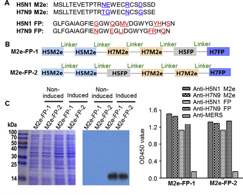
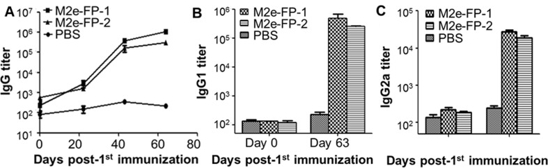
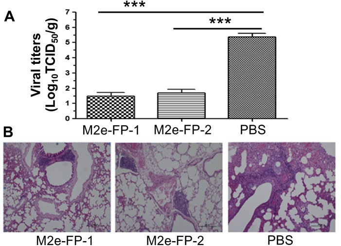
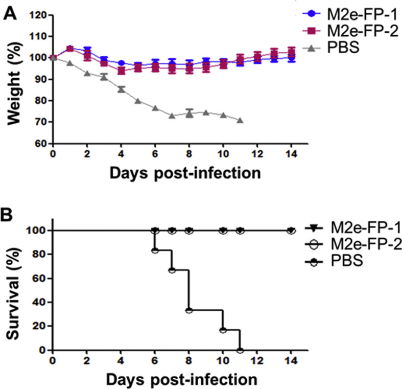
Highly pathogenic influenza viruses continue to cause serious threat to public health due to their pandemic potential, calling for an urgent need to develop effective, safe, convenient, and universal vaccines against influenza virus infection. In this study, we constructed two recombinant protein vaccines, 2H5M2e-2H7M2e-H5FP-H7FP (hereinafter M2e-FP-1) and 2H5M2e-H5FP-2H7M2e-H7FP (hereinafter M2e-FP-2), by respectively linking highly conserved sequences of two molecules of ectodomain of M2 (M2e) and one molecule of fusion peptide (FP) epitope of hemagglutinin (HA) of H5N1 and H7N9 influenza viruses in different orders. The Escherichia coli-expressed M2e-FP-1 and M2e-FP-2 proteins induced similarly high-titer M2e-FP-specific antibodies in the immunized mice. Importantly, both proteins were able to prevent lethal challenge of heterologous H1N1 influenza virus, with significantly reduced viral titers and alleviated pathological changes in the lungs, as well as increased body weight and complete survivals, in the challenge mice. Taken together, our study demonstrates that highly conserved M2e and FP epitope of HA of H5N1 and H7N9 influenza viruses can be used as important targets for development of safe and economical universal influenza vaccines, and that the position of H7N9 M2e and H5N1 HA epitope sequences in the vaccine components has no significant effects on the immunogenicity and efficacy of M2e-FP-based subunit vaccines.

Keywords: Hemagglutinin fusion peptide; Influenza virus; M2e; Protection; Universal vaccines.


Figures

Fig. 1
Fig. 1
Construction and expression of recombinant proteins. (A) Amino acid sequences of H5N1 and H7N9 M2e and FP of HA2 proteins. Amino acids with differences in M2e of H5N1 and H7N9 influenza viruses were shown in blue, and those in FP of H5N1 and H7N9 influenza viruses were in red. (B) Schematic structure of constructed M2e-FP-1 and M2e-FP-2 proteins. Linker sequences, GGGGS. (C) SDS-PAGE (left) and Western blot analysis (middle) of expressed M2e-FP-1 and M2e-FP-2 proteins. The protein molecular weight marker (kDa) is shown on the left. Anti-M2e antibodies (1:1000) were used for the Western blot detection. Shown on the right is the ELISA result for detection of the reactivity of M2e-FP-1 and M2e-FP-2 proteins with sera of mice immunized with M2e or FP peptides of H5N1 and H7N9 influenza viruses (1:2000). Sera of mice immunized with Middle East respiratory syndrome coronavirus (anti-MERS) were included as control.
Fig. 2
Fig. 2
Antibody responses in sera of mice immunized with M2e-FP-1 and M2e-FP-2 proteins. Mice were immunized with M2e-FP-1 and M2e-FP-2 proteins, or PBS as a control, and sera were collected at the indicated time points post-immunization to detect M2e-FP-specific IgG (A), IgG1 (B), and IgG2a (C) antibodies by ELISA. The antibody titers were expressed as the endpoint dilutions that remain positively detectable, and presented as mean ± SD of 5 mice in each group.
Fig. 3
Fig. 3
Viral titers and histopathological changes in lung tissues of challenged mice. The mice immunized with M2e-FP-1 and M2e-FP-2 proteins, respectively, or PBS as a control were challenged with A/PR/8/34(H1N1) influenza virus (103 TCID50), and collected for lung tissues at 5 days p.i. to detect viral titers (A) and histopathological changes (B). The data in (A) are presented as mean ± SD of 5 mice in each group. ***P < 0.001. For (B), Representative images from lung tissues of immunized mice and control mice are shown. The tissue sections were stained by H&E straining, and observed for pathological damages under light microscopy (10× magnification).
Fig. 4
Fig. 4
Survivals and weights of immunized mice challenged with influenza virus. The mice immunized with M2e-FP-1 and M2e-FP-2 proteins, respectively, or PBS as a control were challenged with A/PR/8/34(H1N1) influenza virus (103 TCID50), observed for 14 days p.i., and calculated for percentages of weights (A) and survivals (B). The data in (A) are presented as mean ± SD of 6 mice in each group.

Similar articles

Cited by

References

    1. Fineberg H.V. Pandemic preparedness and response – lessons from the H1N1 influenza of 2009. N Engl J Med. 2014;370:1335–1342. - PubMed
    1. Rewar S., Mirdha D., Rewar P. Treatment and prevention of pandemic H1N1 influenza. Ann Glob Health. 2015;81:645–653. - PubMed
    1. Zhou L., Tan Y., Kang M., Liu F., Ren R., Wang Y. Preliminary epidemiology of human infections with highly pathogenic avian influenza A(H7N9) virus, China, 2017. Emerg Infect Dis. 2017;23:1355–1359. - PMC - PubMed
    1. Wang X., Jiang H., Wu P., Uyeki T.M., Feng L., Lai S. Epidemiology of avian influenza A H7N9 virus in human beings across five epidemics in mainland China, 2013-17: an epidemiological study of laboratory-confirmed case series. Lancet Infect Dis. 2017;17:822–832. - PMC - PubMed
    1. Kang M., Lau E.H.Y., Guan W., Yang Y., Song T., Cowling B.J. Epidemiology of human infections with highly pathogenic avian influenza A(H7N9) virus in Guangdong, 2016 to 2017. Euro Surveill. 2017;22 - PMC - PubMed

Publication types

MeSH terms