. 2017 Jun 27;8(33):54966-54977.
doi: 10.18632/oncotarget.18629. eCollection 2017 Aug 15.

TGF-β2-induced ANGPTL4 expression promotes tumor progression and osteoclast differentiation in giant cell tumor of bone

Affiliations

TGF-β2-induced ANGPTL4 expression promotes tumor progression and osteoclast differentiation in giant cell tumor of bone

Bo Li et al. Oncotarget. .

Abstract

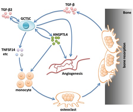
Although emerging studies have implicated that Aiopoietin-like 4 Protein (ANGPTL4) is related to the aggressiveness and metastasis of many tumors, the role of ANGPLT4 in giant cell tumor (GCT) of bone was rarely investigated. The mechanism of ANGPLT4 in tumor-induced osteoclastogenesis still remains unclear. In this study, we first demonstrated that ANGPTL4 was highly expressed in GCT compared to normal tissues, while we showed that TGF-β2 released by osteoclasts induced bone resorption could increase the expression of ANGPTL4 in GCTSCs. By using the luciferase reporter assay, we found that two downstreams of TGF-β2, Smad3 and Smad4, could directly activate the promoter of ANGPTL4, which might explain the mechanism of TGF-β2-induced ANGPLT4 expression. Moreover, knockout of ANGPTL4 by TALENs in GCTSCs inhibited tumor growth, angiogenesis and osteoclastogenesis in GCT in vitro. By using the chick chorio-allantoic membrane (CAM) models, we further showed that inhibition of ANGPTL4 suppressed tumor growth and giant cell formation in vivo. In addition, some new pathways involved in ANGPTL4 application were identified through microarray assay, which may partly explain the mechanism of ANGPTL4 in GCT. Taken together, our study for the first time identified the role of ANGPLT4 in GCT of bone, which may provide a new target for the diagnosis and treatment of GCT.

Keywords: ANGPTL4; angiogenesis; cell proliferation; giant cell tumor of bone; osteoclast differentiation.


Conflict of interest statement

CONFLICTS OF INTEREST All authors declared no conflicts of interest.

Figures

Figure 1
Figure 1. Expression profile of ANGPTL4 in GCT
(A) immunolocalization and H&E staining of ANGPTL4 in human specimens of GCT and para-tumor normal bone formalin-fixed-paraffin-embedded tissues. ANGPTL4 was marked with red arrows. (B and C) western blot analysis and qRT-PCR analysis of ANGPTL4 expression level in GCT samples and para-tumor normal bone tissues. (D and E) western blot analysis and qRT-PCR analysis of ANGPTL4 expression levels of GCTSCs and BMSCs. (F) immunofluorescence staining of ANGPTL4 in GCTSCs and BMSCs, ** p<0.01.
Figure 2
Figure 2. Expression profiles of Foxo1, HIF-1α, PPAR-α and PPAR-γ in GCT
(A-D) qRT-PCR analysis of Foxo1, HIF-1α, PPAR-α and PPAR-γ mRNA levels in GCTSCs and BMSCs. (E) western blot analysis of Foxo1, HIF-1α, PPAR-α and PPAR-γ expression levels in GCTSCs and BMSCs.
Figure 3
Figure 3. ANGPTL4 enhanced by highly expressed TGF-β2 in GCTSCs
(A) immunolocalization of TGF-β in human specimens of GCT and para-tumor normal bone formalin-fixed-paraffin-embedded tissues. (B-E) mRNA and protein levels of TGF-β1, TGF-β2 and TGF-β3 were determined by qRT-PCR and western blot analysis. (F) GCTSCs were treated with TGF-β2, and expression levels of ANGPTL4 were analyzed by western blotting over times as indicated. Actin was used as a protein loading control. (G) GCTSCs and BMSCs were exposed to TGF-β2 and its inhibitor (LY2109761) for 8 hours. ANGPTL4 levels were analyzed by western blotting. (H) the effect of Smad3, Smad4 and HIF-1α on luciferase activity in GCTSCs, * p<0.05, ** p<0.01.
Figure 4
Figure 4. Detailed experimental schematic diagram of ANGPTL4 knockout in GCTSCs (GCTSCsANG-/-) using TALENs method
Figure 5
Figure 5. ANGPTL4 promotes GCTSC induced osteoclast proliferation and angiogenesis in vitro
(A and B) RAW264.7 and BMM cells were treated with indicated concentration of ANGPTL4 for 5 days. The numbers of multinucleated osteoclasts were determined by TRAP staining (A) and the numbers of TRAP-positive multinucleated (>5 nuclei) osteoclasts were counted as treated with indicated concentration of ANGPTL4 in RAW264.7 and BMM cells (B). (C) TRAP staining of BMM cells cultured with conditional mediums of GCTSCsANG-/- and GCTSCsWT, respectively. (D) the numbers of TRAP-positive multinucleated (>5 nuclei) osteoclasts were counted. (E-G) qRT-PCR analysis of NFATC1, TRAP and CTSK expression in BMM cells. (H) rhodamine phalloidin staining of BMM cells from GCTSCsANG-/- and GCTSCsWT group, respectively. (I and J) CD31 staining of BMM cells and analyzing the number of vessel sprouting, * p<0.05.
Figure 6
Figure 6. Heat map and pathway analysis of differentially expressed genes in GCTSCs stimulated with or without recombinant ANGPTL4
Right: Microarray assays showed bone and tumor metabolism asscociated genens in recombinant ANGPTL4 stimulation group compared with control group. (Red box denoted the gene TNFSF14 which could increases osteoclastogenesis and decreases osteoblastogenesis) Left: Patheway analysis showed in GCTSCs stimulated with or without ANGPTL4-AP. (Red box denoted NF-kappa B signaling pathway which could activate mature osteoclasts.)
Figure 7
Figure 7. ANGPTL4 increases tumor proliferation and angiogenesis in vivo
(A) the tumor solute was seed into the plastic ring on the CAM with condition mediums of ANGPTL4 inhibitor or not, after 6 days of incubation. (B) the analysis of tumor sizes in two groups. (C and D) the CD31 staining and the assay of angiogenesis of tumor in two groups, * p<0.05.
Figure 8
Figure 8. ANGPTL4 acting as a critical cytokine modulates the biological cycle formation of GCT-bone microenvironment

References

    1. Zhou W, Yin H, Wang T, Liu T, Li Z, Yan W, Song D, Chen H, Chen J, Xu W, Yang X, Wu Z, Xiao J. MiR-126-5p regulates osteolysis formation and stromal cell proliferation in giant cell tumor through inhibition of PTHrP. Bone. 2014;66:267–276. - PubMed
    1. Xu W, Li X, Huang W, Wang Y, Han S, Chen S, Xu L, Yang X, Liu T, Xiao J. Factors affecting prognosis of patients with giant cell tumors of the mobile spine: retrospective analysis of 102 patients in a single center. Annals of surgical oncology. 2013;20:804–810. - PubMed
    1. Balke M, Schremper L, Gebert C, Ahrens H, Streitbuerger A, Koehler G, Hardes J, Gosheger G. Giant cell tumor of bone: treatment and outcome of 214 cases. Journal of cancer research and clinical oncology. 2008;134:969–978. - PubMed
    1. Balke M, Neumann A, Szuhai K, Agelopoulos K, August C, Gosheger G, Hogendoorn PC, Athanasou N, Buerger H, Hagedorn M. A short-term in vivo model for giant cell tumor of bone. BMC cancer. 2011;11:241. - PMC - PubMed
    1. Larsson SE, Lorentzon R, Boquist L. Giant-cell tumor of bone. A demographic, clinical, and histopathological study of all cases recorded in the Swedish Cancer Registry for the years 1958 through 1968. The Journal of bone and joint surgery American volume. 1975;57:167–173. - PubMed

LinkOut - more resources