. 2017 Jul 21;8(33):55280-55297.
doi: 10.18632/oncotarget.19424. eCollection 2017 Aug 15.

Development of zebrafish medulloblastoma-like PNET model by TALEN-mediated somatic gene inactivation

Affiliations

Development of zebrafish medulloblastoma-like PNET model by TALEN-mediated somatic gene inactivation

Jaegal Shim et al. Oncotarget. .

Abstract

Genetically engineered animal tumor models have traditionally been generated by the gain of single or multiple oncogenes or the loss of tumor suppressor genes; however, the development of live animal models has been difficult given that cancer phenotypes are generally induced by somatic mutation rather than by germline genetic inactivation. In this study, we developed somatically mutated tumor models using TALEN-mediated somatic gene inactivation of cdkn2a/b or rb1 tumor suppressor genes in zebrafish. One-cell stage injection of cdkn2a/b-TALEN mRNA resulted in malignant peripheral nerve sheath tumors with high frequency (about 39%) and early onset (about 35 weeks of age) in F0 tp53e7/e7 mutant zebrafish. Injection of rb1-TALEN mRNA also led to the formation of brain tumors at high frequency (58%, 31 weeks of age) in F0 tp53e7/e7 mutant zebrafish. Analysis of each tumor induced by somatic inactivation showed that the targeted genes had bi-allelic mutations. Tumors induced by rb1 somatic inactivation were characterized as medulloblastoma-like primitive neuroectodermal tumors based on incidence location, histopathological features, and immunohistochemical tests. In addition, 3' mRNA Quanti-Seq analysis showed differential activation of genes involved in cell cycle, DNA replication, and protein synthesis; especially, genes involved in neuronal development were up-regulated.

Keywords: MPNST; PNET; TALEN; medulloblastoma; somatic inactivation.


Conflict of interest statement

CONFLICTS OF INTEREST The authors declare no conflicts of interest.

Figures
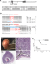
Figure 1
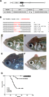
Figure 1. Somatic inactivation of cdkn2a/b gene by the injection of TALENs mRNA leads to MPNSTs in F0 founder zebrafish
(A) The targeted regions of two different TALENs (cdkn2a/b TALENs-1 and cdkn2a/b TALENs-2) are located in exon 1 of cdkn2a/b gene. The binding sequences of TALENs for cdkn2a/b gene are also shown. (B) By the injection of cdkn2a/b TALENs-1 mRNA, 7 different germline mutated alleles of cdkn2a/b gene were recovered at F1 generation. And 9 different alleles were also recovered at F1 generation after injection of cdkn2a/b TALENs-2 mRNA. (C) The large mass of tumor was observed in adjacent to eyes of the cdkn2a/b TALENs mRNA injected tp53 mutant zebrafish at 5.5 month post fertilization. (DF) Histological image of cross sections of tumor tissue was visualized with Hematoxylin-Eosin staining. (D) High magnification image of tumor tissue shows the herringbone pattern that is a typical feature of malignant peripheral nerve sheath tumor. (F) There is no evident abnormality in the neuroepithelial layer of retina adjacent to tumor tissue. (G) Kaplan-Meier survival representation of wild type (AB), tp53 mutant, and cdkn2a/b TALENs mRNA injected AB or tp53 mutant zebrafish. The cdkn2a/b TALENs mRNA injected tp53 mutant zebrafish had only started to exhibit death with tumor bearing as early as about 5 months. Other adult zebrafish such as tp53 mutant and cdkn2a/b TALENs mRNA injected AB zebrafish did not show early death with tumor bearing. All data was acquired from two independent experiments. Scale bars: 2 mm (E) and 100 μm (D and F).
Figure 2
Figure 2. Immunohistochemical analysis of tumors from cdkn2a/b TALENs mRNA injected tp53 mutant zebrafish
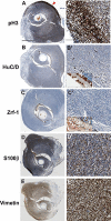
(A) (A’) Immunostaining with anti- phospho-Histone 3 antibidy. The tumor tissue from cdkn2a/b TALENs mRNA injected tp53 mutant zebrafish is highly mitotic. The phospho-Histone 3 positive mitotic cells were more evident in the outer region of tumor tissue (red arrowhead in A). (B) (B’) Neuronal signature that was visualized by immunostaining with anti-HuC/D antibody was scarcely detected in densely cellular region of tumor tissue. (C) (C’) Immunostaining with zrf-1 which detect zebrafish GFAP protein. The tumor tissue induced with cdkn2a/b TALENs mRNA also did not show any glial characteristics. Normal staining of anti-HuC/D and zrf-1 antibody could be observed in retinal layers near to tumor tissues (Inset box in B’ and C’). (D) (D’) Epithelioid MPNST marker, S100β, is strongly expressed in tumor tissue. (E) (E’) Mesenchymal marker, Vimentin, is also positively stained in tumor tissue. Scale bars: 1 mm (A) and 100 μm (A’).
Figure 3
Figure 3. Somatic inactivation of rb1 by the injection of rb1-TALENs mRNA leads to medulloblastoma like PNETs in F0 founder zebrafish
(A) The targeted region of rb1-TALENs is located in 1st exon of zebrafish rb1 locus. The binding sequences of TALENs for rb1 are indicated. (B) After the injection of rb1-TALENs mRNA, 4 different deletion and 1 insertion germline mutated alleles of rb1 were successfully recovered at F1 generation. Hence, 13 independent lines which have frameshift mutation in rb1 could be obtained. (CF) Morphological appearance of tumor bearing in head region of 5 month old zebrafish was presented. Protruding tumor mass were observed in rb1-TALENs mRNA injected zebrafish. The same aged wild type (C) and mutant (D–F) zebrafish are shown for comparison. (G) Kaplan-Meier survival representation of wild type, tp53 mutant, and rb1-TALENs injected wild type or tp53 mutant zebrafish. At about 6 month post fertilization, zebrafish which were injected with rb1-TALENs mRNA at one cell stage had started to die with tumors in the head region in wild type adult zebrafish. The tumor incidence which was induced by the injection of rb1-TALENs mRNA was more evident in tp53 mutant zebrafish. Adult zebrafish that showed abnormal swimming behavior started to die with tumor in head regions as early as about 20 weeks after injection of rb1-TALENs mRNA into one cell stage embryos.
Figure 4
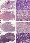
Figure 4. Histopathology of tumors from rb1-TALENs injected tp53 mutant zebrafish
(A) Sagittal section image of H & E staining of wild type zebrafish at 5 month post fertilization. Black arrows and arrow heads indicate forebrain and optic tectum, respectively. Cerebellum and cerebellar granule cells are indicated by white arrows and arrow heads, respectively. (B) High magnitude image of A. Normal cerebellar granule cells have a unique feature that is characterized with small rounded nuclei. (CH) Sagittal section images of H & E staining of tumors from rb1-TALENs injected tp53 mutant zebrafish. (C) Tumors were mainly arising in cerebellum, medulla, and brainstem. (D) High magnitude image of C. Highly cellular tumor cells with wedged nuclei were observed. Rosette like structures was marked by white arrows. (E) Tumors were mainly arising in cerebellum encompassed a majority of medulla, hypothalamus, and brainstem. The forth ventricle which were surrounded with tumor cells were marked by black arrows. (F) High magnitude image of E. Infiltration of tumor cells was indicated by black arrowheads in dorsal brainstem. (H) High magnitude image of G. Homer-Wright rosettes which are seen in PNETs or medulloblastomas were observed distinctly (white arrows). Anterior is left of all images. Scale bars: 500 μm (A, C, E and G), and 50 μm (B, D, F and H).
Figure 5
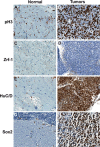
Figure 5. Immunohistochemical analysis of tumors from rb1-TALENs injected tp53 mutant zebrafish
Immunostaining was performed with lineage specific antibody with normal brain tissues (A, C, E, and G) and tumors from rb1-TALENs injected tp53 mutant zebrafish (B, D, F, and H) using anti phospho-Histone 3 (A and B), anti Zrf-1 (C and D), anti HuC/D (E and F) and anti Sox2 (G and H) antibodies. Highly mitotic tumor cells could be detected in tumor tissues from rb1-TALENs mRNA injected tp53 mutant zebrafish (B). Glial marker, Zrf-1 was scarcely stained in densely cellular region of tumor tissue (D). However, HuC/D antigen which is a postmitotic neuronal marker was strongly expressed in densely cellular region of tumor tissue (F). Neural progenitor marker, Sox2 expression was also detected in same cellular region of tumor tissue (H). Scale bars: 50 μm.
Figure 6
Figure 6. Mutational frequency and allele variation of rb1 or cdkn2a/b genes in tumor tissues derived by TALENs injected F0 adult zebrafish
(A) Mutational frequency of rb1 or cdkn2a/b genes was evaluated from sequencing analysis of cloned amplicon from genomic DNA of independent tumor tissues induced by rb1 or cdkn2a/b TALENs injected F0 tp53 mutant zebrafish. Mutational frequency of tumor tissues from rb1 TALENs injected F0 tp53 mutant zebrafish were varied from 66.6% (32/48) to 83.3% (40/48), and that of tumor tissues from cdkn2a/b TALENs injected F0 tp53 mutant zebrafish were varied from 81.3% (39/48) to 91.6% (44/48). (B) Mutation alleles of rb1's exon 1 in tumor tissues from rb1 TALENs injected F0 tp53 mutant zebrafish. (C) Mutation alleles of cdkn2a/b's exon 1 in tumor tissues from cdkn2a/b-TALENs1 or 2 injected F0 tp53 mutant zebrafish.
Figure 7
Figure 7. Gene ontology analysis of the differentially expressed genes in tumors induced by rb1 somatic inactivation
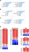
Gene Ontology Analysis was performed with the differentially expressed genes (DEG), after QuantSeq 3′ mRNA sequencing of tumors induced by rb1 somatic inactivation. (AC) -log10 (P value) was indicated at the top. (A) The up or down-regulated in biological processes. The cell cycle, DNA replication, cell division, and translation process were significantly up-regulated however ATP metabolism and vesicle/protein transport were down-regulated. (B) The up or down-regulated in cellular component. The nucleus and ribonucleoprotein complex were up-regulated. (C) The up or down-regulated in molecular function. (D) The featured DEGs in the neurogenesis, WNT, SHH, and NOTCH pathway. Red and blue represent up- and down-regulation of genes with fold change.
Figure 8
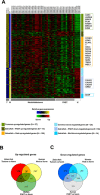
Figure 8. Comparative analysis between gene expression profiles of zebrafish tumors induced by rb1 somatic inactivation and public micro array data of human PNETs and medulloblastomas
(A) Heatmap of gene expression profiles. (T = Tumors, W = Wild type, and N = Normal tissue) The individual samples are indicated at the top of each column and the group of samples is indicated in the bottom. The representative genes are indicated in boxes at the left. (B, C). Venn diagrams showing differently expressed genes. (B) up-regulated and (C) down-regulated genes. (FDR < 0.05) (Norm; Normal sample, Med; medulloblastoma, PNET; primitive neuroectodermal tumor).

Similar articles

Cited by

References

    1. Gopalakrishnan V, Tao RH, Dobson T, Brugmann W, Khatua S. Medulloblastoma development: tumor biology informs treatment decisions. CNS Oncol. 2015;4:79–89. - PMC - PubMed
    1. Northcott PA, Jones DT, Kool M, Robinson GW, Gilbertson RJ, Cho YJ, Pomeroy SL, Korshunov A, Lichter P, Taylor MD, Pfister SM. Medulloblastomics: the end of the beginning. Nat Rev Cancer. 2012;12:818–834. - PMC - PubMed
    1. Goodrich LV, Milenkovic L, Higgins KM, Scott MP. Altered neural cell fates and medulloblastoma in mouse patched mutants. Science. 1997;277:1109–1113. - PubMed
    1. Gibson P, Tong Y, Robinson G, Thompson MC, Currle DS, Eden C, Kranenburg TA, Hogg T, Poppleton H, Martin J, Finkelstein D, Pounds S, Weiss A, et al. Subtypes of medulloblastoma have distinct developmental origins. Nature. 2010;468:1095–1099. - PMC - PubMed
    1. Kawauchi D, Robinson G, Uziel T, Gibson P, Rehg J, Gao C, Finkelstein D, Qu C, Pounds S, Ellison DW, Gilbertson RJ, Roussel MF. A mouse model of the most aggressive subgroup of human medulloblastoma. Cancer Cell. 2012;21:168–180. - PMC - PubMed