Review
. 2017 Sep 29;75(7):ftx093.
doi: 10.1093/femspd/ftx093.

Cellular and molecular mechanisms of viral infection in the human placenta

Affiliations
Review

Cellular and molecular mechanisms of viral infection in the human placenta

Moises León-Juárez et al. Pathog Dis. .

Abstract

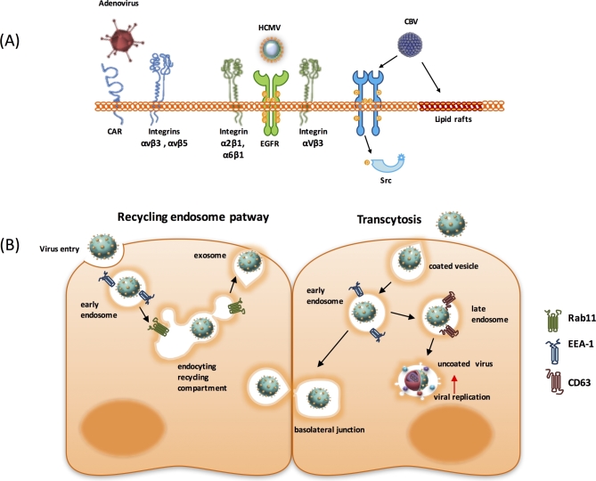
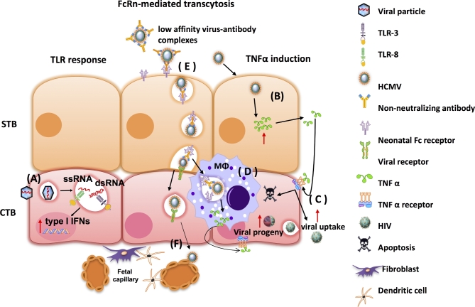
The placenta is a highly specialized organ that is formed during human gestation for conferring protection and generating an optimal microenvironment to maintain the equilibrium between immunological and biochemical factors for fetal development. Diverse pathogens, including viruses, can infect several cellular components of the placenta, such as trophoblasts, syncytiotrophoblasts and other hematopoietic cells. Viral infections during pregnancy have been associated with fetal malformation and pregnancy complications such as preterm labor. In this minireview, we describe the most recent findings regarding virus-host interactions at the placental interface and investigate the mechanisms through which viruses may access trophoblasts and the pathogenic processes involved in viral dissemination at the maternal-fetal interface.

Keywords: maternal–fetal interface; trophoblasts; vertical infection; viral entry; viral pathogenesis; viruses.


Figures

Figure 1.
Figure 1.
Graphical representation of the maternal–fetal interface. The human placenta is formed by chorionic villi, which can be classified into floating villi and anchoring villi. The first participates in the transport of nutrients, gases and waste between the fetus and the mother, and the anchoring villi is associated to the uterine wall forming a support structure between the mother and the fetus. The chorionic villus has a polarized epithelium of CTB anchored to a basal membrane. The floating villi is formed by CTB that fuse to form a group of multinucleated cells known as STB; whereas, the anchoring villi will be formed by aggregates of CTBs which organize into EVT. The EVT invades the maternal decidua and begins the replacement of the endothelial lineage that coats the uterine veins and arteries, inducing the formation of a hybrid of maternal and fetal circulation. Mφ/D = macrophage/dendritic cells; DC = dendritic cells.
Figure 2.
Figure 2.
Viral entry mechanisms in the maternal–fetal interface trophoblasts. (A) For viral entry, different cellular receptors are used. Viral fiber and penton protein of adenovirus interacts with CBV and CAR and integrins αvβ3 and αvβ5, respectively. HCMV entry is mediated by the epidermal growth factor receptor (EGFR) and co-receptors, like integrins αVβ3, α2β1 and α6β1. CBV uses lipid rafts during the entry to the trophoblast and Src tyrosin kinase family plays a key role in this process. (B) Transcytosis as a viral access route. Internalization process of HIV particles employs early endosomes (EEA-1 positive) and accumulate in late endosomes (CD63 positive). The destination could be the degradative pathway or the fusion within the late endosome compartment and beginning of viral replication.
Figure 3.
Figure 3.
Regulation of apoptosis in trophoblast during viral infecton. (A) Induction of apoptosis in trophoblast by HHV-8, HPV-16 and parvovirus B19. The HHV-8 induces DNA fragmentation and releases nucleosomal material in cytoplasm; the HPV-16 through oncoprotein E7 hijacks the RB protein and promotes its degradation by protesome. E5 oncoprotein of HPV, has function like viroporin these protein generates pores in membranes to result in stress osmotic and promoting the apoptosis process. NS1 protein of parvovirus activates the apoptosis by nuclear translocation of this protein and its association with DNA cellular and PARP protein. (B) Inhibition of apoptosis during trophoblast infection with HVB. This virus regulates apoptosis through protein X; this protein translocates to nucleus the infected cell and promotes the overexpression of molecules such as AKT and PI3K; this protein activates others proteins like cyclin D1 p-Smad-2 and 3, which are ways that could control the cell death in trophoblast infected with HBV.
Figure 4.
Figure 4.
Trophoblast immune response in a viral infection. (A) Single-strand RNA (ssRNA) or double-strand RNA (dsRNA), through TLR-8 and TLR-3, respectively, activated type I interferon pro-inflammatory response. (B) CTBs and STBs infected with HCMV can induce the expression and secretion of TNF-α. (C) Paracrine effect of TNF-α promotes the apoptotic mechanisms or a suitable environment for vertical infection. (D) Interaction between the macrophage (Mϕ) and the infected STB stimulates TNF-α secretion, which in turn acts on the infected STB activating viral replication. (E) Low-affinity virus-antibody complexes experience a process of transcytosis in the STB and can be transported to the basal membrane and establish contact with CTBs or captured by Mϕ placental. (F) Focal infection can extend to stromal fibroblasts, capillaries and fetal leukocytes.
Figure 5.
Figure 5.
Effects of viral infection during trophoblast invasion. Trophoblast cells infected with cytomegalovirus (HCMV), adenovirus (AAV-2), HPV and HBV have distinct effects in molecules and process related with trophoblast invasion. HCMV, adenovirus and AAV-2 induce for different mechanisms, low protein and activity levels of MMP-2 and MMP-9, and also, cytopathic effects that produce low invasion. In contrast, HPV and HBV through viral proteins E5, E6/E7 and HBV X protein, respectively, induce a reduction of protein levels of E-cadherin and high invasion. Also HBV X protein increase mesenchymal markers such as vimentin and N-cadherin.

Similar articles

Cited by

References

    1. Aldo P, You Y, Szigeti K et al. . HSV-2 enhances ZIKV infection of the placenta and induces apoptosis in first-trimester trophoblast cells. Am J Reprod Immunol 2016;76:348–57. - PubMed
    1. Aldo PB, Mulla MJ, Romero R et al. . Viral ssRNA induces first trimester trophoblast apoptosis through an inflammatory mechanism. Am J Reprod Immunol 2010;64:27–37. - PMC - PubMed
    1. Ambühl LM, Bændrup U, Dybkær K et al. . Human Papillomavirus infection as a posible cause of spontaneous abortion and spontaneous preterm delivery. Infect Dis Obstet Gynecol 2016;2016:1–19 - PMC - PubMed
    1. Amara A, Mercer J. Viral apoptotic mimicry. Nat Rev Microbiol 2015;13:461–9. - PMC - PubMed
    1. Aplin JD. The cell biological basis of human implantation. Baillieres Best Pract Res Clin Obstet Gynaecol 2000;14:757–64. - PubMed

Publication types