. 2017 Sep 13;14(1):186.
doi: 10.1186/s12974-017-0959-6.

Fluoxetine-enhanced autophagy ameliorates early brain injury via inhibition of NLRP3 inflammasome activation following subrachnoid hemorrhage in rats

Affiliations

Fluoxetine-enhanced autophagy ameliorates early brain injury via inhibition of NLRP3 inflammasome activation following subrachnoid hemorrhage in rats

Jian-Ru Li et al. J Neuroinflammation. .

Abstract

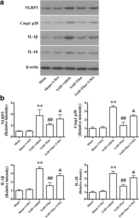
Background: The NLRP3 inflammasome is a multiprotein complex that regulates the innate immune inflammatory response by activating caspase-1 and subsequent IL-1β and IL-18. Fluoxetine has been shown to have the anti-inflammatory properties in many disease models. However, the effects and mechanisms of these effects of fluoxetine in early brain injury after subarachnoid hemorrhage (SAH) have not been defined.

Methods: The SAH model was induced by an endovascular perforation in adult male Sprague-Dawley (SD) rats weighing 300-320 g. N-Ac-Tyr-Val-Ala-Asp-chloromethyl ketone (AC-YVAD-CMK) was injected intraperitoneally (5 mg/kg) 1 h after SAH. Fluoxetine was administered via intravenous route 6 h after SAH. 3-Methyladenine (3-MA) was intracerebroventricularly injected 20 min before SAH. SAH grade, neurological function, brain water content, propidium iodide (PI) staining, western blot, double immunostaining, and transmission electron microscopy were performed.

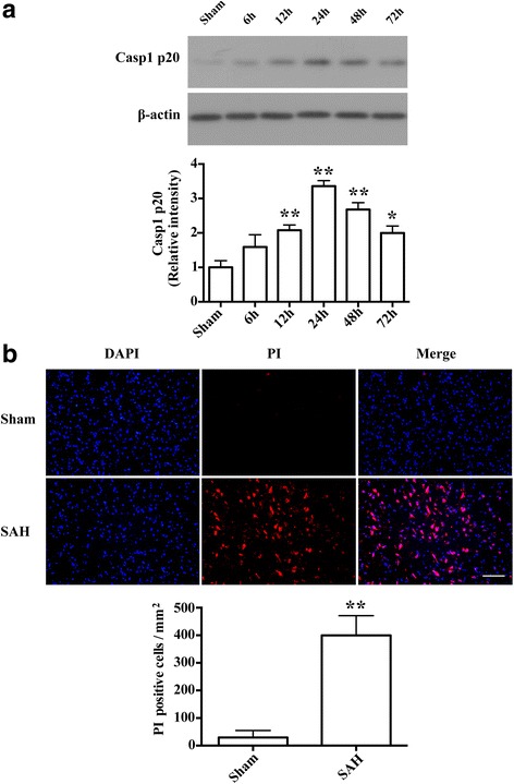
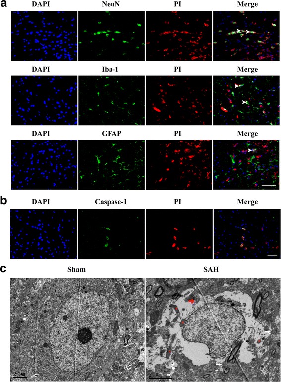
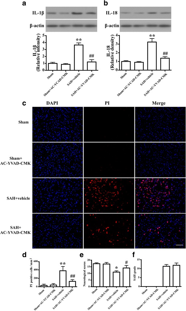
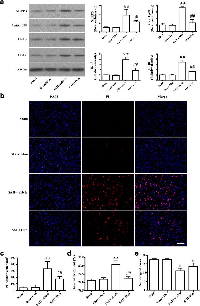
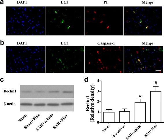
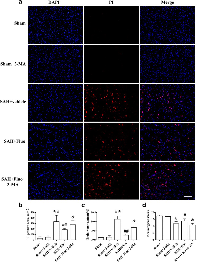
Results: Expression of caspase-1 increased and peaked at 24 h after SAH. Caspase activation was along with the increased necrotic cells, which occurred mainly in neurons. Necrotic cell death of microglia and astrocyte were also found. Administration of AC-YVAD-CMK, a caspase-1 inhibitor, reduced the expression of IL-1β and IL-18 and the number of PI-positive cells, attenuated brain edema, and improved neurological function, which was also observed in fluoxetine-treated rats. Furthermore, fluoxetine treatment significantly decreased the expression of NLRP3 and cleaved caspase-1 and upregulated the expression of beclin-1, a marker for autophagy. Finally, the effects of fluoxetine in NLRP3 inflammasome activation were reversed by additional 3-MA administration.

Conclusions: Together, our present study indicated that NLRP3 inflammasome and caspase-1 activation play a deleterious role in early brain injury and fluoxetine mitigates NLRP3 inflammasome and caspase-1 activation through autophagy activation after SAH, providing a potential therapeutic agent for SAH treatment.

Keywords: Autophagy; Early brain injury; Fluoxetine; Inflammation; NLRP3 inflammasome; Subarachnoid hemorrhage.


Conflict of interest statement

Ethics approval

All animal experiments were approved by the Institutional Animal Care and Use Committee of Zhejiang University.

Consent for publication

Not applicable

Competing interests

The authors declare that they have no competing interests.

Publisher’s Note

Springer Nature remains neutral with regard to jurisdictional claims in published maps and institutional affiliations.

Figures

Fig. 1
Fig. 1
Caspase-1 activation and necrotic cell death were induced in the early stages after SAH. a Representative western blot and the relative band densities of caspase-1 p20 in the ipsilateral cortex at different time points (6, 12, 24, 48, and 72 h) after SAH (n = 4/group). The bar represents the mean ± SD. *P < 0.05 vs sham, **P < 0.01 vs sham. b Representative photomicrographs and the quantification of the PI-positive neural cells in the ipsilateral cortex at 24 h after SAH (n = 3/group). Fluorescence colors: DAPI: blue and PI: red. Scale bar = 100 μm, the quantification of the PI-positive neural cells expressed as positive cells per square millimeter. The bar represents the mean ± SD. **P < 0.01 vs sham
Fig. 2
Fig. 2
Double staining for PI with NeuN (neuron marker), Iba-1(microglia marker), GFAP(astrocyte marker), and caspase-1, and ultrastructural changes of necrotic cells. a Double staining for PI with NeuN (neuron marker), Iba-1(microglia marker), and GFAP(astrocyte marker). Scale bar = 50 μm. b Double staining for PI with caspase-1 in the ipsilateral cortex at 24 h after SAH. Scale bar = 50 μm. c Ultrastructural changes of necrotic cells at 24 h after SAH. Neurons in the left panel appeared normal with nuclei, mitochondria, and cytomembranes. Neurons in the right panel showed fragmented nuclei (black asterisk), swollen mitochondria (red asterisk), disrupted cytomembranes (white arrow), damaged organelles (white arrow), and double-membrane autophagosomes (red arrow). Scale bar = 2 μm
Fig. 3
Fig. 3
Caspase-1 activation contributes to early brain injury after SAH. a Representative western blots and densitometric analyses showing levels of IL-1ß. The bar represents the mean ± SD. **P < 0.01 vs sham. ## P < 0.01 vs vehicle control. b Representative western blots and densitometric analyses showing levels of IL-18 (n = 6/group). The bar represents the mean ± SD. **P < 0.01 vs sham. ## P < 0.01 vs vehicle control. c Representative photomicrographs of PI staining of neural cells in different groups at 24 h after SAH (n = 3/group). Fluorescence colors: DAPI: blue and PI: red. Scale bar = 100 μm. d The quantification of the PI-positive neural cells. The quantification of the PI-positive neural cells expressed as positive cells per square millimeter. The bar represents the mean±SD. **P < 0.01 vs sham, ## P < 0.01 vs vehicle control. e Quantification of neurological scores (n = 9). The bars represent the mean with interquartile range. *P < 0.05 vs sham. # P < 0.05 vs vehicle control. f Quantification of SAH severity (n = 9). The bars represent the mean ± SD. No significant difference was observed between vehicle and SAH+AC-YVAD-CMK group
Fig. 4
Fig. 4
Fluoxetine reduced expression of NLRP3, caspase-1 p20, IL-1ß, and IL-18, decreased the numbers of necrotic cells and brain edema, and improved the neurological scores. a Representative Western blots and densitometric analyses of levels of NLRP3, caspase-1 p20, IL-1ß, and IL-18. The bar represents the mean ± SD. **P < 0.01 vs sham. # P < 0.05 vs vehicle control, ## P < 0.01 vs vehicle control. b Representative photomicrographs of PI-positive neural cells in different groups at 24 h after SAH (n = 3/group). Fluorescence colors: DAPI: blue and PI: red. Scale bar = 100 μm. c The quantification of the PI-positive neural cells. The quantification of the PI-positive neural cells expressed as positive cells per square millimeter. The bar represents the mean ± SD. **P < 0.01 vs sham, ## P < 0.01 vs vehicle control. d Quantification of brain water content (n = 6). The bars represent the mean ± SD. **P < 0.01 vs sham control, ## P < 0.01 vs vehicle control. e Quantification of neurological scores (n=18). The bars represent the mean with interquartile range. *P < 0.05 vs sham control, # P < 0.05 vs vehicle control
Fig. 5
Fig. 5
Autophagy activation was involved in the protective effect of fluoxetine on NLRP3 inflammasome activation. a Double fluorescence of PI staining (red) and cells with autophagy marker LC3 (green). b Representative micrographs showing double immunofluorescence with caspase-1 (red) and LC3 (green). Scale bar = 50 μm. c Representative Western blots and d densitometric analyses of levels of beclin-1 (n = 6). The bars represent the mean ± SD. *P < 0.05 vs sham control, # P < 0.05 vs vehicle control
Fig. 6
Fig. 6
Autophagy inhibitor 3-MA blocks the effects of fluoxetine on expression of NLRP3, caspase-1 p20, IL-1β, and IL-18 after SAH induction. a Representative Western blots of NLRP3, caspase-1 p20, IL-1β, and IL-18. b Densitometric analyses of levels of NLRP3, caspase-1 p20, IL-1β, and IL-18. N = 6. The bar represents the mean ± SD. **P < 0.01 vs the sham group. ## P < 0.01 vs SAH + vehicle group. & P < 0.05 vs SAH + fluo group. Representative photomicrographs and the quantification of the PI-positive neural cells in different groups at 24 h after SAH (n = 3/group). Fluorescence colors: DAPI: blue and PI: red. Scale bar = 100 μm, the quantification of the PI-positive neural cells expressed as a percent of the total DAPI+ cells. The bar represents the mean ± SD. **P < 0.01 vs SAH + vehicle group. ## P < 0.01 vs SAH + fluoxetine group
Fig. 7
Fig. 7
Autophagy inhibitor 3-MA abolishes the neuroprotective effects of fluoxetine on necrotic cell death, brain edema, and neurological function after SAH. a Representative photomicrographs of the PI-positive neural cells in different groups at 24 h after SAH (n = 3/group). Fluorescence colors: DAPI: blue and PI: red. Scale bar = 100 μm. b The quantification of the PI-positive neural cells expressed as positive cells per square millimeter. **P < 0.01 vs the sham group. ## P < 0.01 vs SAH + vehicle group. & P < 0.05 vs SAH + fluo group. c Quantification of brain water content (n = 6). The bars represent the mean ± SD. **P < 0.01 vs the sham group. ## P < 0.01 vs SAH + vehicle group. & P < 0.05 vs SAH + fluo group. d Quantification of neurological scores (n = 15/group). The bars represent the mean with interquartile range. *P < 0.05 vs the sham group. # P < 0.05 vs SAH + vehicle group. & P < 0.05 vs SAH + fluo group

References

    1. Connolly ES, Jr, Rabinstein AA, Carhuapoma JR, Derdeyn CP, Dion J, Higashida RT, Hoh BL, Kirkness CJ, Naidech AM, Ogilvy CS, et al. Guidelines for the management of aneurysmal subarachnoid hemorrhage: a guideline for healthcare professionals from the American Heart Association/american Stroke Association. Stroke. 2012;43:1711–1737. doi: 10.1161/STR.0b013e3182587839. - DOI - PubMed
    1. Nieuwkamp DJ, Setz LE, Algra A, Linn FH, de Rooij NK, Rinkel GJ. Changes in case fatality of aneurysmal subarachnoid haemorrhage over time, according to age, sex, and region: a meta-analysis. Lancet Neurol. 2009;8:635–642. doi: 10.1016/S1474-4422(09)70126-7. - DOI - PubMed
    1. Macdonald RL, Higashida RT, Keller E, Mayer SA, Molyneux A, Raabe A, Vajkoczy P, Wanke I, Bach D, Frey A, et al. Clazosentan, an endothelin receptor antagonist, in patients with aneurysmal subarachnoid haemorrhage undergoing surgical clipping: a randomised, double-blind, placebo-controlled phase 3 trial (CONSCIOUS-2) Lancet Neurol. 2011;10:618–625. doi: 10.1016/S1474-4422(11)70108-9. - DOI - PubMed
    1. Macdonald RL, Higashida RT, Keller E, Mayer SA, Molyneux A, Raabe A, Vajkoczy P, Wanke I, Bach D, Frey A, et al. Randomized trial of clazosentan in patients with aneurysmal subarachnoid hemorrhage undergoing endovascular coiling. Stroke. 2012;43:1463–1469. doi: 10.1161/STROKEAHA.111.648980. - DOI - PubMed
    1. Chen S, Feng H, Sherchan P, Klebe D, Zhao G, Sun X, Zhang J, Tang J, Zhang JH. Controversies and evolving new mechanisms in subarachnoid hemorrhage. Prog Neurobiol. 2014;115:64–91. doi: 10.1016/j.pneurobio.2013.09.002. - DOI - PMC - PubMed

MeSH terms