

Shape, form, function and Leishmania pathogenicity: from textbook descriptions to biological understanding
- PMID: 28903998
- PMCID: PMC5627057
- DOI: 10.1098/rsob.170165
Shape, form, function and Leishmania pathogenicity: from textbook descriptions to biological understanding
Erratum in
-
Correction to 'Shape, form, function and Leishmania pathogenicity: from textbook descriptions to biological understanding'.Open Biol. 2018 Aug;8(8):180134. doi: 10.1098/rsob.180134. Open Biol. 2018. PMID: 30158322 Free PMC article. No abstract available.
Abstract
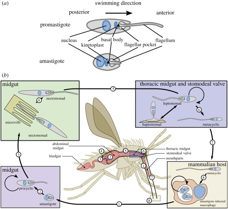
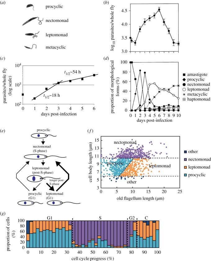
The shape and form of protozoan parasites are inextricably linked to their pathogenicity. The evolutionary pressure associated with establishing and maintaining an infection and transmission to vector or host has shaped parasite morphology. However, there is not a 'one size fits all' morphological solution to these different pressures, and parasites exhibit a range of different morphologies, reflecting the diversity of their complex life cycles. In this review, we will focus on the shape and form of Leishmania spp., a group of very successful protozoan parasites that cause a range of diseases from self-healing cutaneous leishmaniasis to visceral leishmaniasis, which is fatal if left untreated.
Keywords: Leishmania; morphology; parasite; pathogenicity.
© 2017 The Authors.
Conflict of interest statement
The authors declare they have no competing interests.
Figures
References
-
- Matthews KR. 2011. Controlling and coordinating development in vector-transmitted parasites. Science 331, 1149–1153. (doi:10.1126/science.1198077) - DOI - PMC - PubMed
-
- Bates PA. 1994. Complete developmental cycle of Leishmania mexicana in axenic culture. Parasitology 108, 1–9. (doi:10.1017/S0031182000078458) - DOI - PubMed
-
- Shapiro SZ, Naessens J, Liesegang B, Moloo SK, Magondu J. 1984. Analysis by flow cytometry of DNA synthesis during the life cycle of African trypanosomes. Acta Trop. 41, 313–323. - PubMed
-
- Sinden RE, Canning EU, Bray RS, Smalley ME. 1978. Gametocyte and gamete development in Plasmodium falciparum. Proc. R. Soc. Lond. B 201, 375–399. (doi:10.1098/rspb.1978.0051) - DOI - PubMed
-
- Aleman C. 1969. Finestructure of cultured Leishmania brasiliensis. Exp. Parasitol. 24, 259–264. (doi:10.1016/0014-4894(69)90163-5) - DOI - PubMed
Publication types
MeSH terms
Grants and funding
LinkOut - more resources
Full Text Sources
Other Literature Sources
Miscellaneous