. 2017 Sep;5(17):e13437.
doi: 10.14814/phy2.13437.

Assessing the effects of mitofusin 2 deficiency in the adult heart using 3D electron tomography

Affiliations

Assessing the effects of mitofusin 2 deficiency in the adult heart using 3D electron tomography

Siavash Beikoghli Kalkhoran et al. Physiol Rep. 2017 Sep.

Abstract

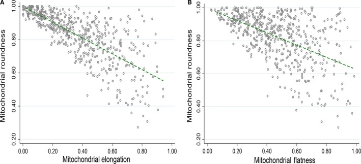
The effects of mitofusin 2 (MFN2) deficiency, on mitochondrial morphology and the mitochondria-junctional sarcoplasmic reticulum (jSR) complex in the adult heart, have been previously investigated using 2D electron microscopy, an approach which is unable to provide a 3D spatial assessment of these imaging parameters. Here, we use 3D electron tomography to show that MFN2-deficient mitochondria are larger in volume, more elongated, and less rounded; have fewer mitochondria-jSR contacts, and an increase in the distance between mitochondria and jSR, when compared to WT mitochondria. In comparison to 2D electron microscopy, 3D electron tomography can provide further insights into mitochondrial morphology and the mitochondria-jSR complex in the adult heart.

Keywords: 3D electron tomography; MFN2 KO; mitochondria–jSR junction.


Figures

Figure 1
Figure 1
3D reconstruction of adult cardiac mitochondria and adjacent SR network. (A) TEM depicts intermyofibrillar mitochondria and adjacent SR network in adult mouse WT heart. (B) The 3D reconstruction of the jSR and T‐tubule network (shown by blue and green color, respectively) as well as their neighboring mitochondria (shown by different intensity of red color).
Figure 2
Figure 2
Measuring the minimum mitochondria–jSR distance. (A) TEM micrograph depicts spatial interaction between intermyofibrillar mitochondria and adjacent jSR and T‐tubule network in adult mouse WT heart (white arrow indicates mitochondria–jSR space without measurement). (B) The perpendicular lines that were drawn between the jSR and one mitochondrion to measure the minimum distance between these two organelles (white arrow indicates the perpendicular lines drawn on mitochondria–jSR space). (C) 3D reconstruction depicts the minimum distances (black arrows) between the mitochondrion (red) and jSR (green) along the interface between these two organelles (yellow arrow). The T‐tubule is represented in blue.
Figure 3
Figure 3
Morphometric parameters of WT and MFN2 KO cardiac mitochondria. This figure shows that MFN2 KO mitochondria have increased 3D volume (A), are equally elongated (B), are less rounded (C), and are flatter (D) when compared to WT mitochondria. Values are mean and confidence interval (CI); 317 WT and 219 MFN2 KO mitochondria (N = 5 WT and MFN2 KO mice). (*, **, and *** denote P ≤ 0.05, P < 0.001, and P < 0.0001, respectively).
Figure 4
Figure 4
Correlation between mitochondrial morphometric parameters. There was a negative correlation between (A) elongation and roundness and (B) flatness and roundness; 317 WT and 219 MFN2 KO mitochondria (N = 5 WT and MFN2 KO mice).
Figure 5
Figure 5
Mitochondria–jSR interaction in WT and MFN2 KO hearts. (A) There was an increase in the minimum distance between mitochondria and jSR in MFN2 KO heart when compared to WT hearts. (B) There was no significant difference in the length of the interface between mitochondria and jSR in WT or MFN2 KO hearts. (C) There were fewer mitochondria–jSR networks in MFN2 KO hearts when compared to WT ones (N = 5 WT and MFN2 KO mice).

Similar articles

Cited by

References

    1. Boncompagni, S. , Rossi A. E., Micaroni M., Hamilton S. L., Dirksen R. T., Franzini‐Armstrong C., et al. 2009. Characterization and temporal development of cores in a mouse model of malignant hyperthermia. Proc. Natl Acad. Sci. USA 106:21996–22001. - PMC - PubMed
    1. Boyce, R. W. , Dorph‐Petersen K.‐A., Lyck L., and Gundersen H. J. G.. 2010. Design‐based stereology: introduction to basic concepts and practical approaches for estimation of cell number. Toxicol. Pathol. 38:1011–1025. - PubMed
    1. de Brito, O. M. , and Scorrano L.. 2008. Mitofusin 2 tethers endoplasmic reticulum to mitochondria. Nature 456:605–610. - PubMed
    1. Cartoni, R. , and Martinou J.‐C.. 2009. Role of mitofusin 2 mutations in the physiopathology of Charcot‐Marie‐Tooth disease type 2A. Exp. Neurol. 218:268–273. - PubMed
    1. Chen, Y. , and Dorn G. W.. 2013. PINK1‐phosphorylated mitofusin 2 is a Parkin receptor for culling damaged mitochondria. Science 340:471–475. - PMC - PubMed

MeSH terms

LinkOut - more resources