. 2017 Sep 13;37(37):9086-9099.
doi: 10.1523/JNEUROSCI.1453-17.2017. Epub 2017 Aug 3.

After Nerve Injury, Lineage Tracing Shows That Myelin and Remak Schwann Cells Elongate Extensively and Branch to Form Repair Schwann Cells, Which Shorten Radically on Remyelination

Affiliations

After Nerve Injury, Lineage Tracing Shows That Myelin and Remak Schwann Cells Elongate Extensively and Branch to Form Repair Schwann Cells, Which Shorten Radically on Remyelination

Jose A Gomez-Sanchez et al. J Neurosci. .

Abstract

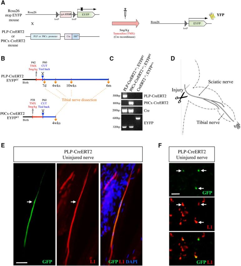
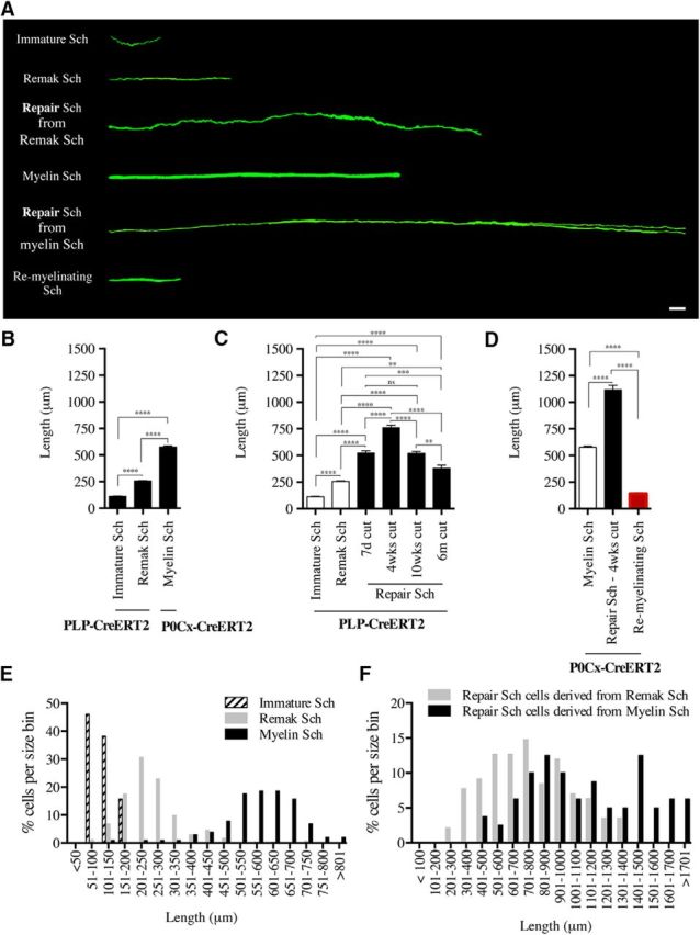
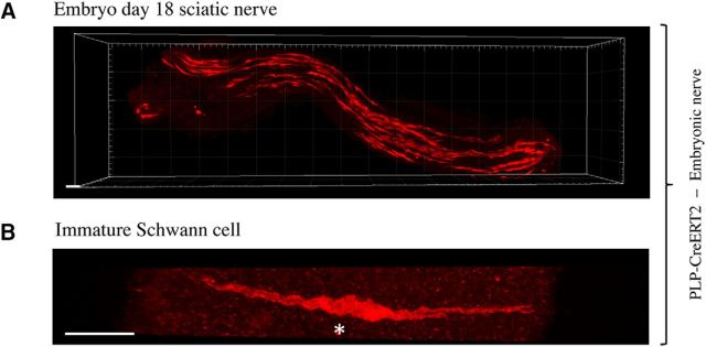
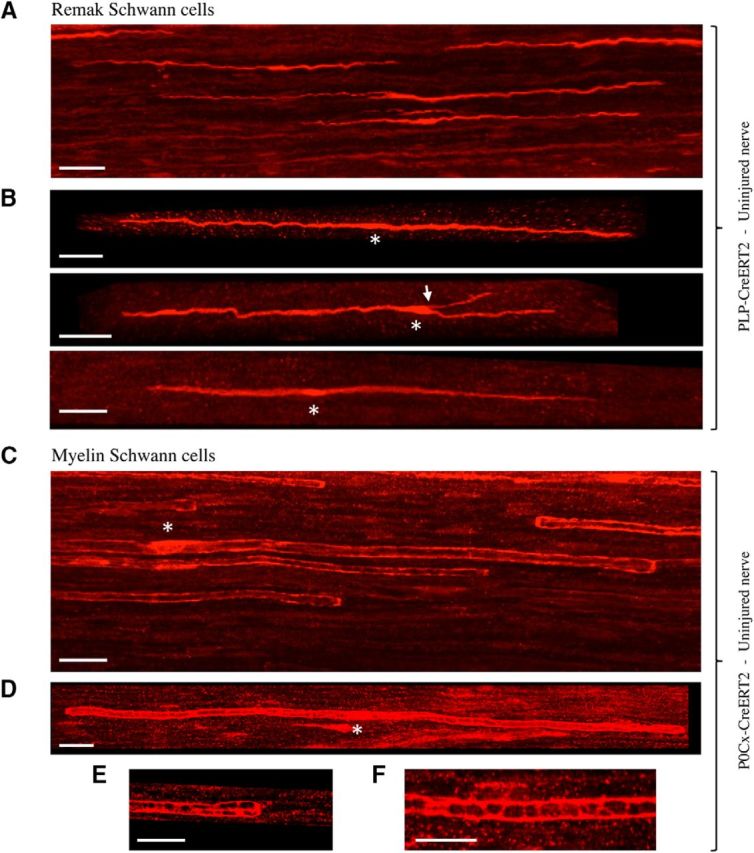
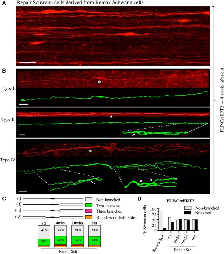
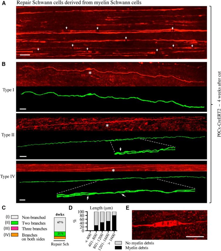
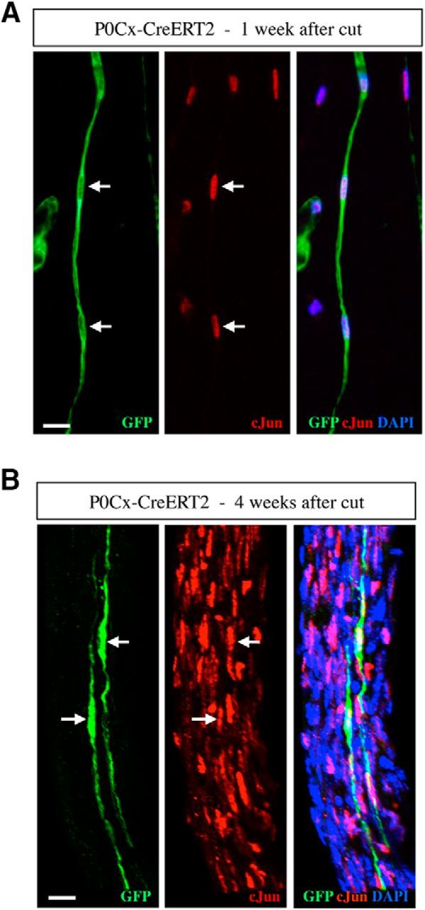
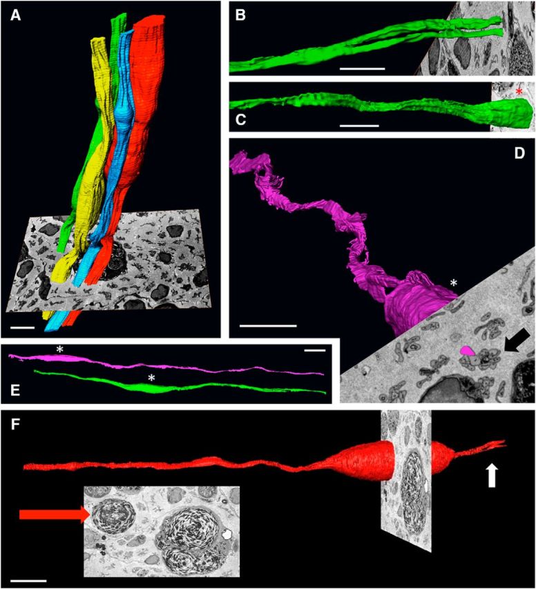
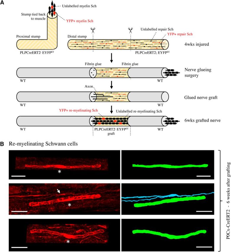
There is consensus that, distal to peripheral nerve injury, myelin and Remak cells reorganize to form cellular columns, Bungner's bands, which are indispensable for regeneration. However, knowledge of the structure of these regeneration tracks has not advanced for decades and the structure of the cells that form them, denervated or repair Schwann cells, remains obscure. Furthermore, the origin of these cells from myelin and Remak cells and their ability to give rise to myelin cells after regeneration has not been demonstrated directly, although these conversions are believed to be central to nerve repair. Using genetic lineage-tracing and scanning-block face electron microscopy, we show that injury of sciatic nerves from mice of either sex triggers extensive and unexpected Schwann cell elongation and branching to form long, parallel processes. Repair cells are 2- to 3-fold longer than myelin and Remak cells and 7- to 10-fold longer than immature Schwann cells. Remarkably, when repair cells transit back to myelinating cells, they shorten ∼7-fold to generate the typically short internodes of regenerated nerves. The present experiments define novel morphological transitions in injured nerves and show that repair Schwann cells have a cell-type-specific structure that differentiates them from other cells in the Schwann cell lineage. They also provide the first direct evidence using genetic lineage tracing for two basic assumptions in Schwann cell biology: that myelin and Remak cells generate the elongated cells that build Bungner bands in injured nerves and that such cells can transform to myelin cells after regeneration.SIGNIFICANCE STATEMENT After injury to peripheral nerves, the myelin and Remak Schwann cells distal to the injury site reorganize and modify their properties to form cells that support the survival of injured neurons, promote axon growth, remove myelin-associated growth inhibitors, and guide regenerating axons to their targets. We show that the generation of these repair-supportive Schwann cells involves an extensive cellular elongation and branching, often to form long, parallel processes. This generates a distinctive repair cell morphology that is favorable for the formation of the regeneration tracks that are essential for nerve repair. Remyelination, conversely, involves a striking cell shortening to form the typical short myelin cells of regenerated nerves. We also provide evidence for direct lineage relationships between: (1) repair cells and myelin and Remak cells of uninjured nerves and (2) remyelinating cells in regenerated nerves.

Keywords: PNS; Schwann cell; injury; nerve; regeneration; remyelination.


Figures

Figure 1.
Figure 1.
Mouse generation, experimental outline, and cell-type specificity of genetic labeling. A, Generation of PLP-Cre ERT2xRosa26stopEYFP and P0Cx-Cre ERT2xRosa26stopEYFP mice. B, Outline of lineage-tracing experiments involving tamoxifen injection of uninjured mice followed by nerve transection without reinnervation. C, PCR analysis showing the presence of the expected PCR products in the mouse lines indicated. D, Diagram showing the experimental nerves and the site of nerve transection. E, F, In uninjured nerves of tamoxifen-treated PLP-Cre ERT2xRosa26stopEYFP mice, GFP expression is essentially restricted to Remak cells. E, Double immunolabeling of a teased nerve with GFP and L1 antibodies. Middle, Schwann cells in a Remak bundle identified by L1 expression. One of the cells expresses GFP (left). Arrow indicates the nucleus. F, Double immunolabeling of transverse cryostat sections using GFP and L1 antibodies. The three GFP-positive cells (top) also express L1 (middle). Scale bars: E, 50 μm; F, 50 μm.
Figure 2.
Figure 2.
Synopsis of the appearance and length of Schwann cells in developing, uninjured, injured, and remyelinating nerves. A, Direct comparison of the cell types visualized in the present work, showing representative examples of each cell. All cells are magnified equally. Scale bar, 30 μm. B, Length of immature, Remak, and myelin Schwann cells in developing and adult uninjured nerves. Measurements of immature and Remak cells are from PLP-Cre ERT2xRosa26stopEYFP mice. Measurements of myelin cells are from P0Cx-Cre ERT2xRosa26stopEYFP mice. Immature cells, n = 89; Remak cells, n = 440; myelin cells, n = 102. ****p < 0.0001, one-way ANOVA, Tukey's comparison. C, Length of denervated Schwann cells (repair cells) derived from Remak cells. The graph shows cell length at different times after injury without reinnervation. Measurements are from PLP-Cre ERT2xRosa26stopEYFP mice. The length of immature and Remak cells in uninjured nerves of the same mouse line is shown for ease of comparison (white columns). Note that, at 4 weeks, repair cells are several-fold longer than immature and Remak cells and that long-term denervation is accompanied by a significant reduction in cell length. Seven-day cut, n = 121; 4-week cut, n = 142; 10-week cut, n = 136; 6-month cut, n = 29. **p < 0.01; ***p < 0.001; ****p < 0.0001, one-way ANOVA, Tukey's comparison. D, Length of repair cells derived from myelin cells in 4-week cut nerves and the length of myelin cells that have remyelinated regenerated axons and are derived from repair cells in the same mouse line, P0Cx-Cre ERT2xRosa26stopEYFP. The length of myelin cells before injury is shown for ease of comparison (white columns). Repair cells, n = 80; remyelinating cells, n = 62. ****p < 0.0001, one-way ANOVA, Tukey's comparison. E, Size distribution of immature Schwann cells (Sch), Remak Schwann cells, and myelin Schwann cells in uninjured nerves. F, Size distribution of repair cells derived from Remak Schwann cells and from myelin Schwann cells.
Figure 3.
Figure 3.
Immature Schwann cells in uninjured nerves. A, Low-magnification view of a nerve segment from an E18 embryo of a PLP-Cre ERT2xRosa26stopEYFP mouse showing numerous immature Schwann cells distributed along the nerve. Scale bar, 30 μm. B, Single E18 Schwann cell showing a typical wavy structure. Asterisk indicates the position of the nucleus. Scale bar, 30 μm.
Figure 4.
Figure 4.
Remak and myelin Schwann cells in uninjured adult nerves. A, Overview showing several scattered Remak cells in the tibial nerve of a PLP-Cre ERT2xRosa26stopEYFP mouse. The cells often have a wavy appearance. Scale bar, 30 μm. B, Higher-magnification view illustrating two typical Remak cells (top and bottom) and a Remak cell with a short branch (arrow, middle). Asterisks indicate position of the nucleus. Scale bar, 30 μm. C, Overview showing a part of a number of myelin cells in the tibial nerve of a P0Cx-Cre ERT2xRosa26stopEYFP mouse. Asterisk indicates position of the nucleus. The cells have a straight appearance and the blunt ends of myelin cells, heminodes, are seen clearly (arrows). As expected, the labeling is strongest in the outer cytoplasmic compartment (longitudinal Cajal bands), generating a railway track appearance. Scale bar, 30 μm. D, Whole myelin cell with heminodes visible at both ends (arrows). Longitudinal Cajal bands are clearly seen, but transverse Cajal bands are indistinct. Asterisk indicates the position of a nucleus. Scale bar, 30 μm. E, Closer view of the end of a myelin cells showing longitudinal and transverse Cajal bands at a heminode. Scale bar, 20 μm. F, Longitudinal and transverse Cajal bands near the middle of a myelin cell. Scale bar, 20 μm.
Figure 5.
Figure 5.
Remak-derived repair cells. A, Overview showing several repair cells derived from Remak cells in 4-week cut nerve of a PLP-Cre ERT2xRosa26stopYFP mouse. Scale bar, 30 μm. B, Three examples of repair cells. Under each confocal image is an artificially colored (green) trace image of the same cell. Asterisks indicate the position of the nucleus. The cells are very narrow and elongated and have an undulating structure with occasional straighter stretches (e.g., type 2; middle). Top, Unbranched cell (type 1). Middle, Cell with two branches lying very close to each other on one side of the nucleus (type 2; enlarged part indicated by interrupted lines; arrow indicates the branch point). The two branches overlap toward the end of the cell. Bottom, Cell with long and irregularly curved branches on both sides of the nucleus (type 4; enlarged part indicated by interrupted lines; arrows indicate branch points). Scale bar, 30 μm. C, Schematic drawings show a classification of repair cells depending on the presence of processes and their disposition. The box chart shows the frequency of each repair cell type in the total population of Remak-derived repair cells (as a percentage of total cells) at different times after injury. D, Total percentage of branched cells among Remak cells and repair cells at different times after injury. Note that branching does not change significantly with time.
Figure 6.
Figure 6.
Myelin-derived repair cells. A, Overview showing parts of a number of repair cells derived from myelin cells in 4-week cut nerve of a P0Cx-Cre ERT2xRosa26stopEYFP mouse. Some cells show swellings, which represent myelin debris and exhibit partial disruption of the fluorescence signal (examples indicated with arrows; see also D, E). Scale bar, 30 μm. B, Three examples of repair cells derived from myelin cells. Under each confocal image is an artificially colored (green) trace image of the same cell. Asterisks indicate the position of the nucleus. The cells are extremely long and show a general appearance that is similar to that of Remak-derived cells. Top, Unbranched cell (type 1; see Fig. 5C). Middle, Cell with two branches on one side of the nucleus (type 2; enlarged part indicated by interrupted lines; arrow indicates the branch-point). Bottom, Cell forming branches on both sides of the nucleus (type 4; enlarged part indicated by interrupted lines; arrows indicate branch points). In both of the branched cells, the branches appear to be separate along a part of the cells only. This is caused by the two closely apposed processes overlapping at this angle of representation. Scale bar, 30 μm. C, Frequency of each repair cell type in 4-week cut nerves. D, Percentage of cells with myelin debris in different length groups of repair cells in 4-week cut nerves. Percentage of myelin-cell-derived repair cells that still contain undigested myelin increases with increased cell length. E, Cose view of a swelling caused by undigested intracellular myelin debris. Scale bar, 20 μm.
Figure 7.
Figure 7.
GFP-labeled repair cells express high levels of c-Jun. A, B, Teased nerve preparations made from 1- and 4-week cut nerves of tamoxifen-treated P0Cx-Cre ERT2xRosa26stopEYFP mice. The nerves are double immunolabeled with GFP and c-Jun antibodies. A, Column (Bungner's band) of GFP-positive repair cells (left) 1 week after cut. The nuclei (arrows) are c-Jun-positive (middle). B, Two GFP-positive repair cells (left). The nuclei (arrows) are c-Jun-positive (middle).
Figure 8.
Figure 8.
Block-face-scanning electron microscopy. A, Tracing of whole Bungner's bands showing parts of four irregular cellular columns running longitudinally along the tibial nerve. Shown also is a 2D image of the block face from which they were generated. This shows transverse sections of several Bungner's bands, some of which cut through Schwann cell nuclei (arrow). Part of a myelin-containing macrophage can be seen behind the colored Bungner's bands. Scale bar, 10 μm. B, Tracing of a single repair cell showing the point at which the cell branches, each branch lying in a separate Bungner's band. Scale bar, 10 μm. C, D, Part of two cells illustrating the spiraling, corkscrew-like structure typical of repair cells. In C, the Bungner's band to which the cell belongs is seen in the 2D image of the block face (arrow). Scale bar, 10 μm. E, Parts of two repair cells illustrating their undulating appearance. These cells belong to two neighboring Bungner's bands (view of the block face not shown). Scale bar, 10 μm. F, Part of a repair cells containing a myelin lump. The scanning block face is shown face-on in the inset. The large red arrow points to the undigested myelin in the Schwann cell. A large macrophage also containing myelin is seen to the right of the Schwann cell. Small white arrow points to the point where the repair cell is starting to branch. Scale bar, 10 μm.
Figure 9.
Figure 9.
Remyelinating Schwann cells derive from repair cells. A, Outline of nerve grafting experiments. B, Three examples of the typically short remyelinating cells. These cells have formed myelin around axons in a 6-week nerve graft. Asterisks indicate position of the nucleus. Right, Artificially colored trace image of the same cells. The myelin cell in the middle panels lies close to a branched repair cell (arrow; blue), which has not converted to a myelin cell. Scale bar, 30 μm.

References

    1. Arthur-Farraj PJ, Latouche M, Wilton DK, Quintes S, Chabrol E, Banerjee A, Woodhoo A, Jenkins B, Rahman M, Turmaine M, Wicher GK, Mitter R, Greensmith L, Behrens A, Raivich G, Mirsky R, Jessen KR (2012) c-Jun reprograms Schwann cells of injured nerves to generate a repair cell essential for regeneration. Neuron 75:633–647. 10.1016/j.neuron.2012.06.021 - DOI - PMC - PubMed
    1. Atanasoski S, Shumas S, Dickson C, Scherer SS, Suter U (2001) Differential cyclin D1 requirements of proliferating Schwann cells during development and after injury. Mol Cell Neurosci 18:581–592. 10.1006/mcne.2001.1055 - DOI - PubMed
    1. Atanasoski S, Boentert M, De Ventura L, Pohl H, Baranek C, Beier K, Young P, Barbacid M, Suter U (2008) Postnatal Schwann cell proliferation but not myelination is strictly and uniquely dependent on cyclin-dependent kinase 4 (cdk4). Mol Cell Neurosci 37:519–527. 10.1016/j.mcn.2007.11.005 - DOI - PubMed
    1. Benito C, Davis CM, Gomez-Sanchez JA, Turmaine M, Meijer D, Poli V, Mirsky R, Jessen KR (2017) STAT3 controls the long-term survival and phenotype of repair Schwann cells during nerve regeneration. J Neurosci 37:4255–4269. 10.1523/JNEUROSCI.3481-16.2017 - DOI - PMC - PubMed
    1. Boerboom A, Dion V, Chariot A, Franzen R (2017) Molecular mechanisms involved in Schwann cell plasticity. Front Mol Neurosci 10:38. 10.3389/fnmol.2017.00038 - DOI - PMC - PubMed

Publication types