. 2017 Sep 13;7(1):11466.
doi: 10.1038/s41598-017-11523-3.

Rapid Discovery of De Novo Deleterious Mutations in Cattle Enhances the Value of Livestock as Model Species

Affiliations

Rapid Discovery of De Novo Deleterious Mutations in Cattle Enhances the Value of Livestock as Model Species

E Bourneuf et al. Sci Rep. .

Abstract

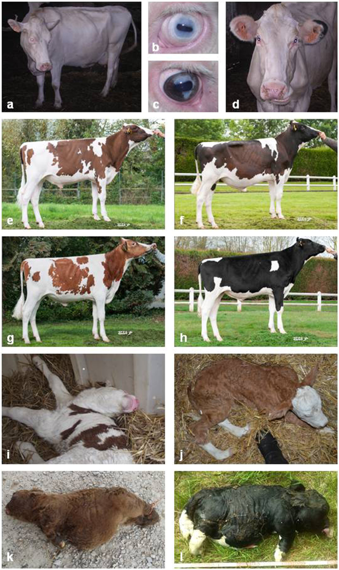
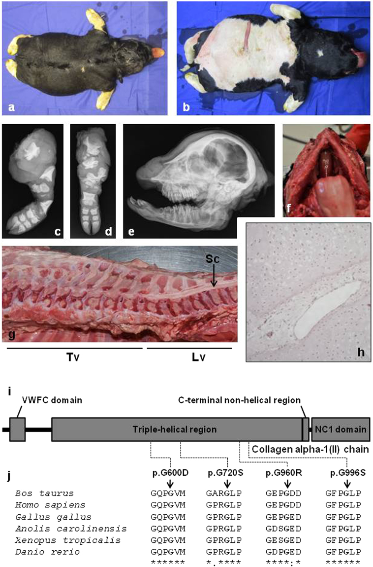
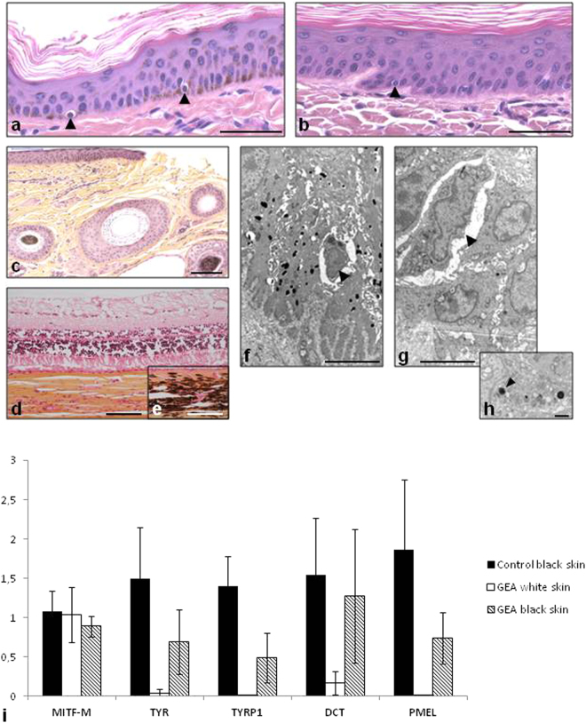
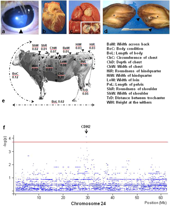
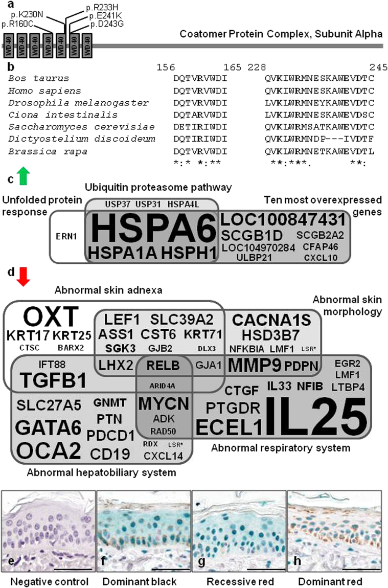
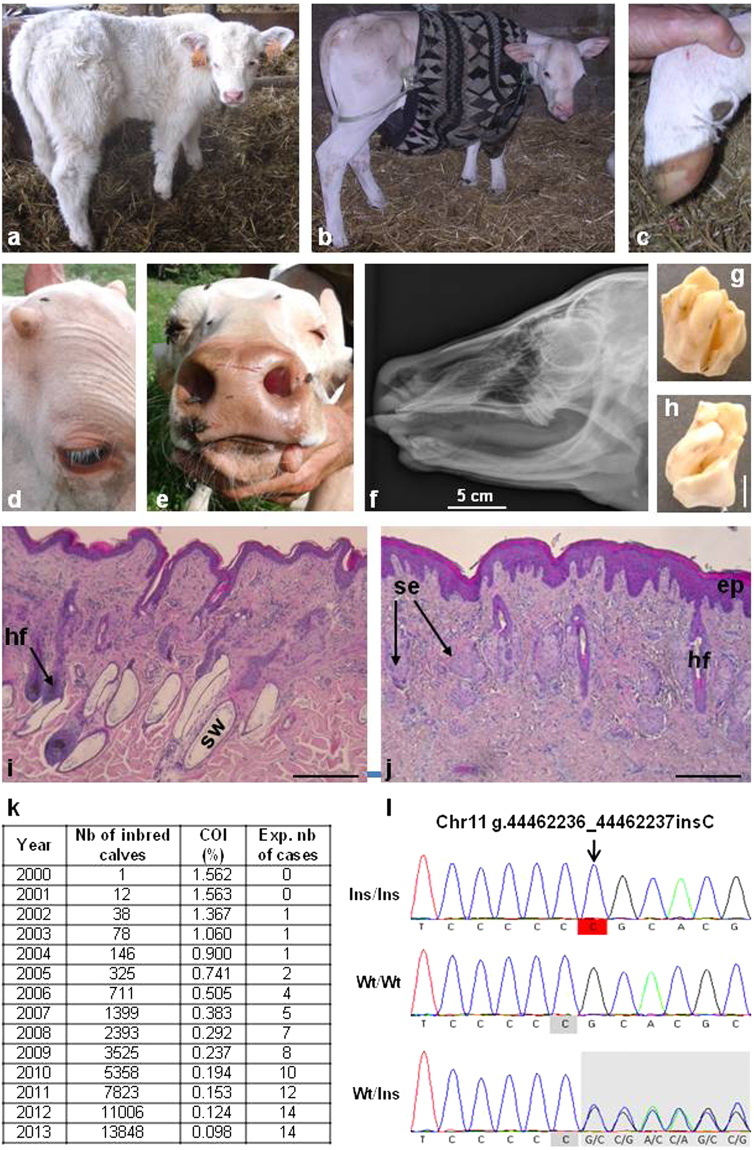
In humans, the clinical and molecular characterization of sporadic syndromes is often hindered by the small number of patients and the difficulty in developing animal models for severe dominant conditions. Here we show that the availability of large data sets of whole-genome sequences, high-density SNP chip genotypes and extensive recording of phenotype offers an unprecedented opportunity to quickly dissect the genetic architecture of severe dominant conditions in livestock. We report on the identification of seven dominant de novo mutations in CHD7, COL1A1, COL2A1, COPA, and MITF and exploit the structure of cattle populations to describe their clinical consequences and map modifier loci. Moreover, we demonstrate that the emergence of recessive genetic defects can be monitored by detecting de novo deleterious mutations in the genome of bulls used for artificial insemination. These results demonstrate the attractiveness of cattle as a model species in the post genomic era, particularly to confirm the genetic aetiology of isolated clinical case reports in humans.


Conflict of interest statement

The authors declare that they have no competing interests.

Figures

Figure 1
Figure 1
Brief overview of the dominant conditions studied. Glass-Eyed Albino (ad) animals trace back to a mutant heifer born in 1994. Clinical features include white coat color (a), deafness, and “glass-eyes” (b). Partial phenotypic reversion consisting in heterochromia irides (c) or black spots on the ears (d) are sometimes observed. Dominant Red (eh) is distinct from the traditional recessive red allele of the Melanocortin-1 receptor (MC1Re) found in Holstein. It emerged in 1980 with the birth of a red mutant heifer (MC1RD/D & DRDR/+) from parents which were homozygous for the dominant black allele (MC1RD/D & DR+/+). In adulthood MC1RD/D & DRDR/+ dominant red animals (e,f) display substantial variations in colour ranging from a light brown which is close to the MC1Re/e & DR+/+ recessive red coat (g), to a dark brown close to MC1RD/D & DR+/+ dominant black coat (h). Neurocristopathy (NC) was observed in half of the progeny of a mildly affected Montbéliarde bull. Its main symptoms include hypotonia (i), lack of balance and coordination in the days following birth, facial abnormalities, heart defects and growth delay. Osteogenesis imperfecta type 2 (OI), is characterized by brittle bones that are prone to fracture (e.g. hind limbs in j). It has been reported in 29% of the progeny of an unaffected Fleckvieh bull. Bulldog (BD) calf syndrome is a lethal form of chondrodysplasia characterized by a generalized shortening of long bones (k,l). BD has been reported in 4% of crossbred calves from a Charolais bull (BD1, k), in 21% of purebred calves from a Holstein sire (BD2, l), and in an isolated case born from Holstein parents (BD3). Pictures e–h were generously provided by the breeding company Origenplus.
Figure 2
Figure 2
Bulldog calves show perfect genotype-phenotype correlations with humans affected by hypochondrogenesis/achondrogenesis type II. (ah) Phenotypic characteristics of a bulldog calf (here the COL2A1 p.G720S/+ calf BD3) which, like humans affected by hypochondrogenesis/achondrogenesis type II, shows abnormal endochondral ossification resulting in a general shortening of long bones. (a) dorsal view and (b) ventral view. Note the extremely short limbs and short head. Radiographs: (c) of the left hind limb and (d) the left front limb showing multiple short dysplastic bones, (e) of the head demonstrating a ventrally rotated and dysplastic splanchnocranium. (f) Cleft palate (palatoschisis). (g) Longitudinal section through columna showing multiple areas of marked spinal cord compression due to abnormal epiphyseal development. (h) The centre of the proximal epiphysis of the femur shows non organized hypertrophic chondrocytes and large vessels. Ossification is not present. (HE 75x). Clinical features for BD1 and BD2 are presented in Supplementary Figs 6 and 7. (i) Domain and region information for the α1 chain of type II collagen obtained from the UniProt database (http://www.uniprot.org/; accession number: P02459). (j) Multispecies alignment of the COL2A1 proteins from different vertebrate species. Note the perfect conservation of the glycine residues of the typical Gly-x-y structural motif, the mutation of which causes hypochondrogenesis/achondrogenesis type II in humans and bulldog calf syndrome in bovine (i.e. p.G600D for BD2, p.G720S for BD3, p.G960R for the cases presented in Daetwyler et al., and p.G996S for BD2). Protein sequences accession numbers in Ensembl are ENSBTAP00000017505, ENSP00000369889, ENSGALP00000035064, ENSACAP00000006225, ENSXETP00000043834 and ENSDARP00000091007.
Figure 3
Figure 3
Histological and gene expression analysis of skin biopsies from GEA and control animals. (ac) Hematoxylin-eosin-stained sections of ear skin biopsies. (a) Black skin from a control black and white Holstein animal. Melanocytes are located on the basal layer of epidermis (indicated by arrowheads) and brown melanin granules are disseminated within keratinocytes. (b) Melanocytes are visible, but not melanin in white skin from a GEA animal. (c) Black skin from a GEA animal with a revertant phenotype. Melanin is present in the epidermis. Note the presence of both black and white hair. (d and e) Hematoxylin-eosin-safran stained sections of eyes from GEA and control animals. Note the complete lack of pigmentation of the choroid from the GEA animal (d) as compared with control (e). (fh) Transmission Electronic Microscopy pictures of skin from wild-type (f) and from a GEA animal (g). Melanocytes (arrowheads) are easily distinguished from the surrounding keratinocytes. Numerous melanosomes lie in the wild-type keratinocytes, while only a few abnormal ones are observed in the mutant skin. (h) Detail of a GEA keratinocyte cytoplasm containing abnormal melanosomes (arrowhead). (i) Relative expression quantification of melanogenic genes in the skin of wild-type and GEA Holstein animals. MITF-M expression is similar in black skin from control animals (n = 3) and in unpigmented (white) and revertant skin (black) from GEA animals (n = 3), showing that the regulation of expression of the melanogenic genes is not a consequence of a downregulation of MITF-M in mutant animals. Melanogenic genes (TYR, TYRP1, and PMEL) are not expressed in GEA white skin, with the exception of DCT which has already been shown to be regulated by MITF-independent mechanisms. In all black skins sampled from GEA revertant animals, the expression of melanogenic genes is partially restored. Scale bars represent 50 µM in (a) and (b); 100 µM in (ce); 5 µM in (f,g); and 1 µM in (h).
Figure 4
Figure 4
A large bovine half-sib pedigree allows the mapping of modifier loci for CHARGE syndromes. (ae) Presentation in CHD7 p.K594AfsX29/+ cattle of the different symptoms which gave rise to the CHARGE acronym in humans (C-coloboma (of the eyes), H-heart disease, A-atresia choanae, R-retarded growth and retarded development and/or central nervous system anomalies, G-genital and/or urinary anomalies, and E-ear anomalies and/or deafness). (a) iris coloboma highlighted by an arrowhead. (b) Heart showing tetralogy of Fallot; note the dextroposition of the aorta. (c) Renal cyst and its section. (d) Ears from an affected heifer showing abnormalities of the right pinna. Atresia choanae is not presented here but was demonstrated by the lack of revulsive reaction of certain animals after smelling alcohol. (e) Picture (at four years old) and scores (at 9 months old) of the mutant sire “Etsar” (MONFRAM002528725202) for 14 morphological traits expressed in percentiles with respect to distributions observed in 467 young Montbéliarde bulls raised by the same breeding company with the same protocol. Other symptoms are presented in Supplementary Note 2, Supplementary Figs 12 and 13 and Supplementary Tables 6–7). (f) Mapping on chromosome 24 of a locus influencing the clinical features of CHARGE syndrome (see Methods). The red line indicates the chromosome-wide empirical significance threshold of 0.05 as determined by 10 000 permutations of phenotype data (see Methods). Seven consecutive windows of ten markers from BTB-00883964 to ARS-BFGL-NGS-1731 (Chr24:29,132,144–29,762,125) had an empirical p-value of 0.03. Information on the gene content of the corresponding region is presented.
Figure 5
Figure 5
Characterization of the effect of bovine p.R160C substitution in COPA. (a) Domain information for COPA obtained from the UniProt database (http://www.uniprot.org/; accession numbers: Q27954 and P53621) and localization of the natural mutations described in cattle (p.R160C) and in human (p.K230N, p.R233H, p.E241K, p.D243G). The coordinates are identical in human and bovine orthologs due to the strong conservation of the WD40 domain that interacts with dilysine motifs. (b) Multispecies alignment of COPA proteins illustrating the strong conservation of the mutated residues among eukaryotes. Protein sequences accession numbers in Ensembl are respectively ENSBTAP0000000567, ENSP00000357048, FBpp0072694, ENSCINP00000018763, YDL145C, EAL73444 and Bra010674.1-P. (c) Ingenuity Pathway Analysis on genes that are markedly upregulated in the skin of dominant red versus dominant black Holstein cattle (fold change ≥2; FDR < 0.05; according to the RNAseq data produced by Dorshorst et al.; Supplementary Data 1) identifies the unfolded protein response and ubiquitin proteasome pathway as the two most significantly enriched canonical pathways (P < 0.001). (d) Functional annotation of downregulated genes in the same RNAseq data using Enrichr, reveals a significant enrichment (P < 0.05) for four “MGI Mammalian Phenotype Level 3′. Results of gene set enrichment analyses are presented in Supplementary Tables 8 and 9. Font size is proportional to the fold change in (c) and to the inverse of the fold change in (d). (eh) Immunohistochemical analysis with rabbit polyclonal antibodies against NFI transcription factors in epidermis from dominant black (MC1RD/D, DR+/+), recessive red (MC1Re/e, DR+/+), and dominant red (MC1RD/D, DRDR/+) Holstein animals. Scale bars correspond to 50 µm. NFI proteins are revealed with HRP-Green system, and all samples have been counterstained with hematoxylin. Staining is absent in negative control (a; recessive red), shows a nuclear and cytoplasmic distribution in dominant black (f) and recessive red (g) skins, while the distribution is only nuclear in dominant red skin (h). This observation was confirmed in samples from different animals (n = 3, 2 and 3 for fh respectively).
Figure 6
Figure 6
Identification of a de novo mutation causing recessive anhidrotic ectodermal dysplasia (AED) before the emergence of this defect in the population. (a) One-month old control calf. (b) One-month old AED affected calf showing generalized hypotrichosis. (c) Hoof of the same AED calf with no notable malformations. Note that in humans most AED patients have normal nails too. (d) Detail of the head of a five-month-old AED calf, showing eyelashes and normal development of horns. (e) Muzzle of the same calf showing normal vibrissae and extreme skin dryness due to the absence of nasolabial glands (see also Supplementary Fig. 19). (f) X-ray of the skull of a one-month old calf showing a complete absence of incisors and the presence of only one upper molar on each side. (g,h) Detail of one upper molar showing abnormal cusps and deep grooves. (i,j) Histological sections of skin biopsies from control (i) and AED (j) one-month-old calves. In AED animals, the epidermis (ep) is acanthotic (i.e. thickened). The number of hair follicles is greatly reduced and deep portions of them are atrophic or absent. Sweat glands (sw) are absent. Sebaceous glands (se) appear normal but a number of them are “orphan” (i.e. without associated hair follicle). Scale bars represent 500 µm. (k) Yearly estimation of the number of calves predicted to be born homozygous for the g.44462236_44462237insC mutation on chromosome 11 which is predicted to cause a frameshift and premature truncation of EDAR. The total numbers of calves with both parents related to primo-mutant bull Invincible, as well as their coefficient of inbreeding (COI) are also presented. (l) Electrophoregrams of a homozygous (Ins/Ins) AED affected calf, a homozygous wild-type (Wt/Wt) animal and a heterozygous carrier of the mutation (Wt/Ins).

References

    1. Simmons D. The Use of Animal Models in Studying Genetic Disease: Transgenesis and Induced Mutation. Nature Education. 2008;1:70.
    1. Rodríguez-Seguí S, Akerman I, Ferrer J. GATA believe it: new essential regulators of pancreas development. J. Clin. Invest. 2012;122:3469–3471. doi: 10.1172/JCI65751. - DOI - PMC - PubMed
    1. Andersson L. Molecular consequences of animal breeding. Curr. Opin. Genet. Dev. 2013;23:295–301. doi: 10.1016/j.gde.2013.02.014. - DOI - PubMed
    1. Daetwyler HD, et al. Whole-genome sequencing of 234 bulls facilitates mapping of monogenic and complex traits in cattle. Nat. Genet. 2014;46:858–865. doi: 10.1038/ng.3034. - DOI - PubMed
    1. Li H, et al. The Sequence Alignment/Map format and SAMtools. Bioinformatics. 2009;25:2078–2079. doi: 10.1093/bioinformatics/btp352. - DOI - PMC - PubMed

Publication types