

Power frequency magnetic field promotes a more malignant phenotype in neuroblastoma cells via redox-related mechanisms
- PMID: 28904402
- PMCID: PMC5597619
- DOI: 10.1038/s41598-017-11869-8
Power frequency magnetic field promotes a more malignant phenotype in neuroblastoma cells via redox-related mechanisms
Abstract
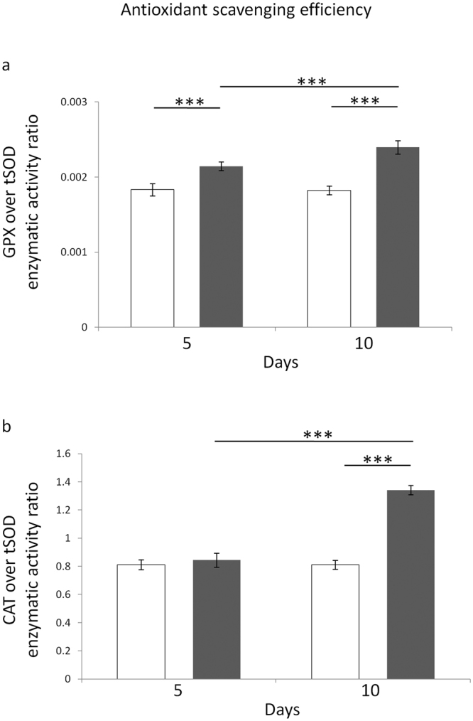
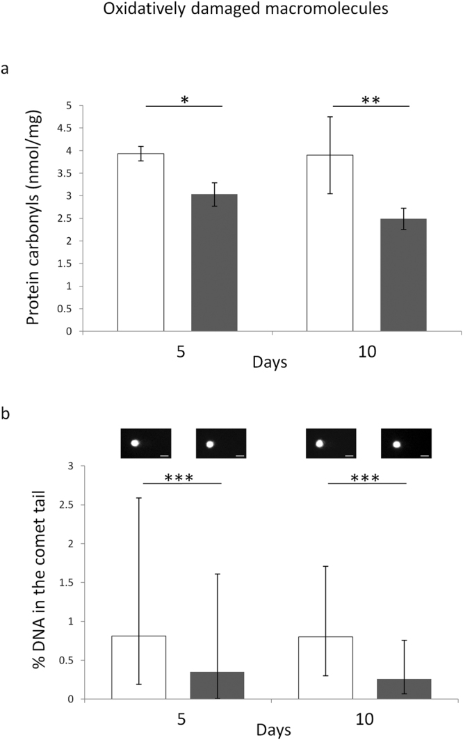
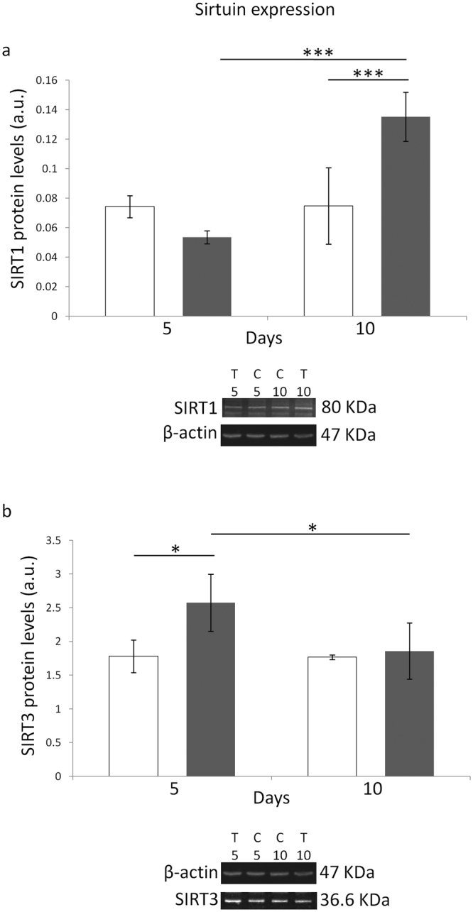
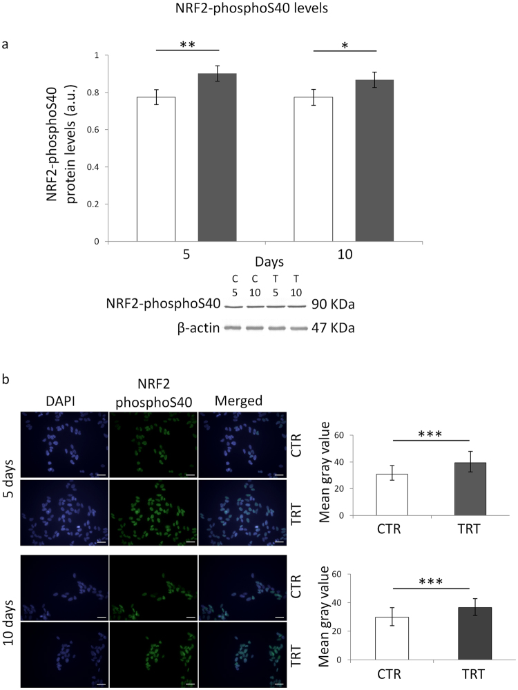
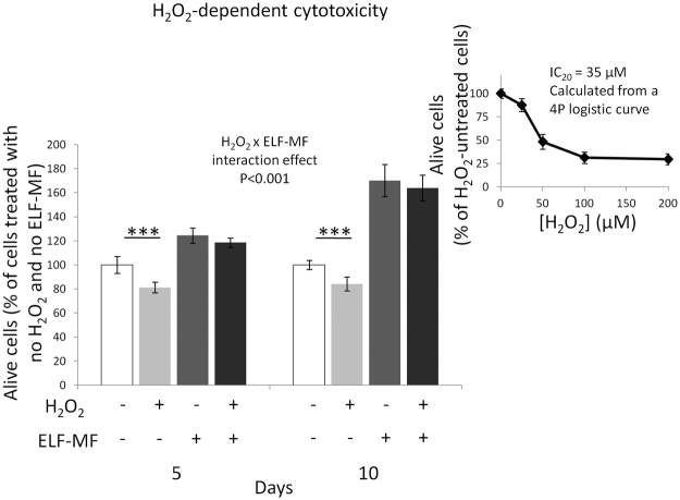
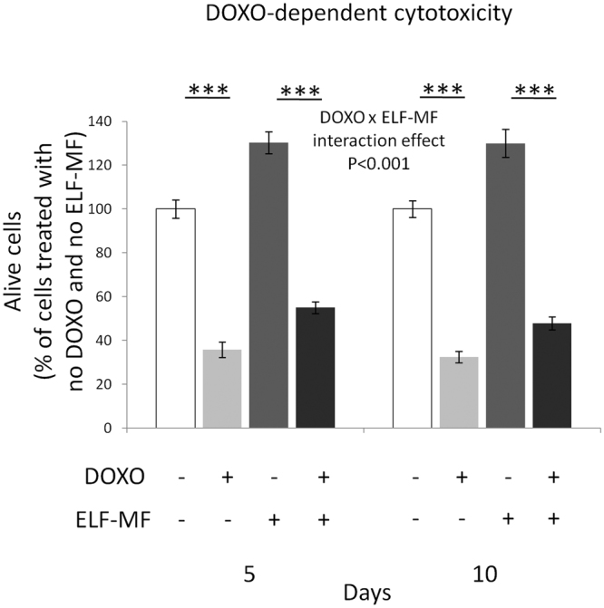
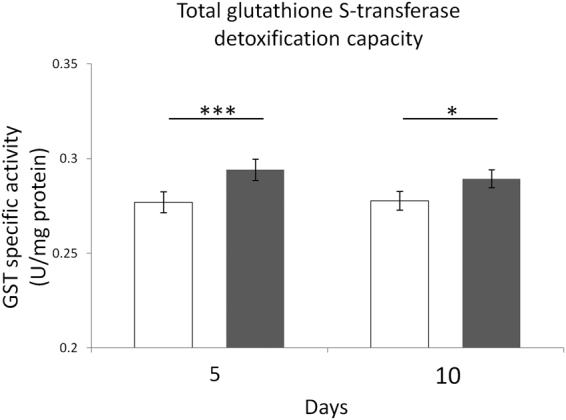
In accordance with the classification of the International Agency for Research on Cancer, extremely low frequency magnetic fields (ELF-MF) are suspected to promote malignant progression by providing survival advantage to cancer cells through the activation of critical cytoprotective pathways. Among these, the major antioxidative and detoxification defence systems might be targeted by ELF-MF by conferring cells significant resistance against clinically-relevant cytotoxic agents. We investigated whether the hyperproliferation that is induced in SH-SY5Y human neuroblastoma cells by a 50 Hz, 1 mT ELF magnetic field was supported by improved defence towards reactive oxygen species (ROS) and xenobiotics, as well as by reduced vulnerability against both H2O2 and anti-tumor ROS-generating drug doxorubicin. ELF-MF induced a proliferative and survival advantage by activating key redox-responsive antioxidative and detoxification cytoprotective pathways that are associated with a more aggressive behavior of neuroblastoma cells. This was coupled with the upregulation of the major sirtuins, as well as with increased signaling activity of the erythroid 2-related nuclear transcription factor 2 (NRF2). Interestingly, we also showed that the exposure to 50 Hz MF as low as 100 µT may still be able to alter behavior and responses of cancer cells to clinically-relevant drugs.
Conflict of interest statement
The authors declare that they have no competing interests.
Figures
Similar articles
-
Extremely Low-Frequency Magnetic Fields and Redox-Responsive Pathways Linked to Cancer Drug Resistance: Insights from Co-Exposure-Based In Vitro Studies.Front Public Health. 2018 Feb 23;6:33. doi: 10.3389/fpubh.2018.00033. eCollection 2018. Front Public Health. 2018. PMID: 29527520 Free PMC article. Review.
-
Improved Mitochondrial and Methylglyoxal-Related Metabolisms Support Hyperproliferation Induced by 50 Hz Magnetic Field in Neuroblastoma Cells.J Cell Physiol. 2016 Sep;231(9):2014-25. doi: 10.1002/jcp.25310. Epub 2016 Jan 28. J Cell Physiol. 2016. PMID: 26757151
-
Induction of genomic instability, oxidative processes, and mitochondrial activity by 50Hz magnetic fields in human SH-SY5Y neuroblastoma cells.Mutat Res. 2014 Feb;760:33-41. doi: 10.1016/j.mrfmmm.2013.12.002. Epub 2013 Dec 26. Mutat Res. 2014. PMID: 24374227
-
Evidences of plasma membrane-mediated ROS generation upon ELF exposure in neuroblastoma cells supported by a computational multiscale approach.Biochim Biophys Acta Biomembr. 2019 Aug 1;1861(8):1446-1457. doi: 10.1016/j.bbamem.2019.06.005. Epub 2019 Jun 11. Biochim Biophys Acta Biomembr. 2019. PMID: 31199897
-
Exposure of the SH-SY5Y Human Neuroblastoma Cells to 50-Hz Magnetic Field: Comparison Between Two-Dimensional (2D) and Three-Dimensional (3D) In Vitro Cultures.Mol Neurobiol. 2021 Apr;58(4):1634-1649. doi: 10.1007/s12035-020-02192-x. Epub 2020 Nov 24. Mol Neurobiol. 2021. PMID: 33230715 Free PMC article.
Cited by
-
Protective effect of 1950 MHz electromagnetic field in human neuroblastoma cells challenged with menadione.Sci Rep. 2018 Sep 5;8(1):13234. doi: 10.1038/s41598-018-31636-7. Sci Rep. 2018. PMID: 30185877 Free PMC article.
-
Cellular stress response to extremely low-frequency electromagnetic fields (ELF-EMF): An explanation for controversial effects of ELF-EMF on apoptosis.Cell Prolif. 2021 Dec;54(12):e13154. doi: 10.1111/cpr.13154. Epub 2021 Nov 6. Cell Prolif. 2021. PMID: 34741480 Free PMC article. Review.
-
Extremely Low-Frequency Magnetic Fields and Redox-Responsive Pathways Linked to Cancer Drug Resistance: Insights from Co-Exposure-Based In Vitro Studies.Front Public Health. 2018 Feb 23;6:33. doi: 10.3389/fpubh.2018.00033. eCollection 2018. Front Public Health. 2018. PMID: 29527520 Free PMC article. Review.
-
Magnetic stimulation of the angiogenic potential of mesenchymal stromal cells in vascular tissue engineering.Sci Technol Adv Mater. 2021 Jun 28;22(1):461-480. doi: 10.1080/14686996.2021.1927834. eCollection 2021. Sci Technol Adv Mater. 2021. PMID: 34248420 Free PMC article.
-
Effects of osteoblast-derived extracellular vesicles on aggressiveness, redox status and mitochondrial bioenergetics of MNNG/HOS osteosarcoma cells.Front Oncol. 2022 Dec 5;12:983254. doi: 10.3389/fonc.2022.983254. eCollection 2022. Front Oncol. 2022. PMID: 36544705 Free PMC article.
References
-
- IARC Working Group on the Evaluation of Carcinogenic Risks to Humans. IARC Monogr. Eval. Carcinog. Risks Hum. 80, 1–395 (2002). - PubMed
-
- Poulletier de Gannes, F., Lagroye, I. & Veyret, B. D3 - Report on the analysis of risks associated to exposure to EMF: in vitro and in vivo (animals) studies. European Health Risk Assessment Network on Electromagnetic Fields Exposure (EFHRAN). Available at http://efhran.polimi.it/docs/IMS-EFHRAN_09072010.pdf (2010).
Publication types
MeSH terms
Substances
LinkOut - more resources
Full Text Sources
Other Literature Sources
Medical
