. 2017 Sep 9:7:47.
doi: 10.1186/s13578-017-0174-2. eCollection 2017.

Genome editing of the HIV co-receptors CCR5 and CXCR4 by CRISPR-Cas9 protects CD4+ T cells from HIV-1 infection

Affiliations

Genome editing of the HIV co-receptors CCR5 and CXCR4 by CRISPR-Cas9 protects CD4+ T cells from HIV-1 infection

Zhepeng Liu et al. Cell Biosci. .

Abstract

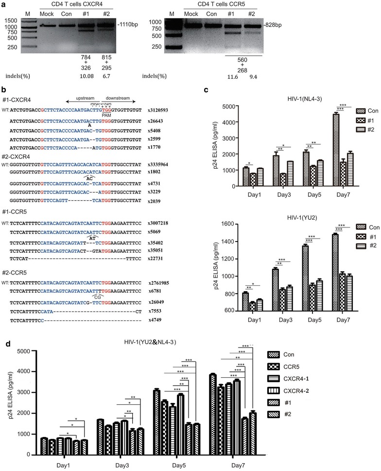
Background: The main approach to treat HIV-1 infection is combination antiretroviral therapy (cART). Although cART is effective in reducing HIV-1 viral load and controlling disease progression, it has many side effects, and is expensive for HIV-1 infected patients who must remain on lifetime treatment. HIV-1 gene therapy has drawn much attention as studies of genome editing tools have progressed. For example, zinc finger nucleases (ZFN), transcription activator like effector nucleases (TALEN) and clustered regularly interspaced short palindromic repeats (CRISPR)-Cas9 have been utilized to successfully disrupt the HIV-1 co-receptors CCR5 or CXCR4, thereby restricting HIV-1 infection. However, the effects of simultaneous genome editing of CXCR4 and CCR5 by CRISPR-Cas9 in blocking HIV-1 infection in primary CD4+ T cells has been rarely reported. Furthermore, combination of different target sites of CXCR4 and CCR5 for disruption also need investigation.

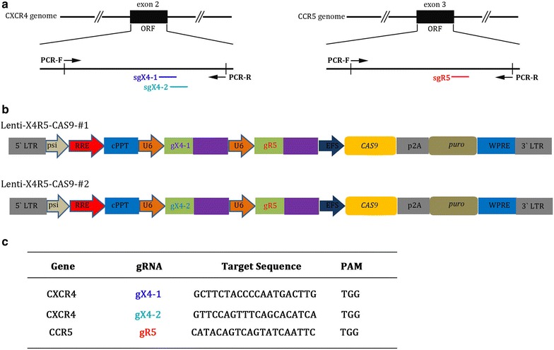
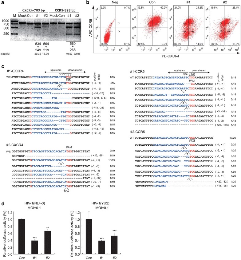
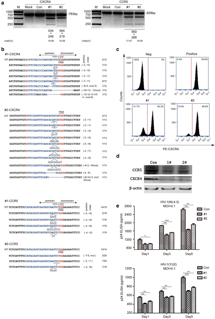
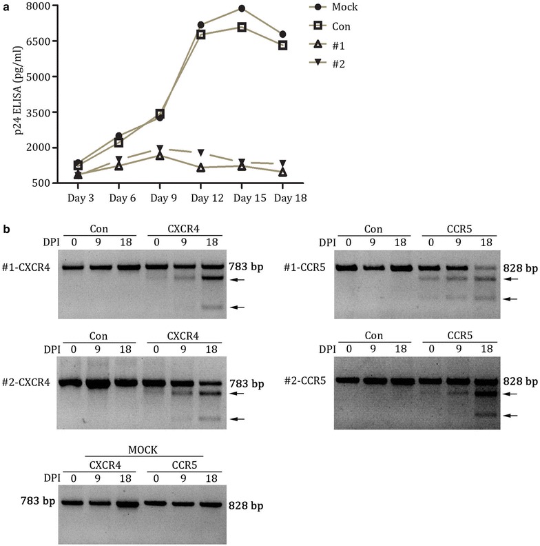
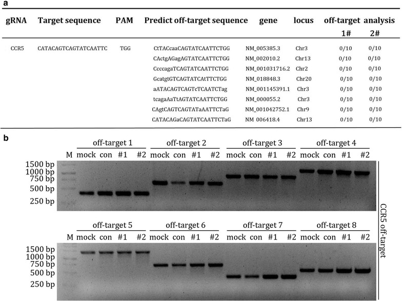
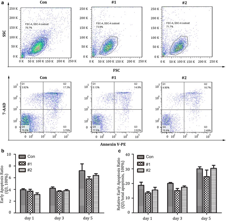
Results: In this report, we designed two different gRNA combinations targeting both CXCR4 and CCR5, in a single vector. The CRISPR-sgRNAs-Cas9 could successfully induce editing of CXCR4 and CCR5 genes in various cell lines and primary CD4+ T cells. Using HIV-1 challenge assays, we demonstrated that CXCR4-tropic or CCR5-tropic HIV-1 infections were significantly reduced in CXCR4- and CCR5-modified cells, and the modified cells exhibited a selective advantage over unmodified cells during HIV-1 infection. The off-target analysis showed that no non-specific editing was identified in all predicted sites. In addition, apoptosis assays indicated that simultaneous disruption of CXCR4 and CCR5 in primary CD4+ T cells by CRISPR-Cas9 had no obvious cytotoxic effects on cell viability.

Conclusions: Our results suggest that simultaneous genome editing of CXCR4 and CCR5 by CRISPR-Cas9 can potentially provide an effective and safe strategy towards a functional cure for HIV-1 infection.

Keywords: AIDS; CCR5 and CXCR4 simultaneous; CRISPR-Cas9; HIV-1.


Figures

Fig. 1
Fig. 1
Schematic diagram of sgRNA of CXCR4 and CCR5 targets and vector construction. a Schematic of the CXCR4 and CCR5 coding region in genomic DNA sequences targeted by lenti-X4R5-Cas9-#1,#2. b Structure of lenti-X4R5-Cas9-#1,#2 vectors expressing Cas9 and dual sgRNA. c gRNA sequences used in lenti-X4R5-Cas9-#1,#2 vectors
Fig. 2
Fig. 2
Disruption of CXCR4 and CCR5 protects TZM-bl cells from HIV-1 infection. a T7E1 assay for genome level cleavage efficacy by lenti-X4R5-Cas9-#1,#2 in TZM-bl. b Expression of CXCR4 or CCR5 in TZM-bl cell line transfected with lenti-X4R5-Cas9 by lipo2000 transfection reagent were analyzed with flow cytometry. c On-target analysis of the cleavage on target sites. d lenti-X4R5-Cas9 transfected TZM-bl cell line challenged with HIV-1NL4-3 or HIV-1YU-2 (3 days post infection). The data shown were the mean ± SD of three independent experiments. *P < 0.05; **P < 0.01; ***P < 0.001; t test
Fig. 3
Fig. 3
Jurkat T cell line modified by X4R5-Cas9 lentivirus antagonized HIV infection. a T7E1 assay identification of packaged X4R5-Cas9 lentivirus mediated cleavage at the genome level. b On-target analysis of each target in Jurkat T cells. c Flow cytometry analysis of CXCR4 expression on the cell surface. Jurkat T cell line was transduced with X4R5-Cas9 lentivirus at MOI = 40. CCR5 surface expression detection was excluded because of its low expression on Jurkat T cells. d Detection of protein level of CXCR4 and CCR5 after Jurkat cells were transduced with X4R5-Cas9 lentivirus. e HIV-1 titer change detected by p24 gag ELISA from day 1 to day 5 post-infection. The data shown were the mean ± SD of three independent experiments. *P < 0.05; **P < 0.01; ***P < 0.001; t test
Fig. 4
Fig. 4
X4R5-Cas9 lentivirus modified Jurkat T cells were enriched after CXCR4-tropic (NL4-3) and CCR5-tropic (YU-2) HIV-1 challenge. a HIV replication in X4R5-Cas9 lentivirus modified as well as mock and control Jurkat T cells infected with X4-tropic and R5-tropic HIV-1 concurrently. Values represent the mean of duplicate infections. b cleavage analysis of CXCR4 and CCR5 by T7 endonuclease 1 in mock, control, lenti-X4R5-Cas9-#1 and lenti-X4R5-Cas9-#2 group at 0, 9 and 18 days after HIV-1 challenge. The lower migrating bands (indicated by arrows) in each lane indicate the disrupted CXCR4 and CCR5 alleles. DPI days post infection
Fig. 5
Fig. 5
lenti-X4R5-Cas9 modified primary CD4+ T cell resists HIV challenge. a T7E1 analysis of CXCR4 and CCR5 disruption. b Deep sequencing analysis of typical NHEJ (indels) of related targets. c lenti-X4R5-Cas9 modified CD4+ T cell challenged with HIV-1NL4-3 or HIV-1YU-2. d lenti-X4R5-Cas9 modified CD4+ T cell exposed to dual-tropic HIV-1 variants (NL4-3 & YU-2, 1:1). The CCR5, CXCR4-1, CXCR4-2 represent single disruption of CCR5 or CXCR4, which use the same corresponding gRNAs used in lenti-X4R5-Cas9-#1 or #2. The data shown were the mean ± SD of three independent experiments. *P < 0.05; **P < 0.01; ***P < 0.001; t test
Fig. 6
Fig. 6
off-target analysis of CCR5. a Mutation frequency analysis at predicted off-target sites of CCR5. The off-target sites were predicted and aligned with the human genome. The sites were amplified and cloned into T-vector and subjected to sequencing. b T7E1 analysis of all predicted off-target sites
Fig. 7
Fig. 7
Apoptosis analysis after lenti-X4R5-Cas9 modification in primary CD4+ T cells. a Annexin V and 7-AAD were utilized to stain modified CD4+T at 1,3 and 5 days post nucleofection with flow cytometry. Necrotic cells (Annexin V-/7AAD +), necrotic or late apoptotic cells (Annexin V +/7AAD +); early apoptotic cells (Annexin V +/7AAD-); viable cells (Annexin V-/7AAD-). b Early apoptosis ratio at day 1,3,5 post gene disruption. c relative early apoptosis ratio (Q3/total none viable cells). The total none viable cells = Q2 + Q3. The data shown were the mean ± SD of three independent experiments. *P < 0.05

References

    1. Update on acquired immune deficiency syndrome (AIDS)–United States. MMWR. 1982;31(37):507-8, 13-4. (Epub 1982/09/24). - PubMed
    1. Bayer R, Levine C, Murray TH. Guidelines for confidentiality in research on AIDS. AIDS Res. 1983;1(4):275–297. doi: 10.1089/aid.1.1983.1.275. - DOI - PubMed
    1. Cunningham AL, Donaghy H, Harman AN, Kim M, Turville SG. Manipulation of dendritic cell function by viruses. Curr Opin Microbiol. 2010;13(4):524–529. doi: 10.1016/j.mib.2010.06.002. - DOI - PubMed
    1. Klasse PJ. The molecular basis of HIV entry. Cell Microbiol. 2012;14(8):1183–1192. doi: 10.1111/j.1462-5822.2012.01812.x. - DOI - PMC - PubMed
    1. Chan DC, Kim PS. HIV entry and its inhibition. Cell. 1998;93(5):681–684. doi: 10.1016/S0092-8674(00)81430-0. - DOI - PubMed