Dermatophagoides pteronyssinus lytFM encoding an NlpC/P60 endopeptidase is also present in mite-associated bacteria that express LytFM variants
- PMID: 28904857
- PMCID: PMC5586350
- DOI: 10.1002/2211-5463.12263
Dermatophagoides pteronyssinus lytFM encoding an NlpC/P60 endopeptidase is also present in mite-associated bacteria that express LytFM variants
Abstract
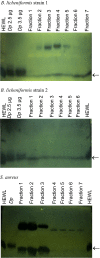
The bodies and faecal pellets of the house dust mite (HDM), Dermatophagoides pteronyssinus, are the source of many allergenic and nonallergenic proteins. One of these, the 14-kDa bacteriolytic enzyme LytFM, originally isolated from the spent HDM growth medium, may contribute to bacteriolytic activity previously detected by zymography at 14 kDa in the culture supernatants of some bacterial species isolated from surface-sterilised HDM. Based on previously reported findings of lateral gene transfer between microbes and their eukaryotic hosts, we investigated the presence of lytFM in the genomes of nine Gram-positive bacteria from surface-sterilised HDM, and the expression by these isolates of LytFM and its variants LytFM1/LytFM2. The lytFM gene was detected by PCR in the genomes of three of the isolates: Bacillus licheniformis strain 1, B. licheniformis strain 2 and Staphylococcus aureus. Expression of the variant LytFM1 was detected in culture supernatants of these bacteria by mass spectrometry (MS) and ELISA, and the bacterial LytFM proteins were shown by zymography to be able to hydrolyse peptidoglycan. Our previous reports of LytFM homologues in other mite species and their phylogenetic analysis had suggested that they originated from a common mite ancestor. The phylogenetic analysis reported herein and the detection of other D. pteronyssinus proteins by MS in the culture supernatants of the three isolates that secreted LytFM1 further support the hypothesis of lateral gene transfer to the bacterial endosymbionts from their HDM host. The complete sequence homology observed between the genes amplified from the microbes and those in their eukaryotic host indicated that the lateral gene transfer was an event that occurred recently.
Keywords: D,L‐endopeptidase; Dermatophagoides pteronyssinus; LytFM; NlpC/P60; host; microbes.
Figures


Similar articles
-
House dust mites possess a polymorphic, single domain putative peptidoglycan d,l endopeptidase belonging to the NlpC/P60 Superfamily.FEBS Open Bio. 2015 Sep 16;5:813-23. doi: 10.1016/j.fob.2015.09.004. eCollection 2015. FEBS Open Bio. 2015. PMID: 26566476 Free PMC article.
-
Skin-associated Bacillus, staphylococcal and micrococcal species from the house dust mite, Dermatophagoides pteronyssinus and bacteriolytic enzymes.Exp Appl Acarol. 2013 Dec;61(4):431-47. doi: 10.1007/s10493-013-9712-8. Epub 2013 Jun 20. Exp Appl Acarol. 2013. PMID: 23783892
-
PCR detection of the 14.5 antibacterial NlpC/P60-like Dermatophagoides pteronyssinus protein in Dermatophagoides farinae (Acari: Pyroglyphidae).J Med Entomol. 2013 Jul;50(4):931-3. doi: 10.1603/me13027. J Med Entomol. 2013. PMID: 23926795
-
Isolation and characterisation of a 13.8-kDa bacteriolytic enzyme from house dust mite extracts: homology with prokaryotic proteins suggests that the enzyme could be bacterially derived.FEMS Immunol Med Microbiol. 2002 Jun 3;33(2):77-88. doi: 10.1111/j.1574-695X.2002.tb00576.x. FEMS Immunol Med Microbiol. 2002. PMID: 12052562
-
A combined transcriptome and proteome analysis extends the allergome of house dust mite Dermatophagoides species.PLoS One. 2017 Oct 5;12(10):e0185830. doi: 10.1371/journal.pone.0185830. eCollection 2017. PLoS One. 2017. PMID: 28982170 Free PMC article.
Cited by
-
Origins of peptidases.Biochimie. 2019 Nov;166:4-18. doi: 10.1016/j.biochi.2019.07.026. Epub 2019 Aug 1. Biochimie. 2019. PMID: 31377195 Free PMC article.
-
Peptidoglycan NlpC/P60 peptidases in bacterial physiology and host interactions.Cell Chem Biol. 2023 May 18;30(5):436-456. doi: 10.1016/j.chembiol.2022.11.001. Epub 2022 Nov 22. Cell Chem Biol. 2023. PMID: 36417916 Free PMC article. Review.
-
Comparative Genomics Reveals Insights into the Divergent Evolution of Astigmatic Mites and Household Pest Adaptations.Mol Biol Evol. 2022 May 3;39(5):msac097. doi: 10.1093/molbev/msac097. Mol Biol Evol. 2022. PMID: 35535514 Free PMC article.
-
The Protozoan Trichomonas vaginalis Targets Bacteria with Laterally Acquired NlpC/P60 Peptidoglycan Hydrolases.mBio. 2018 Dec 11;9(6):e01784-18. doi: 10.1128/mBio.01784-18. mBio. 2018. PMID: 30538181 Free PMC article.
References
-
- Tovey ER, Chapman MD and Platts‐Mills TA (1981) Mite faeces are a major source of house dust allergens. Nature 289, 592–593. - PubMed
-
- Arlian LG, Bernstein IL, Geis DP, Vyszenski‐Moher DL, Gallagher JS and Martin B (1987) Investigations of culture medium‐free house dust mites. III. Antigens and allergens of body and fecal extract of Dermatophagoides farinae . J Allergy Clin Immunol 79, 457–466. - PubMed
-
- Stewart GA, Lake FR and Thompson PJ (1991) Faecally derived hydrolytic enzymes from Dermatophagoides pteronyssinus: physicochemical characterisation of potential allergens. Int Arch Allergy Appl Immunol 95, 248–256. - PubMed
-
- Arlian LG and Platts‐Mills TA (2001) The biology of dust mites and the remediation of mite allergens in allergic disease. J Allergy Clin Immunol 107, S406–S413. - PubMed
-
- Colloff MJ (2009) Dust mites In Identification and Taxonomy, Classification and Phylogeny (Colloff MJ, ed.), pp. 1–44. CSIRO Publishing, Melbourne, Vic., Australia.
LinkOut - more resources
Full Text Sources
Other Literature Sources
Research Materials

